Nuchal test in pregnancy
Nuchal translucency test Information | Mount Sinai
Nuchal translucency screening; NT; Nuchal fold test; Nuchal fold scan; Prenatal genetic screening; Down syndrome - nuchal translucency
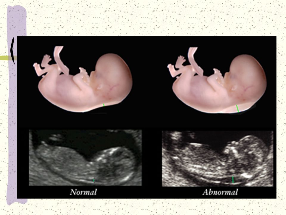
The nuchal translucency test measures the nuchal fold thickness. This is an area of tissue at the back of an unborn baby's neck. Measuring this thickness helps assess the risk for Down syndrome and other genetic problems in the baby.
How the Test is Performed
Your health care provider uses abdominal ultrasound (not vaginal) to measure the nuchal fold. All unborn babies have some fluid at the back of their neck. In a baby with Down syndrome or other genetic disorders, there is more fluid than normal. This makes the space look thicker.
A blood test of the mother is also done. Together, these two tests will tell if the baby could have Down syndrome or another genetic disorder.
How to Prepare for the Test
Having a full bladder will give the best ultrasound picture. You may be asked to drink 2 to 3 glasses of liquid an hour before the test. DO NOT urinate before your ultrasound.
How the Test will Feel
You may have some discomfort from pressure on your bladder during the ultrasound.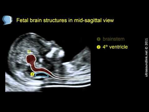 The gel used during the test may feel slightly cold and wet. You will not feel the ultrasound waves.
Why the Test is Performed
Your provider may advise this test to screen your baby for Down syndrome. Many pregnant women decide to have this test.
Nuchal translucency is usually done between the 11th and 14th week of pregnancy. It can be done earlier in pregnancy than amniocentesis. Amniocentesis is another test that checks for birth defects.
Normal Results
A normal amount of fluid in the back of the neck during ultrasound means it is very unlikely your baby has Down syndrome or another genetic disorder.
Nuchal translucency measurement increases with gestational age. This is the period between conception and birth. The higher the measurement compared to babies the same gestational age, the higher the risk is for certain genetic disorders.
The measurements below are considered low risk for genetic disorders:
- At 11 weeks -- up to 2 mm
- At 13 weeks, 6 days -- up to 2.8 mm
What Abnormal Results Mean
More fluid than normal in the back of the neck means there is a higher risk for Down syndrome, trisomy 18, trisomy 13, Turner syndrome, or congenital heart disease. But it does not tell for certain that the baby has Down syndrome or another genetic disorder.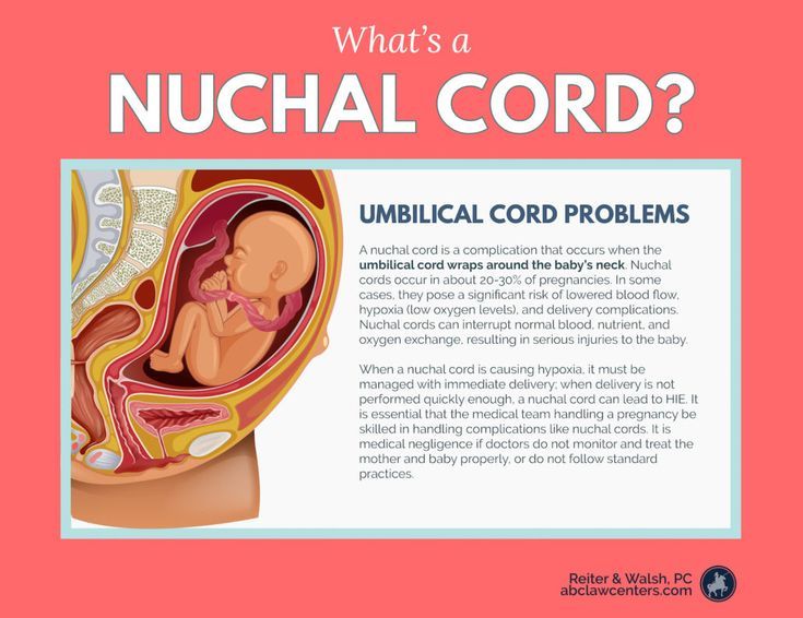
If the result is abnormal, other tests can be done. Most of the time, the other test done is amniocentesis.
Risks
There are no known risks from ultrasound.
Driscoll DA, Simpson JL. Genetic screening and diagnosis. In: Landon MB, Galan HL, Jauniaux ERM, et al, eds. Gabbe's Obstetrics: Normal and Problem Pregnancies. 8th ed. Philadelphia, PA: Elsevier; 2021:chap 10.
Walsh JM, D'Alton ME. Nuchal translucency. In: Copel JA, D'Alton ME, Feltovich H, et al, eds. Obstetric Imaging: Fetal Diagnosis and Care. 2nd ed. Philadelphia, PA: Elsevier; 2018:chap 45.
Wapner RJ, Dugoff L. Prenatal diagnosis of congenital disorders. In: Resnik R, Lockwood CJ, Moore TR, Greene MF, Copel JA, Silver RM, eds. Creasy and Resnik's Maternal-Fetal Medicine: Principles and Practice. 8th ed. Philadelphia, PA: Elsevier; 2019:chap 32.
Last reviewed on: 4/19/2022
Reviewed by: John D. Jacobson, MD, Department of Obstetrics and Gynecology, Loma Linda University School of Medicine, Loma Linda, CA. Also reviewed by David C. Dugdale, MD, Medical Director, Brenda Conaway, Editorial Director, and the A.D.A.M. Editorial team.
First Trimester Screening, Nuchal Translucency and NIPT
First trimester screening (FTS), nuchal translucency (NT) and noninvasive prenatal testing (NIPT) are prenatal tests that provide information on a developing baby’s risk for certain chromosomal differences (anomalies).
The screening is a blood test that evaluates substances in the blood (analytes), and NT is a sonogram that looks at nuchal translucency in the back of the fetal neck.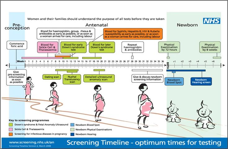 Noninvasive perinatal testing (NIPT) is a newer method that provides a result with a blood test only; a first trimester ultrasound is still recommended.
FTS, NT and NIPT provide data that can help assess if a fetus (developing baby) has one of three genetic anomalies:
- Down syndrome
- Trisomy 13
- Trisomy 18
These tests cannot diagnose these anomalies. But the data they provide help assess the likelihood that a fetus may have one of these conditions.
What You Need to Know
- The first trimester screening test (FTS) is blood work, and the nuchal translucency test is specialized imaging of the fetus using ultrasound.
- When the two tests are performed together, the combined data can help assess the risk of certain genetic conditions, but it cannot diagnose them.
- Noninvasive prenatal testing, or NIPT, is a new option that uses a blood test to look for signs of Down syndrome, trisomy 13 and trisomy 18 by analyzing free fragments of DNA in the bloodstream.

What is combined first trimester screening?
This is available to pregnant people from weeks 11 through 13 of pregnancy. It comprises a maternal blood test and a nuchal translucency test, which is ultrasound imaging of the fetus to look for clues that could affect the chances of certain genetic conditions. The test may be accompanied by genetic counseling.
The nuchal translucency ultrasound portion of combined first trimester screening is performed by specially credentialed sonographers.
FTS is not a diagnostic test, which means it cannot tell you for certain whether the fetus has Down syndrome, trisomy 13 or trisomy 18. Instead, the screening helps measure the probability that a fetus might have one of these conditions. Data from the ultrasound and blood test, together with the mother’s age, can provide information about whether the fetus is at an increased risk for one of these chromosomal disorders.
If the combined first trimester screening data show that there is a 1 in 250 chance ― or greater ― that the developing fetus has one of these conditions, your doctor may recommend further testing to rule them out.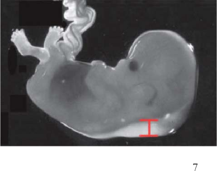
What is noninvasive prenatal testing (NIPT)?
NIPT is a different approach for identifying the risk that a fetus is affected by Down syndrome, trisomy 13 or trisomy 18. It consists of a blood test alone. A nuchal translucency ultrasound can still be performed, and it will not affect the NIPT results. Noninvasive prenatal testing can pick up tiny pieces of DNA in the mother’s bloodstream and analyze them for factors that would raise the risk of the fetus having a chromosomal difference.
In addition to Down syndrome and trisomies 13 and 18, NIPT can detect clues associated with other abnormal chromosomes, such as Turner syndrome, Klinefelter syndrome and triple X syndrome. NIPT can also predict the fetus’s sex with high accuracy.
Nuchal translucency and NIPT: Why are they performed?
Down syndrome, trisomy 13 and trisomy 18 are chromosomal disorders that cause intellectual disability and birth defects in children who are born with them. Anyone can have a baby with these chromosomal abnormalities, but the chance increases with the mother’s age.
Babies with Down syndrome (trisomy 21) have an extra 21st chromosome, which may cause a range of signs and symptoms, including intellectual disability and various medical complications involving the heart, digestive tract and other organ systems.
Trisomy 18 (having an extra 18th chromosome) and trisomy 13 (having an extra 13th chromosome) are more severe disorders that cause profound intellectual disability and severe birth defects in many organ systems. Sadly, few babies with trisomies 13 or 18 survive more than a few months.
Preparing for First Trimester Screening
These screenings include a simple blood test, with or without ultrasound. Neither the blood test nor the ultrasound is invasive, so no special preparations are necessary.
What happens during combined FTS?
The blood test part of the test takes a sample of the mother’s blood. The sample is analyzed to check levels of three chemicals to see if they are higher or lower than average, which can indicate a higher or lower chance of Down syndrome, trisomy 13 or trisomy 18:
- Free beta-human chorionic gonadotropin (hCG)
- Pregnancy-associated plasma protein-A (PAPP-A)
- Alpha-fetoprotein (AFP)
The nuchal translucency will examine:
- The progress of your pregnancy
- The fluid underneath the skin along the back of the fetus’s neck, called the nuchal translucency, or nuchal fold.
 In some pregnancies, when the fetus has Down syndrome, trisomy 13 or trisomy 18, there is extra fluid behind the neck. A larger-than-expected nuchal fold is associated with other birth defects such as congenital heart defects and skeletal problems.
 - Presence of the fetus’s nasal bone. While a nasal bone may be absent in some fetuses with a chromosomal abnormality, most with this finding are normal.
Combining your age-related risk with the nuchal translucency measurement, nasal bone data and bloodwork provides one risk result for Down syndrome and a separate risk result for trisomy 13 or trisomy 18. Your obstetrician will get your screening results in about one week.
How accurate is FTS?
Because these are screening tests, a positive result (showing an increased risk) does not mean that your baby has one of these conditions. It indicates that further diagnostic tests are options for you to consider. Also, a negative or normal result (one that shows a decreased risk) does not mean a chromosomal abnormality is definitely not present.
The combined first trimester screening’s detection rate is approximately 96% for pregnancies in which the baby has Down syndrome, and it is somewhat higher for pregnancies with trisomy 13 or trisomy 18. A nuchal translucency ultrasound can be performed without the bloodwork, but the detection rate is reduced to about 70%.
This screen is not designed to provide information about the possibility of other chromosomal conditions, but it does have limited utility for screening for some other genetic syndromes, genetic disorders and birth defects.
For NIPT, the detection rate depends on the laboratory, but for high-risk mothers pregnant with one baby, the accuracy rate ranges between 90% and 99%, with false positive rates of less than 1%.
What if the screening shows an increased risk for one of the conditions?
If the screening results indicate that your baby is at an increased risk for Down syndrome or trisomy 13 or 18, this does not mean that one of these conditions is present, but this information can help your doctor decide whether further testing is right for you.
A genetic counselor is available to go over your results and to discuss additional screening and testing options, such as chorionic villus sampling (CVS) and amniocentesis. CVS and amniocentesis are more invasive diagnostic procedures that detect a chromosomal abnormality with greater than 99% accuracy.
What is a quad screen?
The second trimester maternal serum screening test, also known as the quad screen, is performed between 16 and 20 weeks, and measures chemicals in the mother’s blood. Like the first trimester screening, results from a second trimester quad screen can be used to statistically adjust a woman’s age-related risk for Down syndrome and trisomy 18 (but not trisomy 13).
In addition, the alpha-fetoprotein (AFP) portion of the screen in the second trimester can identify pregnancies at an increased risk for open neural tube defects such as spina bifida. Quad screening is not recommended if combined first trimester screening has already been performed.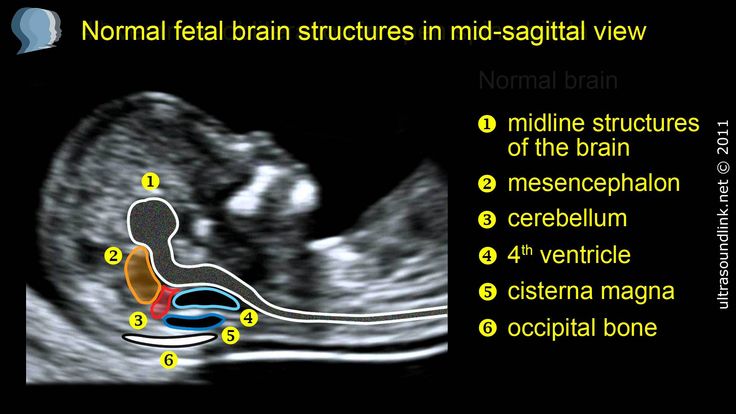 However, AFP can be drawn as an independent test to screen for spina bifida.
3 prenatal screening during pregnancy
Screening examinations conducted at different stages of pregnancy are the most accessible and at the same time comprehensive way to monitor intrauterine development of the fetus, as well as the course of the pregnancy itself. Laboratory data and ultrasound examination, carried out at certain periods of bearing a child, complement each other in terms of information. They allow specialists to make fairly accurate predictions about the risks of developing anomalies in the fetus or violations of the normal course of pregnancy. nine0003
Here are just the main positive aspects of conventional screening:
- Low cost of screening;
- Possibility of mass carrying out with almost 100% coverage of pregnant women;
- Ease of performing the necessary manipulations;
- No risk to mother or fetus;
- For specialists - ease of interpretation of the results;
- Possibility of objective risk assessment.
With such advantages, there is one serious drawback, namely, the insufficient reliability of the data obtained. The accuracy in assessing the risks of fetal anomalies is about 85%. nine0003
Screening data that is very different from normal values is an indication for invasive diagnostics. By the third trimester of pregnancy, these issues are usually resolved in one way or another, and the ultrasound performed at this time makes it possible to clarify the structural features of the fetal skeleton, extraembryonic structures, as well as the characteristics of the course of pregnancy.
In its essence, it is final and often important decisions are made on the tactics of obstetric care based on its results. nine0003
The principle of comparing the obtained results with the "reference" values remains the same with the third screening. Naturally, the evaluation of the results obtained and the interpretation of the data should be carried out exclusively by a specialist. The main indicators of 3 screening during pregnancy are shown in the table below.
| Screening by week of pregnancy | |||||
| 30 week | 31 weeks | 32 week | 33 week | 34 week | |
| Embryonic indicators | |||||
| (DKG) Leg bones length, mm | 42-55 | 43-56 | 45-58 | 47-60 | 48-61 |
| (DBK) Length of the femur, mm | 47-63 | 50-66 | 52-68 | 54-69 | 57-71 |
| (BPR) Biparental head size, mm | 67-83 | 69-85 | 72-87 | 74-89 | nine0044 76-91 |
| (LZR) Fronto-occipital size, mm | 88-108 | 90-110 | 93-113 | 96-115 | 99-117 |
| (DPC) Humerus length, mm | 44-56 | 46-58 | 47-60 | 49-61 nine0042 | 51-63 |
| (DKP) Forearm bone length, mm | 42-50 | 44-52 | 45-53 | 46-54 | 48-56 |
| (OG) Head circumference, mm | 265-305 | 273-315 | 283-325 | 289-333 | 295-339 |
| (OJ) Abdominal circumference, mm | 238-290 | 247-301 | 258-314 | 267-325 | 276-336 |
| Fetal heart rate, bpm | 130-170 with an average of 150 | ||||
| Extraembryonic structures* | |||||
| Placenta | Localization of attachment, structural features, maturity normally corresponds to grade I | ||||
| (PT) Placental thickness, mm | 23. | 24.6-40.6 | 25.3-41.6 | 26-42.7 | 26.8-43.8 |
| Amniotic fluid, calculated index, mm | 82-258 | 79-263 | 77-269 | 74-274 | 72-278 |
| Uterus, cervix | Describes all features of | ||||
The main difference between the third screening during pregnancy is that it does not assess the levels of serum markers, but the ultrasound data are already divided into those associated with the fetus and its development, and those associated with the mother's body and the readiness of her reproductive system for future delivery.
nine0003
Screening of the third trimester evaluates the rate of intrauterine development of the fetus, allows you to identify "minor anomalies", but, most importantly, allows you to determine in advance the optimal tactics of childbirth.
By this time, usually, if there were previously identified risks, the diagnosis has already been verified, but in rare situations, when this did not happen for any reason, the final screening helps to clarify the features of development. At the same time, its information content and accuracy are 90-9five%.
what is it, why, when and how is the second pregnancy screening done
June 3, 2020
267799
0
share
Contents
What is 3rd Trimester Screening
Indications for 3rd Trimester Ultrasound Screening
What Your Doctor Will See at a 3rd Trimester Screening
Timing of 3rd trimester ultrasound
Explanation of results
Why 3rd trimester ultrasound is needed
Screening ultrasounds are the key to the favorable development of the fetus in the womb of the expectant mother.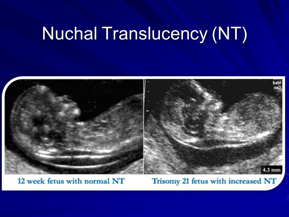 In total, within nine months, the patient is assigned three studies, which, in addition to ultrasound scanning, also include blood tests for biochemistry and specific hormones. It is a mistake to assume that any of the three screenings is optional. At each stage of pregnancy, it is necessary to prevent possible complications and carefully monitor the condition of the patient and the child. Ultrasound at 30 weeks of pregnancy is a mandatory third trimester screening. nine0003
Ultrasound of the 3rd trimester allows the obstetrician-gynecologist to make sure that there are no pregnancy pathologies, to determine the size of the head and the position of the fetus on the eve of childbirth. The full composition of the 3rd screening includes not only ultrasound, but also other studies: cardiotocography, Doppler, biochemistry and blood tests for the hCG hormone, free estriol, and alpha-fetoprotein.
3rd trimester ultrasound screening indications
Three timed ultrasound scans are recommended by WHO for all pregnant women without exception.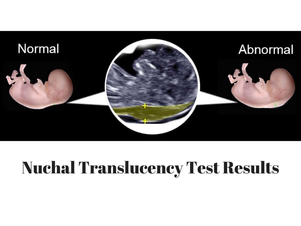 However, there is a risk group whose patients should be screened without fail. nine0003
This group includes the following categories of expectant mothers:
- Living in ecologically disadvantaged regions
- Over 35 years of age
- Previously diagnosed with "miscarriage"
- Having children in the family with genetically determined diseases (syndromes, triploidy, etc.)
- Survivors of infectious diseases in the first trimester
- Treated with antibacterial drugs during pregnancy
- Undergoing high-dose radiation training a few months before conception
- Having a close relative as the father of the child
What the doctor will see at the screening of the third trimester
Diagnosis in the last trimester of pregnancy is important in preparation for childbirth. In the process of scanning with an abdominal sensor, the obstetrician-gynecologist receives a lot of valuable information about the placenta, amniotic fluid and the patient's readiness for childbirth in general.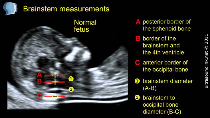 3rd trimester ultrasound provides data on: nine0003
- Amount of amniotic fluid (oligohydramnios, normal, polyhydramnios)
- Maturity of the placenta and the condition of the vessels in it
- The state of the internal organs of the fetus (heart, lungs, liver, kidneys, etc.)
- Fetal parameters: chest diameter, abdominal diameter and circumference, spine parameters, limb length (thigh and humerus
Also, ultrasound at 33 weeks can set the exact date of birth. In addition to ultrasound, the doctor may prescribe such standard studies for pregnant women as dopplerography and cardiotocography. These diagnostic procedures are designed to identify the rate and nature of blood flow in the placenta and assess the state of the fetal cardiovascular system. nine0003
Timing of the 3rd trimester ultrasound
The pre-delivery third screening is performed between the 31st and 33rd weeks of pregnancy. This period is due to the fact that at this time the obstetrician-gynecologist can finally verify the health of the fetus and determine its position in the womb.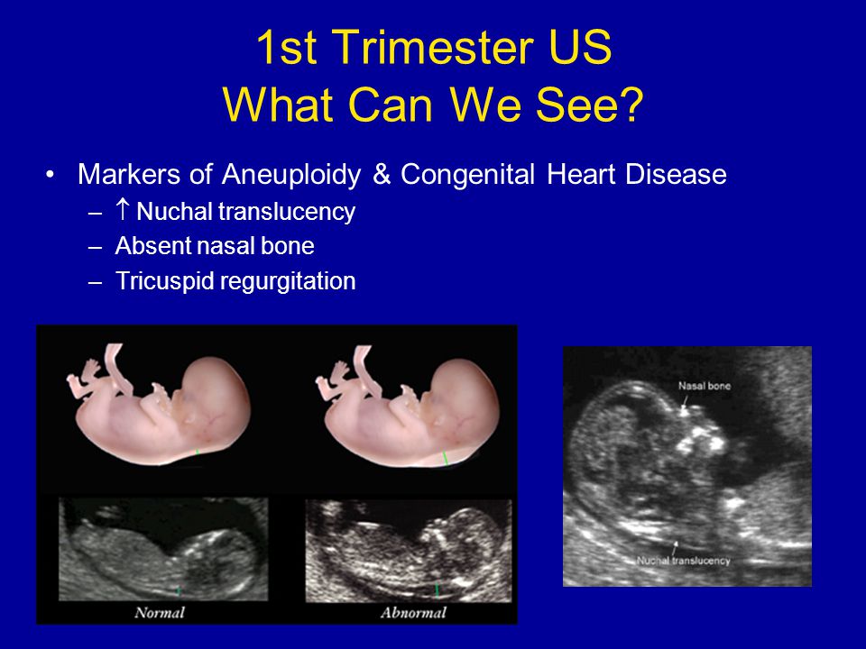 Ultrasound at 32 weeks also allows you to determine the degree of maturation of the placenta. The results will help the doctor navigate the method of the upcoming delivery and recommend either natural childbirth or caesarean section. nine0003
Interpretation of 3rd trimester screening results
Ultrasound examination parameters correlate with the patient's period by week. So, the values for 32 weeks of pregnancy will differ from the same indicators at 34 weeks.
Normal parameters for screening at 32 weeks are:
- placenta: thickness 25.3 to 41.6 mm
- amniotic fluid: 81 to 278 mm
- maturity of the placenta in degrees: first/second
- cervical dimensions (length): from 30 mm
Parameters of the fetal skeleton at the screening of 32-34 weeks of pregnancy:
- biparietal size: 85 to 89 cm;
- weight: from 1790 to 2390 grams - length (height): from 43 to 47 cm;
- head circumference: from 309 to 323 mm;
- fronto-occipital head size: from 102 to 107 mm; nine0008
- forearm (length): 46 to 55 mm;
- shoulder (length): from 55 to 59 mm;
- lower leg (length): from 52 to 57 mm.
 ;
; - thigh (length): 62 to 66 mm;
- abdominal circumference: 266 to 285 mm
Why 3rd trimester ultrasound is needed
Many patients often wonder if screening is necessary at the last stage of bearing a baby. Doctors around the world are unanimous in their opinion: all pregnant women should be examined without exception. This is due to the importance of the scan results for the upcoming birth. Even the most favorable pregnancy can have hidden pathologies. Diagnosis will reveal problems with the placenta, blood flow in the umbilical cord (if any), and the position of the baby in the uterus. nine0003
Ultrasound of the third trimester of pregnancy is the last routine examination for a woman.
The third ultrasound screening allows the attending obstetrician-gynecologist to bring the patient to the final line of pregnancy fully prepared.
(36 ratings, average 4.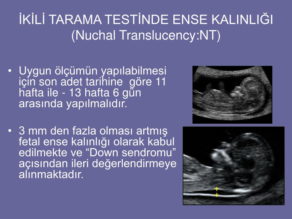
 9-39.5