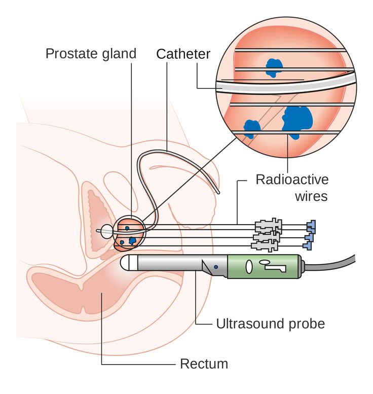
Ultrasound internal probe
Transvaginal ultrasound: MedlinePlus Medical Encyclopedia
URL of this page: //medlineplus.gov/ency/article/003779.htm
To use the sharing features on this page, please enable JavaScript.
Transvaginal ultrasound is a test used to look at a woman's uterus, ovaries, tubes, cervix and pelvic area.
Transvaginal means across or through the vagina. The ultrasound probe will be placed inside the vagina during the test.
You will lie down on your back on a table with your knees bent. Your feet may be held in stirrups.
The ultrasound technician or doctor will introduce a probe into the vagina. It may be mildly uncomfortable, but will not hurt. The probe is covered with a condom and a gel.
- The probe transmits sound waves and records the reflections of those waves off body structures. The ultrasound machine creates an image of the body part.
- The image is displayed on the ultrasound machine. In many offices, the patient can see the image also.
- The provider will gently move the probe around the area to see the pelvic organs.
In some cases, a special transvaginal ultrasound method called saline infusion sonography (SIS) may be needed to more clearly view the uterus.
You will be asked to undress, usually from the waist down. A transvaginal ultrasound is done with your bladder empty or partly filled.
In most cases, there is no pain. Some women may have mild discomfort from the pressure of the probe. Only a small part of the probe is placed into the vagina.
Transvaginal ultrasound may be done for the following problems:
- Abnormal findings on a physical exam, such as cysts, fibroid tumors, or other growths
- Abnormal vaginal bleeding and menstrual problems
- Certain types of infertility
- Ectopic pregnancy
- Pelvic pain
This ultrasound is also used during pregnancy.
The pelvic structures or fetus is normal.
An abnormal result may be due to many conditions. Some problems that may be seen include:
- Birth defects
- Cancers of the uterus, ovaries, vagina, and other pelvic structures
- Infection, including pelvic inflammatory disease
- Benign growths in or around the uterus and ovaries (such as cysts or fibroids)
- Endometriosis
- Pregnancy outside of the uterus (ectopic pregnancy)
- Twisting of the ovaries
There are no known harmful effects of transvaginal ultrasound on humans.
Unlike traditional x-rays, there is no radiation exposure with this test.
Endovaginal ultrasound; Ultrasound - transvaginal; Fibroids - transvaginal ultrasound; Vaginal bleeding - transvaginal ultrasound; Uterine bleeding - transvaginal ultrasound; Menstrual bleeding - transvaginal ultrasound; Infertility - transvaginal ultrasound; Ovarian - transvaginal ultrasound; Abscess - transvaginal ultrasound
- Ultrasound in pregnancy
- Female reproductive anatomy
- Uterus
- Transvaginal ultrasound
Brown D, Levine D. The uterus. In: Rumack CM, Levine D, eds. Diagnostic Ultrasound. 5th ed. Philadelphia, PA: Elsevier; 2018:chap 15.
Coleman RL, Ramirez PT, Gershenson DM. Neoplastic diseases of the ovary: screening, benign and malignant epithelial and germ cell neoplasms, sex-cord stromal tumors. In: Lobo RA, Gershenson DM, Lentz GM, Valea FA, eds. Comprehensive Gynecology. 7th ed. Philadelphia, PA: Elsevier; 2017:chap 33.
Dolan MS, Hill C, Valea FA. Benign gynecologic lesions: vulva, vagina, cervix, uterus, oviduct, ovary, ultrasound imaging of pelvic structures. In: Lobo RA, Gershenson DM, Lentz GM, Valea FA, eds. Comprehensive Gynecology. 7th ed. Philadelphia, PA: Elsevier; 2017:chap 18.
Updated by: John D. Jacobson, MD, Professor of Obstetrics and Gynecology, Loma Linda University School of Medicine, Loma Linda Center for Fertility, Loma Linda, CA. Also reviewed by David Zieve, MD, MHA, Medical Director, Brenda Conaway, Editorial Director, and the A.D.A.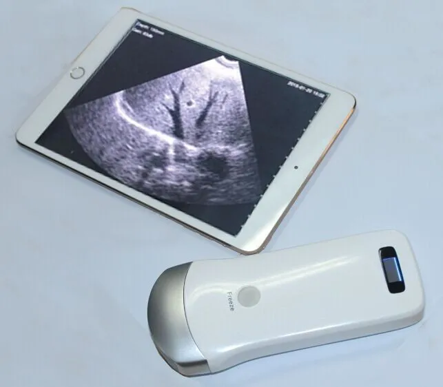 M. Editorial team.
Is it painful, purpose, and results
A transvaginal, or endovaginal, ultrasound is a safe, straightforward way for doctors to examine the internal organs of the female pelvic region.
An ultrasound uses high-frequency sound waves to produce detailed images of internal organs.
Unlike X-rays, ultrasound scanning does not involve radiation, which means that it has no known side effects and is very safe.
In this article, we explain the uses of transvaginal ultrasounds and how to prepare for one. We also describe what to expect during the scan.
A healthcare professional uses an ultrasound to create images of the inside of the body. High-frequency sound waves bounce off the internal organs and form these images.
There are two ways to perform an ultrasound — abdominally and transvaginally.
A transvaginal ultrasound is an internal scan of the female reproductive organs. It involves inserting a small ultrasound probe, called a transducer, into the vagina to produce incredibly detailed images of the organs in the pelvic region.
It may be necessary to use a transvaginal ultrasound to examine the:
- vagina
- cervix
- uterus
- fallopian tubes
- ovaries
- bladder
Transvaginal ultrasounds can check for:
- the shape, position, and size of the ovaries and uterus
- the thickness and length of the cervix
- blood flow through the organs in the pelvis
- the shape of the bladder and any changes
- the thickness and presence of fluids near the bladder or in the:
- fallopian tubes
- myometrium, the muscle tissue of the uterus
- endometrium
Doctors may request a transvaginal ultrasound for a variety of reasons. For example, it might be necessary to identify the cause of:
- pelvic pain
- unexplained vaginal bleeding
- infertility
- abnormal results of a pelvic or abdominal exam
These scans can also help diagnose:
- benign growths, such as fibroids, cysts, and masses
- pelvic inflammatory disease
- endometriosis
- postmenopausal bleeding
In addition, it can check for the presence of an intrauterine contraceptive device.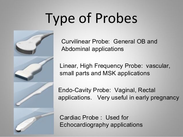
Doctors may request a transvaginal ultrasound during pregnancy, as it can help:
- check the heartbeat of the fetus
- confirm the date of delivery
- assess the condition of the placenta
- check for an ectopic pregnancy
- monitor pregnancies with a higher risk of pregnancy loss
A transvaginal ultrasound is not typically painful, but the insertion of the probe may be uncomfortable.
The healthcare professional performing the scan first covers the probe in a sheath and lubricating gel before inserting it slowly into the vagina to a depth of around 5–8 centimeters (cm). There may be mild pressure or discomfort at this stage.
There are no after-effects of a transvaginal ultrasound, and a person can return to their regular activities afterward.
A pelvic ultrasound is a noninvasive exam that produces images of the internal reproductive organs to help healthcare professionals diagnose certain conditions.
Doctors may use the term “pelvic ultrasound” to describe both a transvaginal and a transabdominal ultrasound.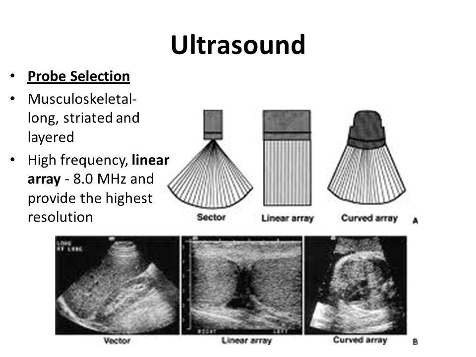 During a transabdominal ultrasound, the person performing the scan uses the probe outside the abdomen.
Another name for a transabdominal ultrasound is an external pelvic ultrasound. A person lies on their back on an examination table, and the healthcare professional applies a warm gel to the lower section of the person’s abdomen. Then, they move a probe over the area.
The person might feel slight pressure on their abdomen, but the scan is not painful. The probe uses sound waves to form an image of the internal organs and structures of the pelvic area.
By comparison, a transvaginal ultrasound can provide more close-up images of the internal organs than an external pelvic ultrasound.
A transvaginal ultrasound is a simple, painless scan that requires very little preparation. A person should be able to eat and drink normally before the appointment.
At the appointment, a healthcare professional will describe any necessary steps. A person needs to empty their bladder right before the scan, and anyone using a tampon should remove it.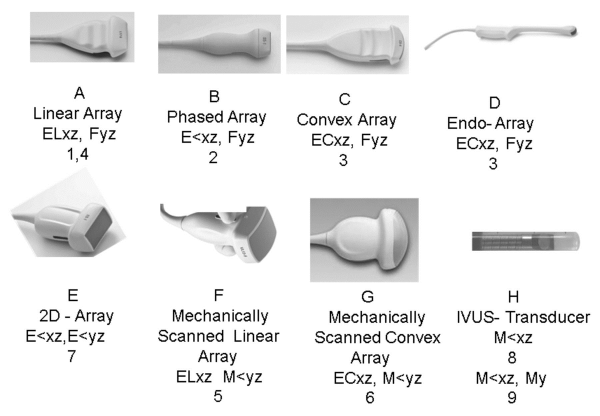
A doctor or a specially trained technician, called a sonographer, performs most transvaginal ultrasounds.
A person needs to undress from the waist down and put on a hospital gown. Next, they lie on an examination table with their knees bent. The healthcare professional uses a sheet to cover the person’s lower body.
The transducer resembles a wand and is slightly larger than a tampon. The sonographer or doctor covers the transducer with a sheath and lubricating gel before inserting it 5–8 cm into the vagina.
Once the transducer is in place, it produces sound waves that bounce off of the internal organs and relay information. To create a complete picture and bring different areas into focus, the sonographer or doctor rotates the transducer. This tool transmits the information directly to a screen.
The images display immediately on the screen, making it possible for the person and the healthcare professional to monitor the scan in real-time.
The whole process may last 15–30 minutes.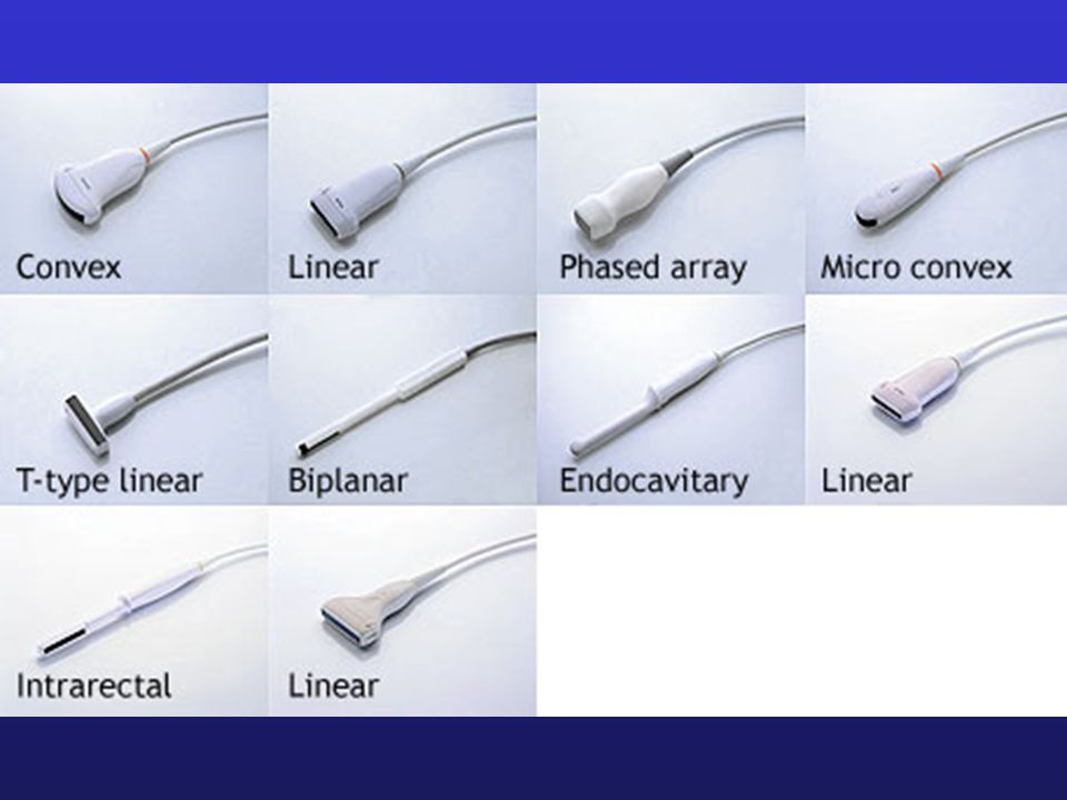
Unlike a traditional X-ray, a transvaginal ultrasound does not use radiation. As a result, it is very safe — there are no known risks.
It is also safe to perform transvaginal ultrasounds during pregnancy — there is no risk to the fetus.
During the insertion of the transducer, there may be some pressure and minimal discomfort. This feeling should go away after the scan.
It is essential to let the sonographer or doctor right away if anything feels particularly uncomfortable.
As the Miscarriage Association confirms, there is no evidence that a transvaginal ultrasound can harm a fetus or cause pregnancy loss.
If a person notices bleeding after a transvaginal ultrasound, this may be because blood has collected higher up in the vagina, and the transducer may have dislodged it.
People may have a transvaginal ultrasound in the first 11–12 weeks of pregnancy. After 11–12 weeks, it may be more common to have a transabdominal ultrasound. However, in some cases, doctors may still request a transvaginal ultrasound, which is a more accurate and detailed scan.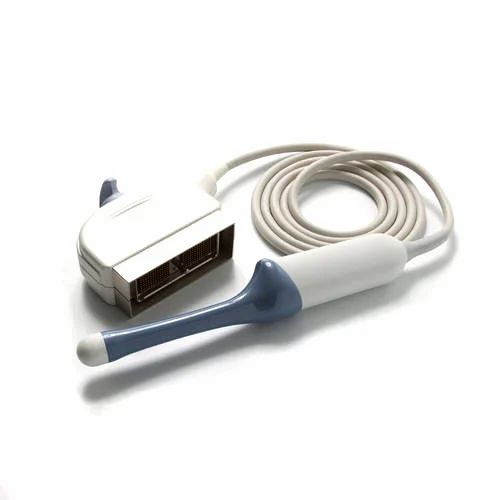
Transvaginal ultrasounds are safe up until a person’s water breaks.
If a specialist is present at the ultrasound, the person may hear the results immediately. If a sonographer does the scan, they send the images to a radiologist for analysis. The radiologist then sends a written report of the results to the person’s doctor.
Either way, it is important to discuss the results with a doctor, who can provide more information. This discussion can take place in person or over the phone.
A transvaginal ultrasound is a safe scan, with no known risks. People might experience some light discomfort during it, but this should go away afterward.
The ultrasound may take 15–30 minutes, and the results may be available immediately or within a few days, depending on whether a doctor was present during the scan.
If a person experiences extreme discomfort during the ultrasound, the doctor or sonographer may perform a transabdominal ultrasound instead. This type does not involve inserting the scanning tool into the vagina.
About ultrasonic transducers (part 6)
2015-07-03
Pepperl+Fuchs offers a wide range of ultrasonic transducers, many of which are equipped with an additional synchronization input
When installing ultrasonic sensors , the minimum separation distances cannot always be observed. To reduce the minimum separation distance and prevent sensor interference, Pepperl+Fuchs offers ultrasonic transducers with synchronization inputs . Sensors with these inputs can be used for internal or external synchronization, as well as for multi-channel modes. By synchronizing transmission cycles, the distance between adjacent sensors can be reduced without the risk of interference.
This article explains how the synchronization input of the
ultrasonic sensors works If the synchronization input remains open, the ultrasonic sensor operates at normal mode .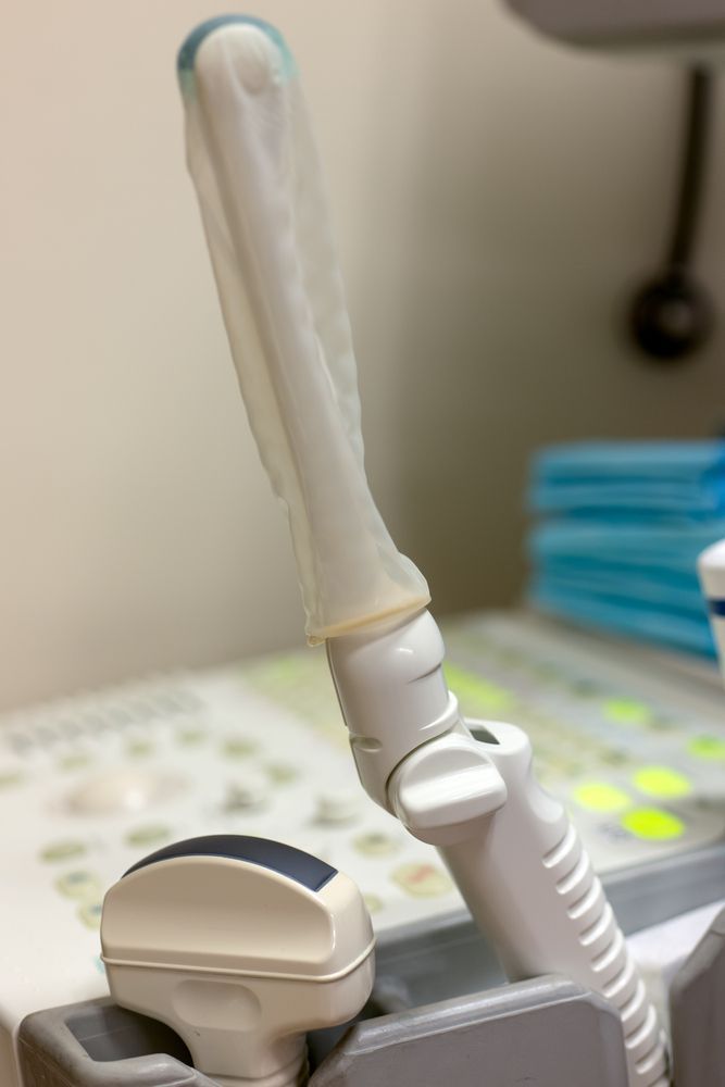 By applying a voltage of a certain value (L+/L-), it is possible to block or enable the sensor using external trigger signal . When the sensor is blocked, it does not emit ultrasonic pulses. In this mode, the sensor outputs (analogue and switching outputs) are blocked. As soon as the sensor is switched on for at least one measuring cycle with a synchronization input, the outputs are updated. This function can be used for external synchronization or multi-channel operation.
In the external synchronization mode , an external clock pulse generator provides communication and control of the synchronization inputs of all sensors (in multi-channel mode, each ultrasonic sensor is switched on separately).
The required signal level, cycle time and the maximum number of sensors are indicated in the specifications of the respective sensor. In the internal synchronization mode , communication and control of the synchronization inputs of all sensors is provided by the sensors themselves.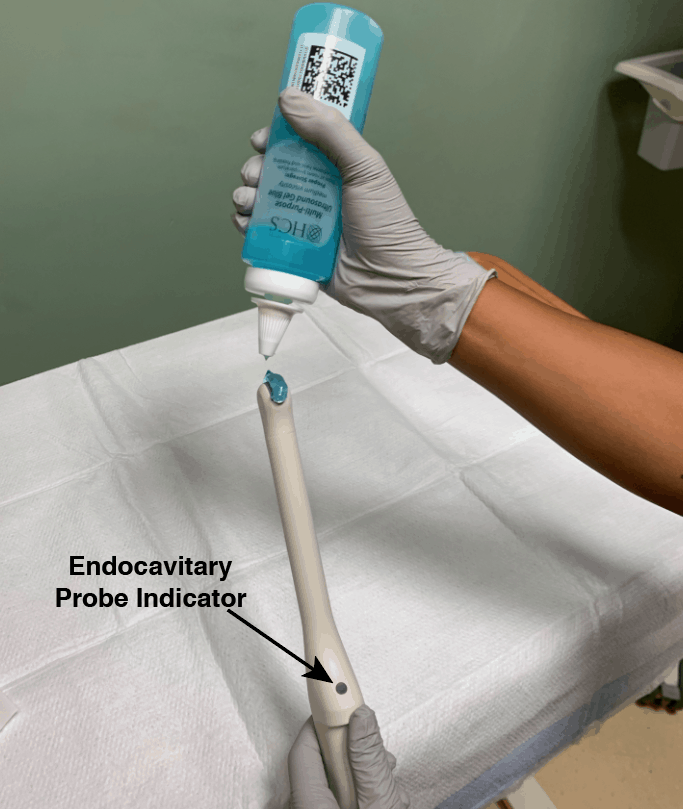 There is no need for an external clock generator.
Synchronization and common mode
In common mode the ultrasonic sensors work in parallel. This means that all sensors simultaneously emit an ultrasonic pulse and wait for the reflected signal to be received from an object in the working area of the sensor. To do this, the synchronization inputs of all sensors must be connected. Depending on the sensor type/family, synchronization mode starts automatically (internal synchronization) or requires an external trigger (external sync) .
Applications:
Several sensors can be combined into the ultrasonic unit for wide area monitoring. In conditions of limited space, the synchronization of sensors allows you to reduce the allowable interval between sensors. When installing the sensors opposite each other, the distances shown in the table below must also be taken into account.
Benefits:
- Reduced wiring costs, connecting affected inputs to each proximity switch
- Quick response because each proximity switch is continuously active
Disadvantages:
- Cannot assign individual object control to a specific proximity switch
Multi-channel
Multi-channel ultrasonic transducers allow ultrasonic pulses to be emitted at time-spaced intervals . This prevents mutual interference between sensors and allows you to reduce the distance between adjacent sensors. However, due to the fact that the sensors are connected in series, the response/response time increases as additional sensors are added to the multi-channel block .
There are differences between internal and external multichannel modes . When using the internal multi-channel mode, it is necessary to connect the synchronization inputs of all sensors. Depending on sensor type/family multichannel mode starts automatically or after assigning an address to the sensor using configuration tool . In external multi-channel operation, the sensors require an external trigger signal, and the synchronized sensor activation sequence must be controlled by the external controller .
Application:
These sensors are used in equipment and machines where there is very little space for installation, closely spaced sensors of the same type are present, or if you want to prevent mutual interference between sensors in different measurement tasks. At the same time, it is not necessary to maintain minimum intervals between the sensors, even if they are located opposite each other.
Benefits:
- Reliable interference prevention
- You can assign a monitored object to the ultrasonic sensor
Disadvantages:
- Additional clock cost when using external multi-channel
- Longer response/response time than in sync mode, as sensors work in series
Downloads: Instructions for use of the
ultrasonic transducers Pepperl+Fuchs offers various literature for ultrasonic transducers for download. In addition to Ultrasound Technology Manual you can also download the new manual for the determination of double materials using ultrasonic probes. Download your free PDF copy of and get useful tips and insights for your daily work!
About ultrasonic sensors, part 5
2015-04-27
Cleaning the ultrasonic sensor
In this case, the ultrasonic sensor is used to detect printed circuit boards. What should be considered when installing and assembling ultrasonic sensors?
The ultrasonic sensors can be installed and operated in any position and are extremely resistant to environmental conditions. However, when adjusting the ultrasonic transducers , there are some points to consider in order to achieve ideal measurement results. For example, when cleaning the ultrasonic transducer , be careful not to damage the transducer surface (separation layer) and the foam around the transducer. Drops of water or dry crust on the reinforcing layer may affect operation ultrasonic sensor . Note. A small amount of dust is not critical.
Direction of action
Objects to be detected can enter the zone of the sound beam from any direction. Suggested detection points for the can be determined using the range and radiation patterns listed in the Ultrasonic Transmitter Specification .
Object surface properties
Ultrasonic sensors can detect solid, liquid and powder objects . To assess the reflected sound of the sensor, the surface properties of the object are of great importance. Flat, smooth surfaces located at right angles to the beam, are characterized by the best sound reflection. For reliable detection, the angular deviation of the measuring plate must not exceed 3°.
Material properties such as transparency, color or surface finish (glossy or matt) do not affect detection reliability. Rough surfaces reflect sound energy in several directions, resulting in a reduced overall sound detection range . On the other hand, rough surfaces allow greater angular deviation due to diffuse reflection of the ultrasonic signal.
This property can be used to determine fill levels or heights of coarse stockpiles up to 45° (reduced range).
The following objects are especially good at detecting:
- all smooth and hard objects located perpendicular to the sound beam;
- all randomly placed solid objects with a rough surface that causes diffuse reflection;
- surfaces of liquids at an angle of <3° relative to the beam axis.
The following materials are difficult to detect :
- materials that absorb ultrasonic signals, such as felt, cotton, rough fabrics and foams;
- materials over 100°C.
To detect such materials, it is recommended to use sensors with separate transmitter and receiver .
Sound beam and gap
The directivity pattern of ultrasonic sensors is referred to as “sound beam” . Objects are detected within the sound beam if they reflect enough sound back to the sensor. The directivity characteristic depends on the reflective properties of the object. Therefore, the technical specifications are sound beam graphics for various standard objects. The sound beam has no well-defined boundaries and can change depending on environmental conditions such as temperature or humidity.
If there are undesirable objects reflecting sound energy in the area of application of the transducer, a gap around the sound beam must be provided . This is the only way to prevent false switching caused by unintended reflections.
Directional pattern 2 (round rod, 25 mm) can be used for orientation detection in the presence of small, round objects and objects with poor reflective properties. It can also be used for smooth surfaces parallel to the ultrasonic transducer beam (container inner walls, pipes). For larger objects with good reflective properties (interfering edges), a gap of at least directional characteristic 1 (flat plate, 100 mm x 100 mm) must be provided.
If a margin is not possible, most Pepperl+Fuchs ultrasonic transducers provide the sound beam reshaping option. This procedure can be performed using the Teaching Mode Buttons or the Programming Interface and Software. The software allows you to selectively suppress many interfering objects within the operating range (suppression of stationary targets).
Ultrasonic Transducers Parallel Mounting Minimum Spacings To prevent interference between ultrasonic transducers installed in parallel and operating in the same range, a minimum distance must be maintained between the transducers as shown in the figures below. The values given in the table are recommended . They are used when the beams are aligned parallel to each other and the object surfaces are at right angles to the beam axis. The actual distance (X) depends on the alignment, type and surface of the target in the area of the sound beam.
Minimum intervals when mounting ultrasonic sensors opposite each other
The intervals given in the table must be observed when mounting sensors opposite each other. If interference occurs, it may be necessary to increase the distance (X) or, if necessary, activate the synchronization or multiplication function . Synchronized and non-synchronized ultrasonic transducers must not be mounted opposite each other.
Downloads: Instructions for use of the
ultrasonic transducers Pepperl+Fuchs offers various literature for ultrasonic transducers for download. In addition to the Ultrasonic Technology Manual , you can also download the new Manual for Dual Material Determination with Ultrasonic Probes.