Internal ultrasound early pregnancy
What To Expect, Purpose & Results
Overview
What is an ultrasound in pregnancy?
A prenatal ultrasound (or sonogram) is a test during pregnancy that checks on the health and development of your baby. An obstetrician, nurse midwife or ultrasound technician (sonographer) performs ultrasounds during pregnancy for many reasons. Sometimes ultrasounds occur to check on your baby and make sure they’re growing properly. Other times your pregnancy care provider orders an ultrasound after they detect a problem.
During an ultrasound, sound waves are sent through your abdomen or vagina by a device called a transducer. The sound waves bounce off structures inside your body, including your baby and your reproductive organs. Then, the sound waves transform into images that your provider can see on a screen. It doesn’t use radiation, like X-rays, to see your baby.
Even though prenatal ultrasounds are safe, you should only have them when it’s medically necessary. If there’s no reason for an ultrasound (for example, if you just want to see your baby), your insurance company might not pay for it.
Prenatal ultrasounds may be called fetal ultrasounds or pregnancy ultrasounds. Your provider will talk to you about when you can expect ultrasounds during pregnancy based on your health history.
Why is a fetal ultrasound important during pregnancy?
An ultrasound is one of the few ways your pregnancy care provider can see and hear your baby. It can help them determine how far along you are in pregnancy, if your baby is growing properly or if there are any potential problems with the pregnancy. Ultrasounds may occur at any time in pregnancy depending on what your provider is looking for.
What can be detected in a pregnancy ultrasound?
A prenatal ultrasound does two things:
- Evaluates the overall health, growth and development of the fetus.
- Detects certain complications and medical conditions related to pregnancy.
In most pregnancies, ultrasounds are positive experiences and pregnancy care providers don’t find any problems.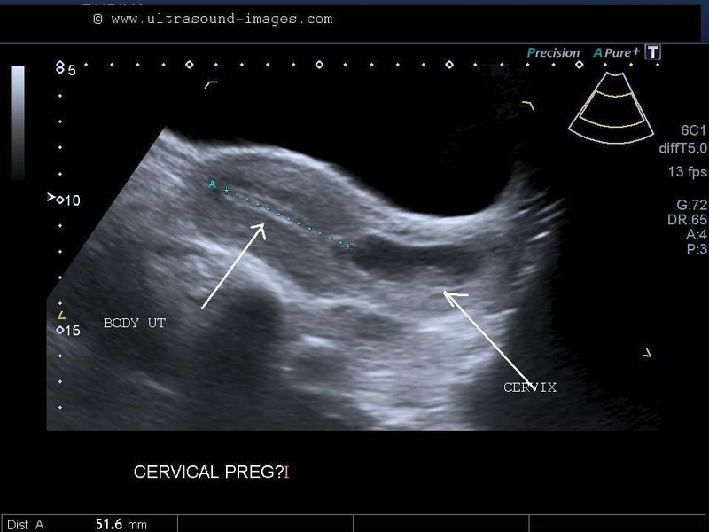 However, there are times this isn’t the case and your provider detects birth disorders or other problems with the pregnancy.
Reasons why your provider performs a prenatal ultrasound are to:
- Confirm you’re pregnant.
- Check for ectopic pregnancy, molar pregnancy, miscarriage or other early pregnancy complications.
- Determine your baby’s gestational age and due date.
- Check your baby’s growth, movement and heart rate.
- Look for multiple babies (twins, triplets or more).
- Examine your pelvic organs like your uterus, ovaries and cervix.
- Examine how much amniotic fluid you have.
- Check the location of the placenta.
- Check your baby’s position in your uterus.
- Detect problems with your baby’s organs, muscles or bones.
Ultrasound is also an important tool to help providers screen for congenital conditions (conditions your baby is born with). A screening is a type of test that determines if your baby is more likely to have a specific health condition. Your provider also uses ultrasound to guide the needle during certain diagnostic procedures in pregnancy like amniocentesis or CVS (chorionic villus sampling).
An ultrasound is also part of a biophysical profile (BPP), a test that combines ultrasound with a nonstress test to evaluate if your baby is getting enough oxygen.
How many ultrasounds do you have during your pregnancy?
Most pregnant people have one or two ultrasounds during pregnancy. However, the number and timing vary depending on your pregnancy care provider and if you have any health conditions. If your pregnancy is high risk or if your provider suspects you or your baby has a health condition, they may suggest more frequent ultrasounds.
When do you have your first prenatal ultrasound?
The timing of your first ultrasound varies depending on your provider. Some people have an early ultrasound (also called a first-trimester ultrasound or dating ultrasound). This can happen as early as seven to eight weeks of pregnancy.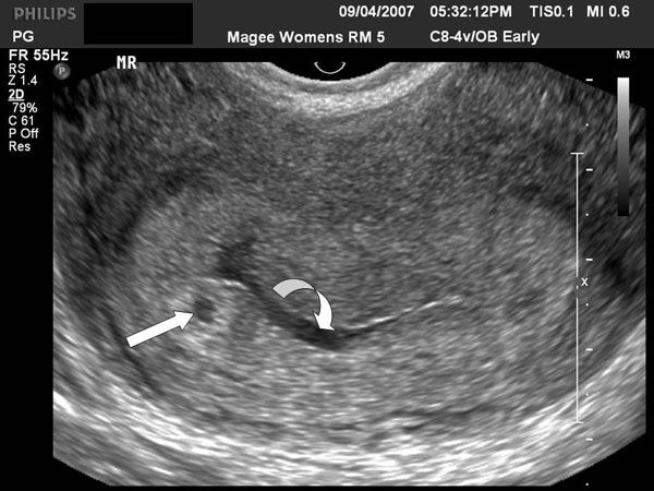 Providers do an early ultrasound through your vagina (transvaginal ultrasound). Early ultrasounds do the following:
- Confirm pregnancy (by detecting a heartbeat).
- Check for multiple fetuses.
- Measure the size of the fetus.
- Help confirm gestational age and due date.
Some providers perform your first ultrasound closer to 12 weeks of pregnancy.
20-week ultrasound (anatomy scan)
You can expect an ultrasound around 18 to 20 weeks in pregnancy. This is known as the anatomy ultrasound or 20-week ultrasound. During this ultrasound, your pregnancy care provider can see your baby’s sex (if your baby is in a good position for viewing their genitals), detect birth disorders like cleft palate or find serious conditions related to your baby’s brain, heart, bones or kidneys. If your pregnancy is progressing well and with no complications, your 20-week ultrasound may be your last ultrasound during pregnancy. However, if your provider detects a problem during your 20-week ultrasound, they may order additional ultrasounds.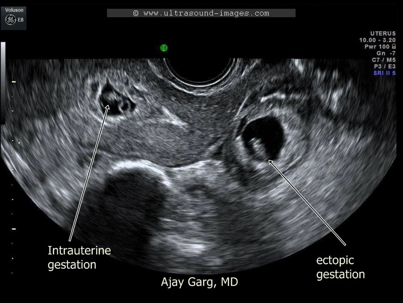
How soon can you see a baby on an ultrasound?
Pregnancy care providers can detect an embryo on an ultrasound as early as six weeks into the pregnancy. An embryo develops into a fetus around the eighth week of pregnancy.
If your last menstrual period isn’t accurate, it’s possible that it may be too early to detect a fetal heart rate.
Which ultrasound is most important during pregnancy?
All ultrasounds during pregnancy are important. Your pregnancy care provider uses ultrasound to tell them important information about your pregnancy.
Test Details
What are the two main types of pregnancy ultrasounds?
The two main types of pregnancy ultrasound are transvaginal ultrasound and abdominal ultrasound. Both use the same technology to produce images of your baby. Your pregnancy care provider performs a transvaginal ultrasound by placing a wand-like device inside your vagina. They perform an abdominal ultrasound by placing a device on the skin of your belly.
Transvaginal ultrasound
During a transvaginal ultrasound, your pregnancy care provider places a device inside your vaginal canal (similar to how you place a tampon). In early pregnancy, this ultrasound helps to detect a fetal heartbeat or determine how far along you are in your pregnancy (gestational age). Images from a transvaginal ultrasound are clearer in early pregnancy as compared to abdominal ultrasound.
Abdominal ultrasound
Your pregnancy care provider performs an abdominal ultrasound by placing a transducer directly on your skin. Then, they move the transducer around your belly (abdomen) to capture images of your baby. Sometimes slight pressure has to be applied to get the best views. Providers use abdominal ultrasounds after about 12 weeks of pregnancy.
Traditional ultrasounds are 2D. More advanced technologies like 3D or 4D ultrasound can create better images. This is helpful when your provider needs to see your baby’s face or organs in greater detail. Not all providers have 3D or 4D ultrasound equipment or specialized training to conduct this type of ultrasound.
Your provider may recommend other types of ultrasounds. Examples of additional ultrasounds are:
- Doppler ultrasound: This type of ultrasound checks how your baby’s blood flows through its blood vessels. Most Doppler ultrasounds occur later in pregnancy.
- Fetal echocardiogram: This type of ultrasound looks at your baby’s heart size, shape, function and structure. Your provider may use it if they suspect your baby has a congenital heart condition, if you had another child that had a heart condition or if you have certain health conditions that warrant taking a closer look at the heart.
How do I prepare for the test?
There’s no special preparation for an ultrasound. Some pregnancy care providers ask that you come with a full bladder and don’t use the restroom before the test. This helps them view your baby better on the ultrasound. You can bring a support person, but bringing children is discouraged as this is an important test that requires complete focus.
You may be asked to change into a hospital gown, but this isn’t usually required for abdominal ultrasounds. If your provider is performing a transvaginal ultrasound in your first trimester, you’ll put on a hospital gown or undress from the waist down.
What should I expect during a prenatal ultrasound?
You’ll lie on a padded examining table during the test. Most ultrasounds occur in a dimly lit room, which helps your ultrasound technician (or sonographer) see the screen. Your sonographer applies a small amount of water-soluble gel to the skin of your belly. The gel doesn’t harm your skin or stain your clothes, but it may feel cold. This gel helps transmit sound waves more efficiently.
Next, the sonographer places a transducer on the skin of your abdomen. The transducer sends sound waves into your body, which reflect off internal structures, including your baby. The sound waves that reflect back create pictures on a screen. Your sonographer uses these images to take important measurements such as your baby’s head circumference and length. You may see them making lines on the screen or clicking a button to “freeze” certain angles.
There’s virtually no discomfort during a prenatal ultrasound. You may feel mild discomfort if you have to pee. The ultrasound test takes about 30 minutes to complete.
If you have a transvaginal ultrasound, the process is only different in that the transducer is inside your vagina and not on your belly.
What should I expect after a pregnancy ultrasound?
If you had an abdominal ultrasound, your sonographer wipes the gel off your belly. They may print off some ultrasound pictures for you to take home with you.
In most cases, your sonographer won’t discuss the results of your test with you. If your obstetrician performs your ultrasound, they may discuss what they see as they go along.
If a sonographer performs your ultrasound, an obstetrician will look at the images, then discuss their findings with you at your next appointment. Most practices schedule your appointment right after your ultrasound so you get your results the same day.
What are the risks of prenatal ultrasounds?
Studies have shown ultrasounds are safe during pregnancy. There are no harmful side effects to you or your baby.
Is it safe to do an ultrasound every month during pregnancy?
While ultrasounds are safe for you and your baby, most major medical associations recommend that pregnancy care providers should only do ultrasounds when the tests are medically necessary. If your ultrasounds are normal and your pregnancy is uncomplicated or low risk, repeat ultrasounds aren’t necessary.
Results and Follow-Up
What results do you get on a pregnancy ultrasound?
Your ultrasound results will be normal or abnormal. A normal result means your pregnancy care provider didn’t find any problems and that your baby is growing and developing normally. An abnormal result means your provider noticed something irregular. If they do, your provider will order additional ultrasounds or diagnostic tests to determine if something is wrong.
Occasionally, the ultrasound is incomplete if there’s difficulty seeing all the structures needed for that particular ultrasound. Your baby’s position or movement sometimes makes it difficult to see everything your provider needs to see. If this is the case, you’ll need a repeat ultrasound and they’ll try again.
There are some limitations to ultrasounds, so your provider may not find certain abnormalities until after birth.
What are reasons you need more ultrasounds during pregnancy?
There are several reasons your pregnancy care provider may order additional ultrasounds during your pregnancy. Some of these reasons include:
- Problems with your ovaries, uterus, cervix or other pelvic organs.
- Your baby is measuring small for their gestational age or your provider suspects IUGR (intrauterine growth restriction).
- Problems with the placenta like placenta previa or placental abruption.

- You’re pregnant with twins, triplets or more.
- Your baby is breech.
- You have too much amniotic fluid (polyhydramnios).
- You have too little amniotic fluid (oligohydramnios).
- You have a condition like gestational diabetes or preeclampsia.
- Your baby has a congenital disorder.
Normal results on pregnancy ultrasounds can vary. Generally, a normal result means your baby appears healthy and your provider didn’t find any issues.
Why do some pregnancy providers schedule ultrasounds differently?
The number of ultrasounds you’ll have and when you have them can vary between providers. Every practice operates differently and some providers do things differently based on your health history or symptoms.
When does a pregnancy ultrasound determine sex?
Your baby’s sex isn’t visible on an ultrasound until about 18 to 20 weeks. Be sure to tell your pregnancy care provider whether or not you want to know the sex of your baby before your ultrasound.
A note from Cleveland Clinic
An ultrasound during pregnancy can be both exciting and terrifying. Your pregnancy care provider uses ultrasound to get a better idea of how your baby is growing and developing. There are different types of ultrasounds, and the exact timing may vary depending on your provider. Most pregnant people have two ultrasounds — one in the first trimester and one in the second trimester. However, if there’s a potential complication or medical reason for more ultrasounds, your provider will order more as a precaution. Talk to your provider about the ultrasound schedule during pregnancy and what you can expect.
Vaginal ultrasound | Pregnancy Birth and Baby
beginning of content3-minute read
Listen
A vaginal ultrasound is an ultrasound scan taken by a probe inserted into the vagina. It gives a clear picture of the fetus, cervix and placenta. It is also called an internal ultrasound or a transvaginal ultrasound.
What does a vaginal ultrasound test for?
A vaginal ultrasound lets the doctor or ultrasound technologist (sonographer) see and measure the fetus. You might even discover if you are pregnant with twins or triplets.
It also allows the doctor or sonographer to look at your vagina, placenta, cervix, fallopian tubes, uterus and ovaries.
A vaginal ultrasound can be used as well as, or instead of, the standard abdominal ultrasound in which the probe is put on your belly.
Why is a vaginal ultrasound recommended?
A vaginal ultrasound will be recommended when your doctor or midwife wants a clearer diagnostic image than the one they can get through a standard abdominal ultrasound.
When is it used during pregnancy?
A vaginal ultrasound can confirm you are pregnant as it can detect the heartbeat very early in your pregnancy. It can also record the location and size of the fetus and determine if you are pregnant with 1 baby or more.
A vaginal ultrasound can also be used to diagnose problems or potential problems, including to:
- detect an ectopic pregnancy
- measure the cervix to determine the risk of a premature birth, which will allow any necessary interventions
- detect abnormalities in the placenta or cervix
- to determine the source of any bleeding
How is a vaginal ultrasound done?
A vaginal ultrasound is done by your doctor or sonographer in a hospital, clinic or consulting room. It uses a probe that is slightly wider than your finger.
You’ll probably be asked to empty your bladder (pee) before the ultrasound starts. If you’re using a tampon, you’ll need to take it out.
You’ll be asked to take off the lower half of your clothing and lie back on the examination table with your knees bent. There might be stirrups, or your hips might be slightly raised.
The probe is covered by a sheath, which is then covered in lubricating gel. It will be inserted slowly for about 5 to 8cm into your vagina. That usually doesn’t hurt, but you will feel pressure and it can be uncomfortable.
The probe will be moved around to get the best view of what is being examined. The examination usually takes 15 to 30 minutes.
If you are not comfortable having a male perform the examination, you can ask for a female sonographer. You can also ask for a female health worker to accompany you for support, or have a family member with you.
If you live in Western Australia, you must give written consent to a vaginal ultrasound.
When will I get the results?
Often you can see the ultrasound images on a monitor while you have your scan. If your specialist is there, they might discuss the results with you straight away.
If a specialist isn’t there, the sonographer is usually not allowed to discuss what they see with you. Your doctor or midwife will see the images after they have been processed. It usually takes a day or two to get the results.
Are there any risks involved?
Vaginal ultrasounds are safe for you and your baby, as long as your waters haven’t broken.
If you are allergic to latex, let the sonographer know so they can use a latex-free sheath on the probe.
There are no after-effects of the procedure, so you can get back to your normal activities, including driving yourself home if you wish.
Sources:
Australasian Society for Ultrasound in Medicine (Guidelines for the performance of first trimester ultrasound), Inside Radiology (Transvaginal ultrasound), Royal Australian and New Zealand College of Obstetricians and Gynaecologists (Screening in early pregnancy for adverse perinatal outcomes), The Royal Women's Hospital (Bleeding in early pregnancy)Learn more here about the development and quality assurance of healthdirect content.
Last reviewed: June 2020
Back To Top
Related pages
- Pregnancy checkups, screenings and scans
- Ultrasound scans during pregnancy
This information is for your general information and use only and is not intended to be used as medical advice and should not be used to diagnose, treat, cure or prevent any medical condition, nor should it be used for therapeutic purposes.
The information is not a substitute for independent professional advice and should not be used as an alternative to professional health care. If you have a particular medical problem, please consult a healthcare professional.
Except as permitted under the Copyright Act 1968, this publication or any part of it may not be reproduced, altered, adapted, stored and/or distributed in any form or by any means without the prior written permission of Healthdirect Australia.
Support this browser is being discontinued for Pregnancy, Birth and Baby
Support for this browser is being discontinued for this site
- Internet Explorer 11 and lower
We currently support Microsoft Edge, Chrome, Firefox and Safari. For more information, please visit the links below:
- Chrome by Google
- Firefox by Mozilla
- Microsoft Edge
- Safari by Apple
You are welcome to continue browsing this site with this browser. Some features, tools or interaction may not work correctly.
Ultrasound of the internal organs during pregnancy in St. Petersburg
When observing pregnant women, ultrasound is of great importance. During the examination of the internal organs, various anomalies are detected that interfere with the normal development of the fetus. If you do not conduct an ultrasound during this period, there is a risk of not noticing the pathology and not taking action in time.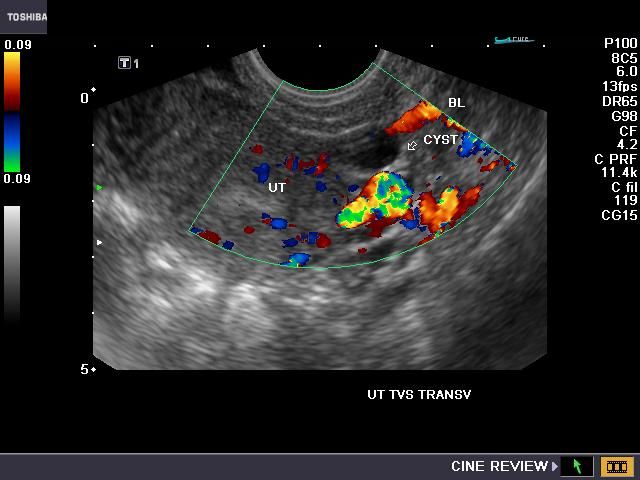
Since ultrasound examination is harmless, doctors advise to undergo it as a preventive measure. Thanks to this, it is possible to detect many diseases at an early stage and not allow them to go into the acute stage.
Ultrasound of the heart during pregnancy
Ultrasound of the heart during pregnancy is mandatory in several cases:
- shortness of breath;
- chest pain;
- tachycardia;
- heart disease;
- undergone heart surgery.
Color Doppler ultrasound is able to track blood flow disturbances. The health of the heart and vascular system is of great importance for the fetus, and in case of any deviations, the doctor will prescribe a course of treatment, preventing the development of the disease.
Ultrasound of the abdominal organs during pregnancy
Examination of the abdominal cavity also plays an important role in the health of the child and mother. Ultrasound of the abdominal organs during pregnancy includes a set of studies - while studying the spleen, gallbladder, liver and other organs.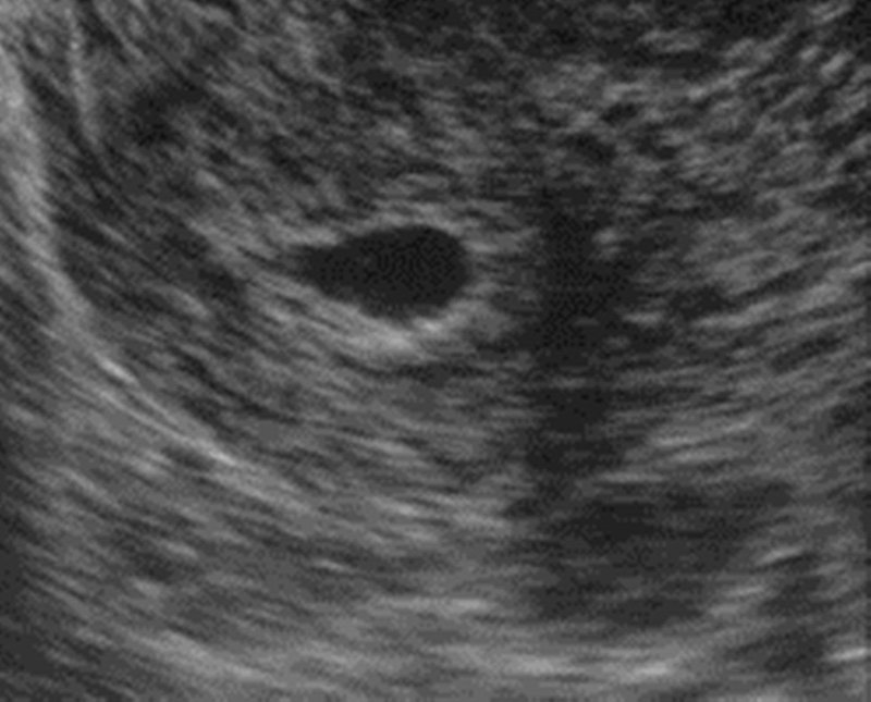
Any abnormalities such as liver stones, tumors, ulcers and cysts are noted during the procedure. If incipient or chronic diseases are detected, the doctor, after making a diagnosis, will prescribe a treatment that will help get rid of the disease, but at the same time will not harm the fetus.
Ultrasound of the kidneys during pregnancy
In the examination during pregnancy, special attention is paid to the ultrasound examination of the kidneys.
In the presence of kidney stones various symptoms appear:
- renal colic;
- blood in urine;
- nausea and vomiting;
- chills and high fever.
Ultrasound of the kidneys during pregnancy will allow you to identify the disease before the onset of alarming symptoms and cure it in time.
In addition to the examination of the kidneys during pregnancy, it is recommended to undergo an examination of the genitourinary system, mammary glands, adrenal glands and thyroid gland.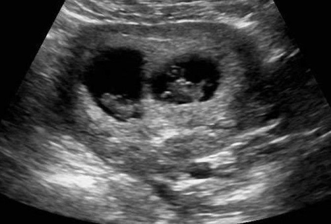 All this will show the general condition of the organs and, based on this information, will allow drawing conclusions and prescribing the correct treatment.
As the fetus grows, the abdominal organs move slightly in late pregnancy. This sometimes leads to misdiagnosis.
To prevent this from happening, the examination should be carried out by experienced diagnosticians using modern ultrasound scanners.
Medicenter clinics are equipped with modern Japanese diagnostic equipment, and doctors have been trained in the best medical universities in the country and have been working in the field of ultrasound diagnostics for many years.
Our clinics in St. Petersburg
You can get detailed information and make an appointment by calling +7 (812) 640-55-25
Make an appointment
Early pregnancy ultrasound
Modern high-tech equipment provides high accuracy and informativeness of the method even with the smallest size of the ovum. Ultrasound in the early stages helps to fully control the most vulnerable period of pregnancy.
- the latest device Mindray DC-70 PRO
- specialists with more than 15 years of experience
- detailed description of the examination and doctor’s comments during the diagnostic process . The procedure is carried out by experienced obstetrician-gynecologists who are certified in the field of ultrasound diagnostics and understand all its intricacies. Our specialists are guided by international standards for the provision of medical care, regularly improve their professionalism and apply a personalized approach to each patient.
Indications
First trimester ultrasound screening is performed from the 11th to the end of the 13th week. However, in special situations, the doctor may recommend an ultrasound at an earlier date, namely:
- From the 4th-5th week. The study is carried out to confirm the fact of pregnancy, with negative test results or the inability to conduct laboratory diagnostics (analysis for hCG).
 Ultrasound in the early stages is prescribed to confirm pregnancy resulting from IVF. The procedure allows you to clarify the location of the fetal egg and exclude ectopia, which is especially important in the presence of cases of ectopic pregnancy in history.
 - From 6-7 weeks. Ultrasound of the fetus in the early stages may be relevant for assessing its viability. During the study, the location, size, area of attachment of the embryo, as well as the presence and speed of the heartbeat, are evaluated. The procedure is especially relevant in the presence of a history of miscarriages, in case of abdominal pain or spotting in a pregnant woman.
- From the 9th week. In this period, with the help of ultrasound, it is possible to assess the anatomy of the embryo and diagnose the first signs of developmental anomalies. Early diagnosis is relevant in the presence of genetic diseases in one or both parents.
Early pregnancy ultrasound is not a substitute for first trimester screening.
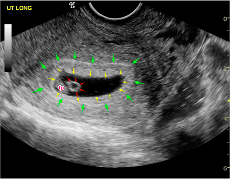 Even after conducting several studies in the first weeks of gestation, the procedure is repeated from the 11th to the 13th week, because. diagnostic capabilities of the method are different.
 How early ultrasound is performed
In the first trimester, when the fetus is small, ultrasound is performed transvaginally (through the vagina). The study is carried out using a vaginal sensor using a disposable condom. It should be noted that the introduction of equipment into the pelvic cavity cannot harm the health of the mother and fetus, or provoke complications during pregnancy. The procedure is painless and takes no more than 10 minutes.
Make an appointment
Our administrator will call you back to set the time and answer your questions
Preparation for the procedure
No specific preparation is required. On the day of the study, a hygienic shower is shown. Immediately before entering the diagnostic room, you need to empty your bladder.
 Only with a transabdominal scan should the bladder be full. This method is used for heavy bleeding.
 Early ultrasound contraindications
One of the main advantages of ultrasound diagnostics is the absolute absence of contraindications. Ultrasound has no effect on the condition of the uterus and fetus. The procedure has no restrictions regarding the multiplicity and frequency of its implementation. If necessary, ultrasound is done in dynamics until the threat of abortion disappears.
Where can ultrasound be done to determine pregnancy? Residents of St. Petersburg can undergo an ultrasound to determine pregnancy, monitor the vital activity of the fetus and an initial assessment of its anatomy as prescribed by a doctor or on their own initiative. The clinic also performs screenings of the first, second and third trimesters, Doppler, ultrasound in 3D and 4D. The high professionalism of doctors and the availability of expert equipment guarantee high accuracy of the results.

- From the 4th-5th week. The study is carried out to confirm the fact of pregnancy, with negative test results or the inability to conduct laboratory diagnostics (analysis for hCG).


