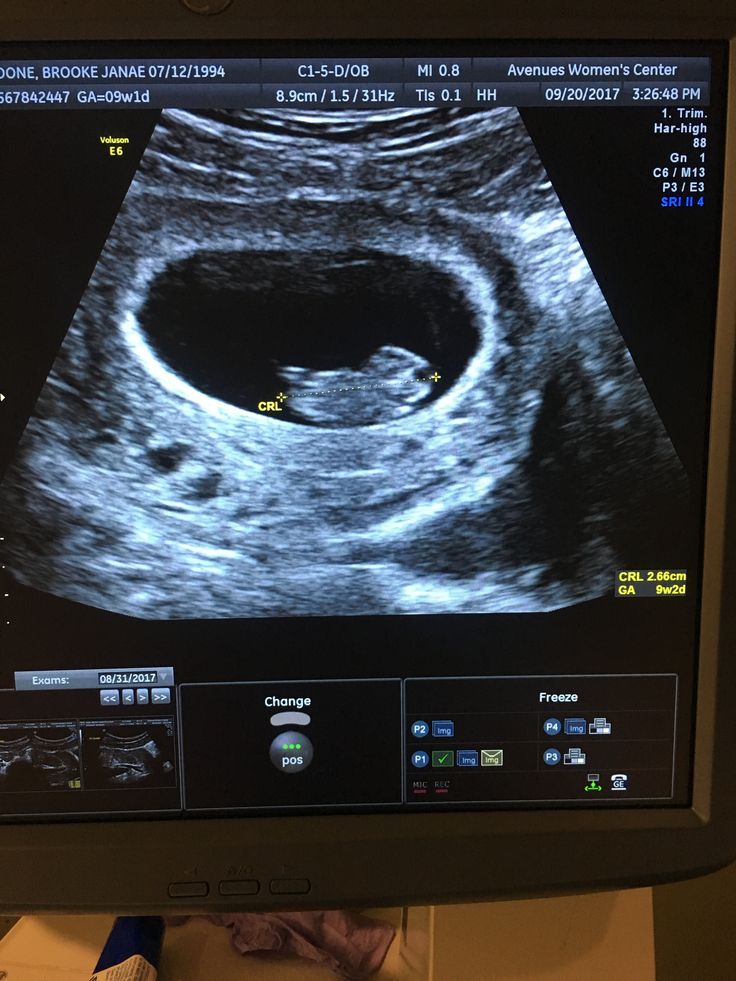
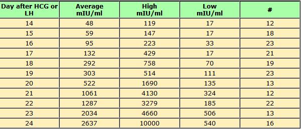
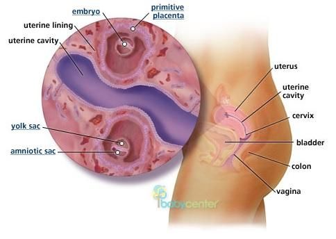
Internal ultrasound 8 weeks
What Can I See On an 8-Week Ultrasound?
In the early days of pregnancy, those initial appointments can feel surreal — especially if this is your first pregnancy. Those first appointments are usually designed to get a baseline understanding of your health going into pregnancy and to make sure things are progressing properly.
One major milestone is the 8-week ultrasound. So, why do you have an ultrasound this early into pregnancy, and what can you expect at your 8-week ultrasound? We’ll answer those questions and more.
Even though you can get a positive pregnancy test result roughly 2 weeks after conception, it can be a while until that tiny ball of cells demonstrates physical changes that confirm your pregnancy is progressing. Specifically, a healthcare professional will want to confirm that your fetus has a heartbeat — a clear sign that it’s alive.
In some cases, a heartbeat may be detected as early as 6 weeks. When you have a positive pregnancy test, contact a physician or healthcare professional to see if you need to come in for an ultrasound.
Transvaginal vs. abdominal ultrasounds
When most of us think of ultrasounds, we think of a technician passing a wand over a person’s stomach that’s covered in gel. This is known as an abdominal ultrasound. In most situations, early ultrasounds generally take less than half an hour.
But a transvaginal ultrasound is when a wand is inserted into the vagina. Often, this is used during early pregnancy to get a closer look at the fetus.
Beyond heartbeat, the technician or physician will be able to immediately identify key features such as the gestational sac and your fetus’s crown-rump length. These can help determine gestational age and pinpoint your due date.
Share on PinterestSuzanne Tucker/Shutterstock
This is going to be your first peek at your growing bundle of joy! Don’t expect to see a lot of definition or details this early in the game.
For now, you’ll see a little figure that looks something like an oblong bean. If there are twins, you might see two figures. The head is still nearly the same size as the rest of the body.
You’ll also see the gestational sac, the fluid-filled space around your baby(s). Within it, you can also see the yolk sac, which is a bubble-like structure. Depending on the location, you might even get to hear their heartbeat, too.
The main reasons for the 8-week ultrasound may be to confirm a pregnancy, determine a due date, and confirm the baby’s heartbeat. First, your doctor or technician will look for key physical indicators, like a gestational sac and a fetal pole, to verify the pregnancy is in the uterus. This is may be your first indication of twins.
Once they’ve confirmed that you’re pregnant, the next step is to verify your projected due date. Even though you might have initially received a projected due date at an earlier appointment, it’s not always accurate. The initial due date is determined by confirming the first day of your last period, deducting 3 months, and then adding 1 year and 7 days. But because not every person’s menstrual cycle is the same length of time, these projections can be off.
With an ultrasound, a physician or technician can determine gestational age and due date by measuring the size of your fetus. The accepted method for early pregnancy dating is the crown-rump length (CRL) measurement because it’s the most accurate (within about 5 to 7 days) in the first trimester.
When you can’t see the baby or a heartbeat
Sometimes you can’t see the fetus or hear a heartbeat — but that doesn’t always mean the worst. Sometimes it means that your calculations on the conception date were off.
If you ovulated and conceived later than you initially assumed, you might be getting an ultrasound too early to get a physical confirmation. In other scenarios, you might have large fibroids or anatomic issues with the uterus that can make screening your uterus more difficult.
But in some situations, it might not be the news you hoped for. Occasionally, the absence of a visible fetus in the uterus could mean you have an ectopic pregnancy, where the embryo implanted outside of the uterine cavity.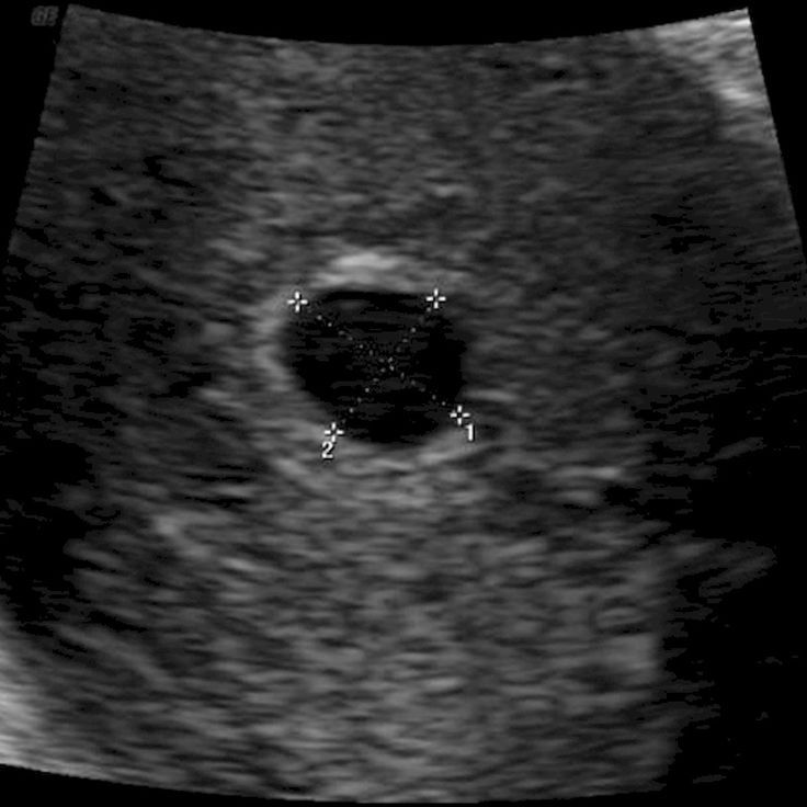
Other times, you might have experienced a blighted ovum — when the embryo fails to develop or stops developing, yet a gestational sac remains. Or, unfortunately, you might have miscarried.
Your doctor will be able to give you insights into what is happening in your specific case and when, if desired, you can try for another pregnancy.
The first trimester is a busy time for your little one. This is when all of the core building blocks of their body are being developed.
At 8 weeks, your fetus is about the size of a kidney bean and could be nearly half an inch long. While they still don’t look like the bouncing bundle of joy you’ll give birth to, they look more human and less otherworldly.
Now they have arm and leg buds, and although they’re webbed, they have fingers and toes. Other essential bodily infrastructure — like bones, muscles, and skin — are also developing, but their skin is still see-through at this point. They’re a busy little thing that’s constantly moving right now!
The first trimester can be a rollercoaster, and not just because you’re excited to be pregnant. The first trimester can throw some rough symptoms at you, and around 8 weeks is when they can kick into high gear. Common symptoms include:
- fatigue
- sore or tender breasts
- morning sickness
- nausea that can last all day
- difficulty sleeping
- frequent urination
- heartburn
When you first know you’re pregnant (via a pregnancy test), you should contact a doctor or a healthcare professional to see when you should come in for an evaluation and ultrasound. This is often done to confirm the pregnancy, verify your expected due date, and ensure that your baby — or babies — has a healthy heartbeat.
Your 8-week appointment may include a transvaginal or abdominal ultrasound, which is low risk but can offer the first glimpse at your baby. However, it’s important to know that when it’s this early in the pregnancy, you might not be able to identify a heartbeat or see your fetus just yet.
What Can I See On an 8-Week Ultrasound?
In the early days of pregnancy, those initial appointments can feel surreal — especially if this is your first pregnancy.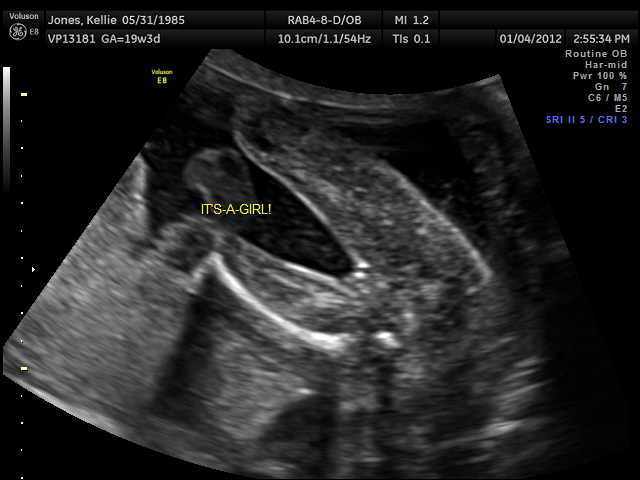 Those first appointments are usually designed to get a baseline understanding of your health going into pregnancy and to make sure things are progressing properly.
One major milestone is the 8-week ultrasound. So, why do you have an ultrasound this early into pregnancy, and what can you expect at your 8-week ultrasound? We’ll answer those questions and more.
Even though you can get a positive pregnancy test result roughly 2 weeks after conception, it can be a while until that tiny ball of cells demonstrates physical changes that confirm your pregnancy is progressing. Specifically, a healthcare professional will want to confirm that your fetus has a heartbeat — a clear sign that it’s alive.
In some cases, a heartbeat may be detected as early as 6 weeks. When you have a positive pregnancy test, contact a physician or healthcare professional to see if you need to come in for an ultrasound.
Transvaginal vs. abdominal ultrasounds
When most of us think of ultrasounds, we think of a technician passing a wand over a person’s stomach that’s covered in gel. This is known as an abdominal ultrasound. In most situations, early ultrasounds generally take less than half an hour.
But a transvaginal ultrasound is when a wand is inserted into the vagina. Often, this is used during early pregnancy to get a closer look at the fetus.
Beyond heartbeat, the technician or physician will be able to immediately identify key features such as the gestational sac and your fetus’s crown-rump length. These can help determine gestational age and pinpoint your due date.
Share on PinterestSuzanne Tucker/Shutterstock
This is going to be your first peek at your growing bundle of joy! Don’t expect to see a lot of definition or details this early in the game.
For now, you’ll see a little figure that looks something like an oblong bean. If there are twins, you might see two figures. The head is still nearly the same size as the rest of the body.
You’ll also see the gestational sac, the fluid-filled space around your baby(s). Within it, you can also see the yolk sac, which is a bubble-like structure. Depending on the location, you might even get to hear their heartbeat, too.
The main reasons for the 8-week ultrasound may be to confirm a pregnancy, determine a due date, and confirm the baby’s heartbeat. First, your doctor or technician will look for key physical indicators, like a gestational sac and a fetal pole, to verify the pregnancy is in the uterus. This is may be your first indication of twins.
Once they’ve confirmed that you’re pregnant, the next step is to verify your projected due date. Even though you might have initially received a projected due date at an earlier appointment, it’s not always accurate. The initial due date is determined by confirming the first day of your last period, deducting 3 months, and then adding 1 year and 7 days. But because not every person’s menstrual cycle is the same length of time, these projections can be off.
With an ultrasound, a physician or technician can determine gestational age and due date by measuring the size of your fetus.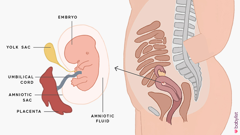 The accepted method for early pregnancy dating is the crown-rump length (CRL) measurement because it’s the most accurate (within about 5 to 7 days) in the first trimester.
When you can’t see the baby or a heartbeat
Sometimes you can’t see the fetus or hear a heartbeat — but that doesn’t always mean the worst. Sometimes it means that your calculations on the conception date were off.
If you ovulated and conceived later than you initially assumed, you might be getting an ultrasound too early to get a physical confirmation. In other scenarios, you might have large fibroids or anatomic issues with the uterus that can make screening your uterus more difficult.
But in some situations, it might not be the news you hoped for. Occasionally, the absence of a visible fetus in the uterus could mean you have an ectopic pregnancy, where the embryo implanted outside of the uterine cavity.
Other times, you might have experienced a blighted ovum — when the embryo fails to develop or stops developing, yet a gestational sac remains. Or, unfortunately, you might have miscarried.
Your doctor will be able to give you insights into what is happening in your specific case and when, if desired, you can try for another pregnancy.
The first trimester is a busy time for your little one. This is when all of the core building blocks of their body are being developed.
At 8 weeks, your fetus is about the size of a kidney bean and could be nearly half an inch long. While they still don’t look like the bouncing bundle of joy you’ll give birth to, they look more human and less otherworldly.
Now they have arm and leg buds, and although they’re webbed, they have fingers and toes. Other essential bodily infrastructure — like bones, muscles, and skin — are also developing, but their skin is still see-through at this point. They’re a busy little thing that’s constantly moving right now!
The first trimester can be a rollercoaster, and not just because you’re excited to be pregnant. The first trimester can throw some rough symptoms at you, and around 8 weeks is when they can kick into high gear. Common symptoms include:
- fatigue
- sore or tender breasts
- morning sickness
- nausea that can last all day
- difficulty sleeping
- frequent urination
- heartburn
When you first know you’re pregnant (via a pregnancy test), you should contact a doctor or a healthcare professional to see when you should come in for an evaluation and ultrasound. This is often done to confirm the pregnancy, verify your expected due date, and ensure that your baby — or babies — has a healthy heartbeat.
Your 8-week appointment may include a transvaginal or abdominal ultrasound, which is low risk but can offer the first glimpse at your baby. However, it’s important to know that when it’s this early in the pregnancy, you might not be able to identify a heartbeat or see your fetus just yet.
prices for tests for pregnant women
During the development of the fetus, ultrasound is one of the key methods of examination. There is no exact schedule for ultrasound, so the doctor focuses primarily on the condition of the patient, the condition of the fetus, the presence of somatic pathology. If we take a physiologically occurring pregnancy, then ultrasound is performed in each of the trimesters.
Ultrasound at 8 weeks is the first time that this study is recommended. The eighth week is the first critical period, therefore, it is most rational to carry out diagnostics during this period.
Since it is the 8th week of pregnancy, a transvaginal examination is performed more often, which is more reliable. At this time, transabdominal ultrasound is used less and less. The transvaginal method does not pose any threat to either the mother or the fetus.
Ultrasound at 8 weeks can confirm
- Normal uterine pregnancy;
- Dimensions of the gestational sac;
- Whether the pregnancy is multiple;
- Place of attachment of the fetus;
- Exclude the presence of malformations;
- Exclude various obstetric complications.
If the doctor confirms a physiologically proceeding pregnancy on an ultrasound examination, does not reveal complications, the fetus develops according to age norms, then the next ultrasound is prescribed at 12 weeks.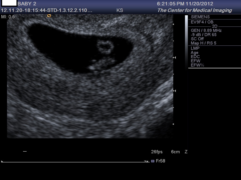 This period is another critical period when it is necessary to assess the condition of the woman and the unborn baby.
If the first two critical periods are mainly related to hormonal changes, then the next period - 22 weeks, is most often associated with sexual infections. Therefore, we also perform an ultrasound at 22 weeks. Pregnancy can provoke the development of latent infections, which, in turn, can lead to certain complications.
Tests at 8 weeks of pregnancy
There are no exact recommendations for registering pregnant women, but doctors recommend starting monitoring at the antenatal clinic before 12 weeks. There is a mandatory list of tests in accordance with the order of the Ministry of Health of the Russian Federation, and an additional one, which is appointed by the gynecologist individually.
Tests at 8 weeks of gestation:
- CBC;
- OAM;
- Swab;
- Determination of blood group and Rh factor;
- Tests for major venereal diseases.

In addition, the 8th week of pregnancy is the period of visiting the following doctors:
- Therapist;
- Optometrist;
- ENT;
- Dentist.
Accordingly, if any pathology is detected, any of these specialists has the right to prescribe an additional analysis in their direction. If this pregnancy proceeds with complications, or if there are previous complications in the anamnesis, then the laboratory and instrumental complex may expand.
You will have to take tests regularly, so you should prepare in advance for the next tests at week 12.
Ultrasound at 12 weeks pregnant
As we mentioned earlier, the next critical period is the 12th week of pregnancy. Therefore, the next ultrasound is performed at this time. Here we are not talking about a simple study, but about screening, which allows you to identify intrauterine malformations of the fetus.
In contrast to the ultrasound at 8 weeks, this is predominantly a transabdominal examination.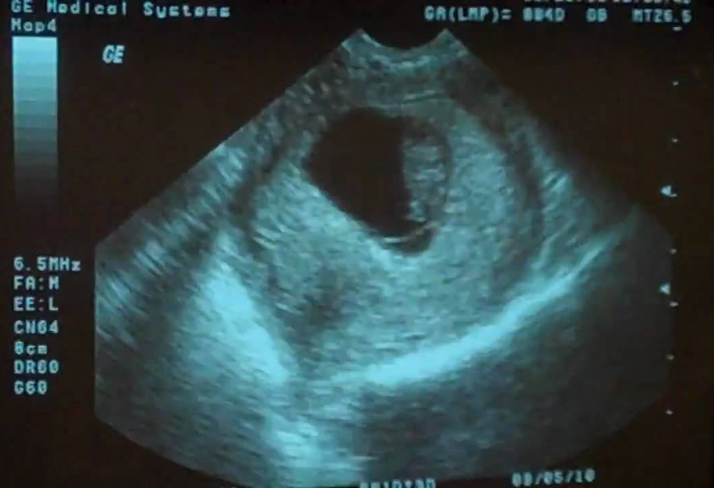 Transvaginal ultrasound is performed only for certain indications:
- Low insertion of the placenta;
- Isthmic-cervical insufficiency;
- Pelvic inflammation;
- Atypical location of myomatous nodes;
- Malposition of the fetus, in which transabdominal ultrasound at 12 weeks of gestation is uninformative.
So, what information does an ultrasound give a gynecologist?
- Detection of deviations in the growth and development of the fetus;
- Possibility to suspect congenital anomalies;
- Exact gestational age;
- Palpitation;
- Cord entanglement;
- Characteristics of amniotic fluid;
- Condition of the placenta.
It must be clearly understood that ultrasound during pregnancy is only one of the methods of examination, therefore, if there are any deviations in the indicators, you should not panic, you should consult a specialist. The doctor, interpreting the results, always relies on many factors, which allows him to adequately assess the true state of the future mother and fetus.
Even if deviations in some indicators are confirmed, doctors still only talk about a certain probability of developing a particular pathology. In this case, a second screening may be prescribed, or a similar study is carried out at a later date. In any case, the final decision regarding the continuation of the pregnancy is made exclusively by a specialist.
Tests at 12 weeks of pregnancy
As we have already said, 12 weeks is the time for screening. For this, an ultrasound examination is prescribed, the external indicators of the woman are evaluated, and a blood test is performed.
Particular attention is paid to two coefficients: β-hCG and PAPP-A. It is believed that in the presence of deviations according to these parameters, one can judge the presence of fetal pathologies. According to the level of hCG, doctors predict the further management of pregnancy and the possibility of miscarriage or premature birth.
Accordingly, according to the screening results, it is possible to identify:
- The baby has Down syndrome;
- Multiple pregnancy;
- Specify term;
- Detect toxicosis;
- Determine the risk of miscarriage;
- Check for other chromosomal abnormalities.

Thus, tests at the 12th week of pregnancy are decisive for solving a number of issues related to the further development of the fetus. It should be clearly understood that screening is not 100% reliable, therefore, if you receive any deviations from normal results, you should immediately contact a specialist who can correctly assess such changes in indicators.
Ultrasound at 22 weeks gestation
The next scheduled ultrasound is performed at 22 weeks of gestation. Here, for the doctor, it is not the condition of the mother or the size of the fetus that is important, but the assessment of the formation of various organs and systems, as well as bones.
So, the 22nd week - the fetus has already formed all the main organs and systems, and on the monitor screen the child acquires the outlines familiar to future parents. Accordingly, the following indicators come to the fore:
- All internal organs should form. During an ultrasound, the doctor evaluates their location, how they function;
- The spine is also studied, the presence of bones and their sizes are determined;
- Assessing the brain and its activity;
- Assess the condition of the placenta, umbilical cord vessels;
- Composition of amniotic fluid;
- Condition of the cervix.

Unlike ultrasound at 8 weeks, ultrasound at this time is performed transabdominally. 22 weeks is already the second trimester, and right now the question is being decided whether there are serious deviations in the development of the fetus, and further actions of obstetricians. The question of whether to continue a further pregnancy is decided not by one doctor, but by a whole council, based on numerous research results.
Tests at 22 weeks pregnant
Week 22 is when the second screening is more common. This analysis is prescribed in the period from 18 to 24 weeks, but this time is considered optimal. Accordingly, during repeated screening, the doctor already makes final conclusions about the condition and development of the fetus, the presence of deviations.
In addition to ultrasound, the doctor prescribes mandatory tests to monitor the woman's health. The expectant mother again takes a general blood and urine test, where the most important are a number of indicators: hemoglobin, erythrocytes, leukocytes, ESR, protein in the urine.
During the examination, the gynecologist necessarily measures the woman's height, weight, blood pressure level, and measures the abdomen. Tests at 22 weeks of gestation are designed to assess the likelihood of developing preeclampsia. Here, the role is played not by hormonal changes, but by the exacerbation of chronic pathology. Therefore, timely screening and assessment of the woman's condition allows you to plan further tactics for managing pregnancy.
You can find out the cost of an appointment with a gynecologist by phone number +7 495 478 10 03. Also during the planning of pregnancy in our center there is an opportunity to undergo a comprehensive examination of the body for women, and for future fathers - a comprehensive examination of men.
The author of the text: Sokolov Alexander Aleksandrovich
Position: Ultrasound diagnostic doctor
Total experience: 6 years
90,000 conducting an ultrasound at 8 weeks0186Ultrasound at the 8th obstetric week of pregnancy
For ultrasound in the first trimester of , certain terms are set - 10-13 weeks. During the gestation period of this particular duration, the fetus will already begin laying the main internal organs, the skeleton, and the brain. With the help of ultrasound, all these parameters can be seen and evaluated.
However, it happens that the doctor prescribes an ultrasound at the 8th week of pregnancy, when it is too early to judge the presence or absence of pathologies in the fetus. Such early research has goals.
Why is ultrasound done at 8 weeks pregnant?
Ultrasound at the eighth week is done for:
- Confirmation of uterine pregnancy.
- Clarification of the position of the ovum (it is necessary to exclude a pathological ectopic pregnancy).
- Detection of the number of embryos in the fetal egg (at this time it will already be possible to detect a multiple pregnancy).
- Identification of pathologies of the female reproductive system (for example, a short cervix can cause miscarriage or premature birth).

- Clarification of the gestational age.
- Identification of threats of miscarriage or its fading.
Fetal development at 8 weeks of gestation
By the eighth week, the fetus reaches a length of two centimeters. He has already formed the circulatory system, the first veins and the main internal organs, which will continue to develop throughout the entire period of pregnancy. The fetal heart is already beating, and this can be seen if an ultrasound is performed.
Fingers are already appearing on the hands and feet of the baby, and the upper limbs are increasing in length. Shoulders and knees appeared. The inner ear, that is, the center of balance, begins to form.
The eyes of the fetus are forming, they are already closed by thin eyelids.
Preparation for ultrasound at 8 weeks
Ultrasound at the 8th obstetric week of pregnancy does not require special preparation. Women are simply advised 2-3 days before the study to refrain from food that provokes excessive gas formation in the intestines, as well as from carbonated drinks. You need to come to the doctor's appointment calm and in a good mood.
How is an ultrasound done at the eighth week of pregnancy?
At the eighth week, ultrasound can be performed in two ways:
- Transvaginally using a special probe inserted into the vaginal cavity.
- Transabdominally , i.e. through the skin of the abdomen of a pregnant woman.
The first method will give more accurate results, but may cause some discomfort to the woman. In addition, transvaginal ultrasound is not recommended if a woman complains of abdominal pain or bloody discharge.
After the ultrasound, the doctor may take a photo showing the tiny embryo.
What will the ultrasound show?
Ultrasound at the eighth week of pregnancy, with its correct course, visualizes a fetal egg with a live embryo inside. Also, according to the results of the study, it will be possible to judge the place of attachment of the chorion, the position of the fetal egg in the uterine cavity.
Ultrasound at 8 weeks should show the fetal heartbeat and the frequency can be assessed.
At a period of 8 weeks, it is already possible to identify some pathologies in the development of the fetus, in particular in the work of his heart. At the same time, specialists have not yet made unambiguous conclusions about the presence of heart disease.
Ultrasound will also show how the neural tube of the fetus develops, how limbs are formed.
Embryo examination
A large proportion of the total ultrasounds at the eighth week of is the study of the embryo. Its size, location, structure is important. It is also necessary to evaluate the motor activity of the embryo, which should be present even at such a short time. At the eighth week, the embryo should be clearly visible. If it is not there, most likely its development has froze or there is an anembryonic fact, that is, the initial absence of an embryo in a fetal egg (or its death immediately after conception).
Yolk sac examination
When conducting an ultrasound scan at 8-9 obstetric weeks of pregnancy, it is required to assess not only the state of the embryo itself, but also the organs outside its structure. These organs include the yolk sac.
Its presence in the ovum is temporary. The pouch appears approximately 15-16 days after fertilization and disappears by the end of the first trimester.
The yolk sac plays an important role in the development of the embryo - it produces germ cells, forms embryonic red blood cells for the first time, produces proteins necessary for development, helps to form immunity, and so on.
At the eighth week, the ultrasound should show the yolk sac. Its size should be approximately 4-5 mm. An increase in the size of the yolk sac can be the first sign of pathological development of the fetus.
Ultrasound norms at the 8th week of pregnancy
At the eighth week of gestation, an ultrasound is done and they look at how the parameters of the developing fetus correlate with the norm for a particular gestational age.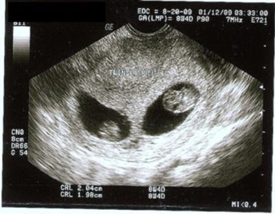 The main indicators of fetal development include:
- The size of the embryo from the coccyx to the crown (KTP). At 8 weeks for KTP, the norm is 14-20 mm.
- Heart rate (HR). The norm for the 8th week is 110-150 beats / min.
- Bipaterial head size (BDP). The norm is 6 mm.
- Yolk sac. This extraembryonic organ should be visualized and reach 4-5 mm in diameter.
A deviation from the norm of one or several parameters at once can be a signal that the fetus is developing with pathology. Although doctors do not make unambiguous conclusions on such a short period of time and, as a rule, prescribe a second ultrasound in 1-2 weeks.
Sign up for a consultation or diagnostic today!
You can make an appointment by phone: +7 (812) 901-03-03
Or leave a request
full name
Phone number
By clicking the "Make an appointment" button, I accept the terms of the Personal Data Processing and Security Policy and consent to the processing of my personal data.
Our medical centers
Making an appointment
Patient's last name *
Incorrect first name
Name *
Middle name
Contact phone *
E-mail *
By clicking the "Make an appointment" button, I accept the terms of the Personal Data Processing and Security Policy and consent to the processing of my personal data.
Registration and payment for repeated online appointment
Patient's last name *
Incorrect first name
Name *
Middle name *
Contact phone *
E-mail *
By clicking the "Submit request" button, I accept the terms of the Personal Data Processing and Security Policy and consent to the processing of my personal data.