When do you have a ultrasound during pregnancy
Ultrasound during pregnancy | March of Dimes
A prenatal ultrasound uses sound waves and a computer screen to show a picture of your baby inside the womb.
Ultrasounds can help your health care provider see how your baby is growing and developing.
Your provider also may use ultrasounds to see if other tests need to be done to check on your baby’s health.
There are several types of ultrasounds and they are safe for you and your baby when done by a trained health care provider.
What is an ultrasound?
Ultrasound (also called sonogram) is a prenatal test offered to most pregnant women. It uses sound waves to show a picture of your baby in the uterus (womb). Ultrasound helps your health care provider check on your baby’s health and development.
Ultrasound can be a special part of pregnancy—it’s the first time you get to “see” your baby! Depending on when it’s done and your baby’s position, you may be able to see his hands, legs and other body parts. You may be able to tell if your baby’s a boy or a girl, so be sure to tell your provider if you don’t want to know!
Most women get an ultrasound in their second trimester at 18 to 20 weeks of pregnancy. Some also get a first-trimester ultrasound (also called an early ultrasound) before 14 weeks of pregnancy. The number of ultrasounds and timing may be different for women with certain health conditions like as asthma and obesity.
Talk to your provider about when an ultrasound is right for you.
What are some reasons for having an ultrasound?
Your provider uses ultrasound to do several things, including:
- To confirm (make sure) you’re pregnant
- To check your baby’s age and growth. This helps your provider figure out your due date.
- To check your baby’s heartbeat, muscle tone, movement and overall development
- To check to see if you’re pregnant with twins, triplets or more (also called multiples)
- To check if your baby is in the heads-first position before birth
- To examine your ovaries and uterus (womb).
 Ovaries are where eggs are stored in your body.
Your provider also uses ultrasound for screening and other testing. Screening means seeing if your baby is more likely than others to have a health condition; it doesn’t mean finding out for sure if your baby has the condition. Your provider may use ultrasound:
- To screen for birth defects, like spina bifida or heart defects. After an ultrasound, your provider may want to do more tests, called diagnostic tests, to see for sure if your baby has a birth defect. Birth defects are health conditions that a baby has at birth. Birth defects change the shape or function of one or more parts of the body. They can cause problems in overall health, in how the body develops, or in how the body works.
- To help with other prenatal tests, like chorionic villus sampling (also called CVS) or amniocentesis (also called amnio). CVS is when cells from the placenta are taken for testing.
 The placenta is tissue that provides nutrients for your baby. Amnio is a test where amniotic fluid and cells are taken from the sac around your baby.
 - To check for pregnancy complications, including ectopic pregnancy, molar pregnancy and miscarriage.
Are there different kinds of ultrasound?
Yes. The kind you get depends on what your provider is checking for and how far along you are in pregnancy. All ultrasounds use a tool called a transducer that uses sound waves to create pictures of your baby on a computer. The most common kinds of ultrasound are:
- Transabdominal ultrasound. When you hear about ultrasound during pregnancy, it’s most likely this kind. You lay on your back on an exam table, and your provider covers your belly with a thin layer of gel. The gel helps the sound waves move more easily so you get a better picture. Then he moves the transducer across your belly. You may need to drink several glasses of water about 2 hours before the exam to have a full bladder during the test.
 A full bladder helps sound waves move more easily to get a better picture. Ultrasound is painless, but having a full bladder may be uncomfortable. The ultrasound takes about 20 minutes.
 - Transvaginal ultrasound. This kind of ultrasound is done through the vagina (birth canal). You lay on your back on an exam table with your feet in stirrups. Your provider moves a thin transducer shaped like a wand into your vagina. You may feel some pressure from the transducer, but it shouldn’t cause pain. Your bladder needs to be empty or just partly full. This kind of ultrasound also takes about 20 minutes.
In special cases, your provider may use these kinds of ultrasound to get more information about your baby:
- Doppler ultrasound. This kind of ultrasound is used to check your baby’s blood flow if he’s not growing normally. Your provider uses a transducer to listen to your baby’s heartbeat and to measure the blood flow in the umbilical cord and in some of your baby’s blood vessels.
 You also may get a Doppler ultrasound if you have Rh disease. This is a blood condition that can cause serious problems for your baby if it’s not treated. Doppler ultrasound usually is used in the last trimester, but it may be done earlier.
 - 3-D ultrasound. A 3-D ultrasound takes thousands of pictures at once. It makes a 3-D image that’s almost as clear as a photograph. Some providers use this kind of ultrasound to make sure your baby’s organs are growing and developing normally. It can also check for abnormalities in a baby’s face. You also may get a 3-D ultrasound to check for problems in the uterus.
- 4-D ultrasound. This is like a 3-D ultrasound, but it also shows your baby’s movements in a video.
Does ultrasound have any risks?
Ultrasound is safe for you and your baby when done by your health care provider. Because ultrasound uses sound waves instead of radiation, it’s safer than X-rays. Providers have used ultrasound for more than 30 years, and they have not found any dangerous risks.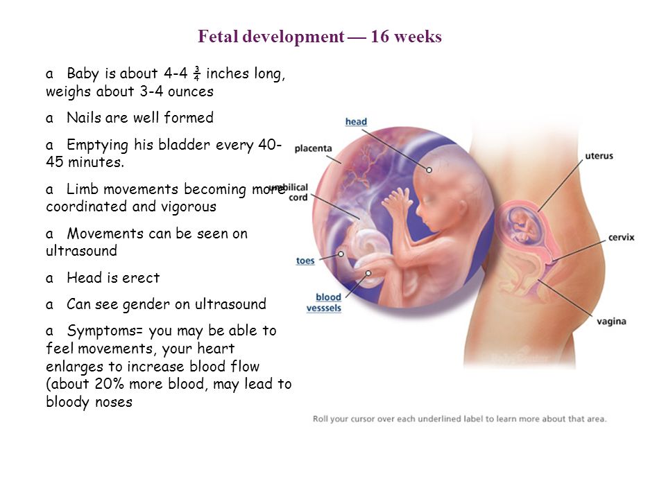
If your pregnancy is healthy, ultrasound is good at ruling out problems, but it can’t find every problem. It may miss some birth defects. Sometimes, a routine ultrasound may suggest that there is a birth defect when there really isn’t one. While follow-up tests often show that the baby is healthy, false alarms can cause worry for parents.
You may know of some places, like stores in a mall, that aren’t run by doctors or other medical professionals that offer “keepsake” 3-D or 4-D ultrasound pictures or videos for parents. The American College of Obstetricians and Gynecologists (ACOG), the Food and Drug Administration (FDA) and the American Institute of Ultrasound in Medicine (AIUM) do not recommend these non-medical ultrasounds. The people doing them may not have medical training and may give you wrong or even harmful information.
What happens after an ultrasound?
For most women, ultrasound shows that the baby is growing normally. If your ultrasound is normal, just be sure to keep going to your prenatal checkups.
Sometimes, ultrasound may show that you and your baby need special care. For example, if the ultrasound shows your baby has spina bifida, he may be treated in the womb before birth. If the ultrasound shows that your baby is breech (feet-down instead of head-down), your provider may try to flip your baby’s position to head-down, or you may need to have a cesarean section (also called c-section). A c-section is surgery in which your baby is born through a cut that your doctor makes in your belly and uterus.
No matter what an ultrasound shows, talk to your provider about the best care for you and your baby.
Last reviewed: October, 2019
What To Expect, Purpose & Results
Overview
What is an ultrasound in pregnancy?
A prenatal ultrasound (or sonogram) is a test during pregnancy that checks on the health and development of your baby. An obstetrician, nurse midwife or ultrasound technician (sonographer) performs ultrasounds during pregnancy for many reasons. Sometimes ultrasounds occur to check on your baby and make sure they’re growing properly. Other times your pregnancy care provider orders an ultrasound after they detect a problem.
During an ultrasound, sound waves are sent through your abdomen or vagina by a device called a transducer. The sound waves bounce off structures inside your body, including your baby and your reproductive organs. Then, the sound waves transform into images that your provider can see on a screen. It doesn’t use radiation, like X-rays, to see your baby.
Even though prenatal ultrasounds are safe, you should only have them when it’s medically necessary. If there’s no reason for an ultrasound (for example, if you just want to see your baby), your insurance company might not pay for it.
Prenatal ultrasounds may be called fetal ultrasounds or pregnancy ultrasounds. Your provider will talk to you about when you can expect ultrasounds during pregnancy based on your health history.
Why is a fetal ultrasound important during pregnancy?
An ultrasound is one of the few ways your pregnancy care provider can see and hear your baby. It can help them determine how far along you are in pregnancy, if your baby is growing properly or if there are any potential problems with the pregnancy. Ultrasounds may occur at any time in pregnancy depending on what your provider is looking for.
What can be detected in a pregnancy ultrasound?
A prenatal ultrasound does two things:
- Evaluates the overall health, growth and development of the fetus.
- Detects certain complications and medical conditions related to pregnancy.
In most pregnancies, ultrasounds are positive experiences and pregnancy care providers don’t find any problems. However, there are times this isn’t the case and your provider detects birth disorders or other problems with the pregnancy.
Reasons why your provider performs a prenatal ultrasound are to:
- Confirm you’re pregnant.
- Check for ectopic pregnancy, molar pregnancy, miscarriage or other early pregnancy complications.
- Determine your baby’s gestational age and due date.

- Check your baby’s growth, movement and heart rate.
- Look for multiple babies (twins, triplets or more).
- Examine your pelvic organs like your uterus, ovaries and cervix.
- Examine how much amniotic fluid you have.
- Check the location of the placenta.
- Check your baby’s position in your uterus.
- Detect problems with your baby’s organs, muscles or bones.
Ultrasound is also an important tool to help providers screen for congenital conditions (conditions your baby is born with). A screening is a type of test that determines if your baby is more likely to have a specific health condition. Your provider also uses ultrasound to guide the needle during certain diagnostic procedures in pregnancy like amniocentesis or CVS (chorionic villus sampling).
An ultrasound is also part of a biophysical profile (BPP), a test that combines ultrasound with a nonstress test to evaluate if your baby is getting enough oxygen.
How many ultrasounds do you have during your pregnancy?
Most pregnant people have one or two ultrasounds during pregnancy. However, the number and timing vary depending on your pregnancy care provider and if you have any health conditions. If your pregnancy is high risk or if your provider suspects you or your baby has a health condition, they may suggest more frequent ultrasounds.
When do you have your first prenatal ultrasound?
The timing of your first ultrasound varies depending on your provider. Some people have an early ultrasound (also called a first-trimester ultrasound or dating ultrasound). This can happen as early as seven to eight weeks of pregnancy. Providers do an early ultrasound through your vagina (transvaginal ultrasound). Early ultrasounds do the following:
- Confirm pregnancy (by detecting a heartbeat).
- Check for multiple fetuses.
- Measure the size of the fetus.
- Help confirm gestational age and due date.
Some providers perform your first ultrasound closer to 12 weeks of pregnancy.
20-week ultrasound (anatomy scan)
You can expect an ultrasound around 18 to 20 weeks in pregnancy. This is known as the anatomy ultrasound or 20-week ultrasound. During this ultrasound, your pregnancy care provider can see your baby’s sex (if your baby is in a good position for viewing their genitals), detect birth disorders like cleft palate or find serious conditions related to your baby’s brain, heart, bones or kidneys. If your pregnancy is progressing well and with no complications, your 20-week ultrasound may be your last ultrasound during pregnancy. However, if your provider detects a problem during your 20-week ultrasound, they may order additional ultrasounds.
How soon can you see a baby on an ultrasound?
Pregnancy care providers can detect an embryo on an ultrasound as early as six weeks into the pregnancy. An embryo develops into a fetus around the eighth week of pregnancy.
If your last menstrual period isn’t accurate, it’s possible that it may be too early to detect a fetal heart rate.
Which ultrasound is most important during pregnancy?
All ultrasounds during pregnancy are important. Your pregnancy care provider uses ultrasound to tell them important information about your pregnancy.
Test Details
What are the two main types of pregnancy ultrasounds?
The two main types of pregnancy ultrasound are transvaginal ultrasound and abdominal ultrasound. Both use the same technology to produce images of your baby. Your pregnancy care provider performs a transvaginal ultrasound by placing a wand-like device inside your vagina. They perform an abdominal ultrasound by placing a device on the skin of your belly.
Transvaginal ultrasound
During a transvaginal ultrasound, your pregnancy care provider places a device inside your vaginal canal (similar to how you place a tampon). In early pregnancy, this ultrasound helps to detect a fetal heartbeat or determine how far along you are in your pregnancy (gestational age). Images from a transvaginal ultrasound are clearer in early pregnancy as compared to abdominal ultrasound.
Abdominal ultrasound
Your pregnancy care provider performs an abdominal ultrasound by placing a transducer directly on your skin. Then, they move the transducer around your belly (abdomen) to capture images of your baby. Sometimes slight pressure has to be applied to get the best views. Providers use abdominal ultrasounds after about 12 weeks of pregnancy.
Traditional ultrasounds are 2D. More advanced technologies like 3D or 4D ultrasound can create better images. This is helpful when your provider needs to see your baby’s face or organs in greater detail. Not all providers have 3D or 4D ultrasound equipment or specialized training to conduct this type of ultrasound.
Your provider may recommend other types of ultrasounds. Examples of additional ultrasounds are:
- Doppler ultrasound: This type of ultrasound checks how your baby’s blood flows through its blood vessels. Most Doppler ultrasounds occur later in pregnancy.
- Fetal echocardiogram: This type of ultrasound looks at your baby’s heart size, shape, function and structure. Your provider may use it if they suspect your baby has a congenital heart condition, if you had another child that had a heart condition or if you have certain health conditions that warrant taking a closer look at the heart.

How do I prepare for the test?
There’s no special preparation for an ultrasound. Some pregnancy care providers ask that you come with a full bladder and don’t use the restroom before the test. This helps them view your baby better on the ultrasound. You can bring a support person, but bringing children is discouraged as this is an important test that requires complete focus.
You may be asked to change into a hospital gown, but this isn’t usually required for abdominal ultrasounds. If your provider is performing a transvaginal ultrasound in your first trimester, you’ll put on a hospital gown or undress from the waist down.
What should I expect during a prenatal ultrasound?
You’ll lie on a padded examining table during the test. Most ultrasounds occur in a dimly lit room, which helps your ultrasound technician (or sonographer) see the screen. Your sonographer applies a small amount of water-soluble gel to the skin of your belly. The gel doesn’t harm your skin or stain your clothes, but it may feel cold. This gel helps transmit sound waves more efficiently.
Next, the sonographer places a transducer on the skin of your abdomen. The transducer sends sound waves into your body, which reflect off internal structures, including your baby. The sound waves that reflect back create pictures on a screen. Your sonographer uses these images to take important measurements such as your baby’s head circumference and length. You may see them making lines on the screen or clicking a button to “freeze” certain angles.
There’s virtually no discomfort during a prenatal ultrasound. You may feel mild discomfort if you have to pee. The ultrasound test takes about 30 minutes to complete.
If you have a transvaginal ultrasound, the process is only different in that the transducer is inside your vagina and not on your belly.
What should I expect after a pregnancy ultrasound?
If you had an abdominal ultrasound, your sonographer wipes the gel off your belly. They may print off some ultrasound pictures for you to take home with you.
In most cases, your sonographer won’t discuss the results of your test with you. If your obstetrician performs your ultrasound, they may discuss what they see as they go along.
If a sonographer performs your ultrasound, an obstetrician will look at the images, then discuss their findings with you at your next appointment. Most practices schedule your appointment right after your ultrasound so you get your results the same day.
What are the risks of prenatal ultrasounds?
Studies have shown ultrasounds are safe during pregnancy. There are no harmful side effects to you or your baby.
Is it safe to do an ultrasound every month during pregnancy?
While ultrasounds are safe for you and your baby, most major medical associations recommend that pregnancy care providers should only do ultrasounds when the tests are medically necessary. If your ultrasounds are normal and your pregnancy is uncomplicated or low risk, repeat ultrasounds aren’t necessary.
Results and Follow-Up
What results do you get on a pregnancy ultrasound?
Your ultrasound results will be normal or abnormal. A normal result means your pregnancy care provider didn’t find any problems and that your baby is growing and developing normally. An abnormal result means your provider noticed something irregular. If they do, your provider will order additional ultrasounds or diagnostic tests to determine if something is wrong.
Occasionally, the ultrasound is incomplete if there’s difficulty seeing all the structures needed for that particular ultrasound. Your baby’s position or movement sometimes makes it difficult to see everything your provider needs to see. If this is the case, you’ll need a repeat ultrasound and they’ll try again.
There are some limitations to ultrasounds, so your provider may not find certain abnormalities until after birth.
What are reasons you need more ultrasounds during pregnancy?
There are several reasons your pregnancy care provider may order additional ultrasounds during your pregnancy. Some of these reasons include:
- Problems with your ovaries, uterus, cervix or other pelvic organs.
- Your baby is measuring small for their gestational age or your provider suspects IUGR (intrauterine growth restriction).
- Problems with the placenta like placenta previa or placental abruption.
- You’re pregnant with twins, triplets or more.
- Your baby is breech.
- You have too much amniotic fluid (polyhydramnios).
- You have too little amniotic fluid (oligohydramnios).
- You have a condition like gestational diabetes or preeclampsia.
- Your baby has a congenital disorder.
Normal results on pregnancy ultrasounds can vary. Generally, a normal result means your baby appears healthy and your provider didn’t find any issues.
Why do some pregnancy providers schedule ultrasounds differently?
The number of ultrasounds you’ll have and when you have them can vary between providers. Every practice operates differently and some providers do things differently based on your health history or symptoms.
When does a pregnancy ultrasound determine sex?
Your baby’s sex isn’t visible on an ultrasound until about 18 to 20 weeks. Be sure to tell your pregnancy care provider whether or not you want to know the sex of your baby before your ultrasound.
A note from Cleveland Clinic
An ultrasound during pregnancy can be both exciting and terrifying. Your pregnancy care provider uses ultrasound to get a better idea of how your baby is growing and developing. There are different types of ultrasounds, and the exact timing may vary depending on your provider. Most pregnant people have two ultrasounds — one in the first trimester and one in the second trimester. However, if there’s a potential complication or medical reason for more ultrasounds, your provider will order more as a precaution. Talk to your provider about the ultrasound schedule during pregnancy and what you can expect.
When to do the first ultrasound during pregnancy - "happy days" for screening of the 1st trimester
The world of a modern woman is always full of important events, meetings and an endless series of pre-planned things. When pregnancy occurs, many planned events can be moved from “expected” to “impossible” or simply postponed indefinitely. It is quite difficult to worry in advance and reschedule an important event at the most appropriate time, because planning events against the background of pregnancy is a very difficult science.
Therefore, when pregnancy occurs, it is advisable to draw up at least an approximate “life plan for 9 months”. For example, it is known that maternity leave is most often preceded by unspent regular leave, and registration for pregnancy is preceded by the fact that a pregnancy has occurred and is developing normally.
Many events will now depend on how the new life develops. So, when a multiple pregnancy is diagnosed, not only the estimated due date will change, but maternity leave will come 14 days earlier, and the beginning of unspent leave will move with it. Therefore, one of the first steps in the event of a desired pregnancy is the passage of an ultrasound examination (ultrasound). Scientific and technological progress is rapidly changing the whole world, including the state of the prenatal ultrasound diagnostic service. Opportunities are changing, and with them the timing of the passage of research, which I would like to dwell on in more detail.
When to do an ultrasound scan during pregnancy
In 2012, the regulation “Decree of the Health Committee of the Government of St. Petersburg No. 39-r”1 came into force in St. Petersburg. Based on the above document, for a full-fledged ultrasound screening, it is desirable to undergo a study in “... the first trimester: 11 + 0-13 + 6 weeks; 18+0-20+6 weeks; and 32+0-34+6 weeks of pregnancy.” However, these terms apply only to uncomplicated singleton pregnancies. At the same time, early registration is limited to up to 12 weeks of pregnancy with the ensuing consequences (payment of a lump-sum allowance to women registered in medical institutions in the early stages of pregnancy, clause 2-c of Decree of the Government of the Russian Federation of October 15, 2001 N 727 (as amended on 04. 09.2012).
Therefore, at 11 weeks, a pregnant woman can be registered when establishing the fact of a developing pregnancy , for which ultrasound "... in the first trimester" is recommended. Most doctors recommend doing it at terms 7-8 weeks of pregnancy , because it is at this time that the heartbeat of a developing embryo is always determined as a sign of the physiological development of pregnancy.
The next obligatory steps in the ultrasound are screening. The entire pregnancy is divided into three periods (trimesters) and ultrasound plays an important role in each.
What is the diagnostic window for ultrasound during pregnancy
The first screening study is carried out from 11 weeks 0 days of pregnancy to 13 weeks 6 days of pregnancy. These limits are adopted for the timely detection of pathological conditions that determine the prognosis for the health of the fetus. Theoretically, any pregnant woman can apply for an ultrasound both at the beginning of the eleventh and at the end of the thirteenth week - the entire period is screening. However, among doctors who have dedicated their lives to prenatal ultrasound diagnostics, there is an opinion about the most preferable period in each screening period - the so-called " diagnostic window" or "happy days" .
Terms of fetal ultrasound examination
| Legally regulated period (order KZ SPb No. 39-r dated 02/01/12) | Optimal timing/happy days |
| - | 7-8 |
| 11 weeks 0 days – 13 weeks 6 days | 12 weeks 2 days - 12 weeks 4 days |
| 18 weeks 0 days – 20 weeks 6 days | 20 weeks 0 days - 20 weeks 6 days |
| 32 weeks 0 days – 34 weeks 6 days | 32 weeks 0 days - 33 weeks 3 days |
Diagnostic window in the 1st trimester of pregnancy
For the first screening study, these days include the interval from 12 weeks to 12 weeks. 2 days to 12 weeks 4 days . It is in this interval that the fetus has already grown enough to evaluate the smallest organs (eye lenses, heart), and the probability of ascertaining the most important morphological changes is significantly higher than at 11 weeks 0 days. On the other hand, each day lived by the baby increases not only the height and weight of the body, but the quality of the picture on the ultrasound machine.
According to various authors, the frequency of congenital pathology reaches 5%, and for patients in this group it is especially important to identify the problem at an early stage. In such special cases, it may be necessary to expand the range of diagnostic procedures, including prenatal karyotyping (obtaining samples of fetal tissue or amniotic structures in order to determine its karyotype). It takes time to carry out these procedures, from preparing the necessary tests for a pregnant woman, ending with directly invasive diagnostics and obtaining results about the health of the fetus.
In some cases, the established disease of the fetus raises the question of the impossibility of prolonging the pregnancy. According to the current legislation of the Russian Federation, the procedure of medical termination of pregnancy by the decision of the woman can be performed "... no later than the end of the twelfth week of pregnancy", but "... not earlier than 48 hours from the moment the woman applied to the medical organization for artificial termination of pregnancy" (clause 3.1, clause 3-b of article 56. Federal Law No. 323 of November 21, 2012)2. In other words, if a patient goes for an ultrasound scan at 12 weeks 5 days, if a serious pathology is detected, she is no longer sent for an artificial medical abortion, but for an abortion, which is a more complex and traumatic procedure. Therefore, when asked about the best rock for the first screening, almost any practitioner will answer: "up to 12 weeks 4 days" .
Diagnostic window in the 2nd trimester of pregnancy
The second screening period occurs at 18 weeks. 0 days and ends at 20 weeks 6 days pregnant . "Lucky days" is considered the entire twentieth week: 20 weeks. 0 days - 20 weeks 6 days . Not all the subtleties of the architectonics of the main organs can be considered so successfully, especially in pregnant women with increased body weight at 18 weeks of pregnancy. If so-called "markers of chromosomal problems" are detected, it may be necessary to perform prenatal karyotyping (obtaining fetal blood or amniotic fluid), which may take some time.
The collection of the material itself takes several minutes, but the preparation of the pregnant woman (examination, obtaining the results of blood and urine tests, etc.), transporting the material to the laboratory, examination and obtaining the results can take several days. If serious deviations in the state of health of the fetus are detected, in order to resolve the issue of further management tactics, the pregnant woman is sent to undergo a prenatal consultation consisting of an ultrasound doctor, a specialist in the field to which the identified disease belongs (for example, a surgeon, neurosurgeon, cardiologist, etc. ) .
Prior to the new regulation, the second screening period ranged from 18-22 weeks, even earlier than 18-24 weeks of pregnancy. According to WHO recommendations, the fetus becomes viable from the 22nd week of pregnancy, therefore, before this period it is very important to obtain all possible information about its condition and form a prognosis for health and later life. That is why now there is a restriction regulated by law, so that if serious problems with the health of the fetus are detected, all additional diagnostic procedures should be carried out in a timely manner and, if necessary, terminate the pregnancy without violating the Legislation of the Russian Federation2.
Diagnostic window in the 3rd trimester of pregnancy
The third screening period (32 weeks 0 days - 34 weeks 6 days of pregnancy) has two main objectives: the exclusion of congenital malformations with late manifestation and assessment of the fetal condition. Issuance of a referral for the passage of the third ultrasound along with maternity leave of 30 weeks. 0 days of pregnancy potentiates untimely early appeal of pregnant women for the third screening ultrasound before the period of 32 weeks 0 days, which in turn may require a second planned ultrasound in the "scheduled time". A later turnout (after 34 weeks) reduces the quality of the ultrasound picture obtained by changing the relationship between the amount of amniotic fluid and the volume of the fetal body towards the latter. Therefore " happy days" the third trimester can be considered the period 32 weeks 0 days - 33 weeks 3 days of pregnancy .
Unscheduled ultrasound at any stage is required, as a rule, only in case of complicated pregnancy, therefore, it is prescribed only according to indications and is performed regardless of the gestational age.
Notes: 1 - Order of the Health Committee of the Government of St. Petersburg dated February 1, 2012 N 39-r "On measures to reduce hereditary and congenital diseases in children in St. Petersburg".
2- Article 56 "Artificial termination of pregnancy". Federal Law of the Russian Federation of November 21, 2011 N 323-FZ "On the basics of protecting the health of citizens in the Russian Federation", entered into force: November 22, 2011, published on November 23, 2011 in "RG" - Federal issue No. 5639
When an ultrasound is done during pregnancy - Timing
Ultrasound during pregnancy is a mandatory procedure that allows you to monitor the condition of the fetus. Each registered woman must undergo an ultrasound examination at least three times. What is the timing of the examination? Does it carry any harm? These and other questions will be discussed in the article.
Book an ultrasound for pregnancy
What kind of ultrasound is done during pregnancy?
Ultrasound during pregnancy is a versatile, non-invasive and convenient method of examination. The procedure allows doctors to obtain reliable information about such parameters as:
- gestational age,
- fetal condition,
- its parameters,
- functioning of the umbilical cord, placenta and uterus.
The mechanism of the method consists in the analysis of differences in the reflection of sound waves from structures of different densities. During the study, ultrasound with a frequency of 2 to 10 MHz is used.
When is an ultrasound done during pregnancy?
A pregnant woman is referred by a gynecologist for a routine examination three times in nine months - at the end of each trimester.
- The first study is scheduled for a period of 10-14 weeks. It is this period that is important, since it allows you to most accurately determine the presence or absence of congenital pathologies.
- The second - in the period from 20 to 23 weeks. At this stage, the doctor can make a preliminary conclusion about the rate of fetal development.
As a rule, it is on the second ultrasound that parents find out the gender of the unborn child.
- The final, third, is held at 32-34 weeks and provides information necessary for planning childbirth, namely: the position of the baby, the amount of water, the state of the placenta and much more.
If necessary, the gynecologist in charge of the pregnancy may order the woman in labor to undergo additional examinations at any time.
When to do 3D ultrasound during pregnancy?
Three-dimensional ultrasound is the most modern, improved diagnostic method than the usual 2D. The advantage of 3D ultrasound is that:
- it gives a three-dimensional image of the fetus
- allows you to see the features of the crumbs and even facial expressions
- the quality of the resulting picture on the screen is much higher
A 3D exam takes longer, about 40-50 minutes.
This procedure is best performed during the second scheduled ultrasound, when the main features of the fetus are already formed, and he himself has grown up and learned to make faces. Thanks to this, you can get great pictures for memory. These services are provided by most private medical clinics.
Is it harmful to have an ultrasound during pregnancy?
Some women are afraid to have an ultrasound scan because they are worried that it will negatively affect their baby's development. We can say with certainty that this opinion is erroneous. A number of medical studies confirm the absolute safety of the procedure both for the health of the expectant mother and for the prenatal well-being of the baby. Ultrasound during pregnancy is recognized throughout the world, because it does not have teratogenic (harmful) properties.
Ultrasound during pregnancy should be done by every woman who cares about her own health and the health of her unborn child. To obtain the most complete information, it is better to contact a paid clinic that is ready to provide modern equipment, high-quality service without queues, the ability to choose the most convenient time, and receive an immediate consultation with a gynecologist in case of questions.

