What is average size of uterus
Average Uterus Size & How Fibroids Change It
Thursday, May 19th, 2022
Average Uterus SizeIf your doctor has talked to you about having an average uterus size or discussed the concern of an enlarged uterus, you may wonder about the definition of “average.” It may help you better understand your uterus and any health concerns if you realize how this organ is measured and what makes it average.
The uterus measurements are found in three ways. First, it is measured by the length, from the fundus of the uterus to the outside opening. The fundus is the part of the uterus farthest from the opening. It may also be measured by the width of the uterus, which is measured across the fundus. The third measurement is the thickness of the uterus.
The uterus grows at puberty when it becomes pear-shaped. At this point, it has reached its full size, but the volume will continue to change during a woman’s reproductive years. It changes throughout the menstrual cycle, ranging from 75 ccs to 200 ccs.
The normal size of the uterus is about three inches in length and two inches at the widest part. The uterus can expand up to five times its normal size during pregnancy. Compare this change in the uterus as if it were going from a lemon to the size of a watermelon.
If your uterus has changed size and you experience other symptoms of uterine fibroids, you can seek treatment with a fibroid specialist through Uterine Fibroid Embolization (UFE). UFE is a minimally invasive procedure which can help your uterus go back to its original size and shape.
Schedule a consultation
Uterus Size with FibroidsUterine fibroids are benign tumors that can grow inside or on the uterus. The fibroids in your uterus can continue to grow and change the shape and size of the average uterus size. How much the fibroids increase the size of the uterus will depend on how many fibroids are present, where they are located, and how big they grow.
Fibroids can have a wide range of sizes, with the ability to change the average uterus size by causing it to expand. They can start small, about the size of a small seed, and grow to be quite large, tor the size of a grapefruit. Large fibroids are classified as those that range from a grapefruit to a watermelon or more than 10 cm in diameter. Even medium fibroids can be large enough to increase the size of the uterus. These fibroids may range from the size of a plum to an orange.
If you have multiple fibroids, they can cause the average size uterus to expand and make you look like you’re pregnant. They can cause the uterus to expand to the point that it reaches the rib cage. As the fibroids grow, they can cause discomfort. You may be concerned about how they will impact your uterus.
What Do Fibroids Do to Your Uterus?Fibroids can cause the average size of the uterus to change shape and expand as they grow. They can also cause the uterus to press on other organs, such as the bladder, making it difficult to empty the bladder or cause you to feel the need to urinate more often.
Fibroids often cause heavy bleeding because the pressure makes the endometrial tissue bleed more than normal. They may prevent the uterus from contracting properly to stop menstrual bleeding as expected with a normal period.
Submucosal Fibroids
Submucosal fibroids are one type of uterine fibroid located in the uterus’s inner lining, also known as the endometrium. Submucosal fibroids can grow alone or in a cluster. They often cause heavy menstrual bleeding, which can lead to anemia. They may cause pain in the lower back and fatigue and dizziness, which is often attributed to anemia.
Subserosal Fibroids
Subserosal fibroids are another type of growth which may not change the average uterus size because they are found on the outside of the organ. They can impact nearby organs, such as the bladder. These fibroids can vary in size and location in different areas around the uterus. They may cause abdominal pain, cramping, feeling full or heavy, and frequent urination.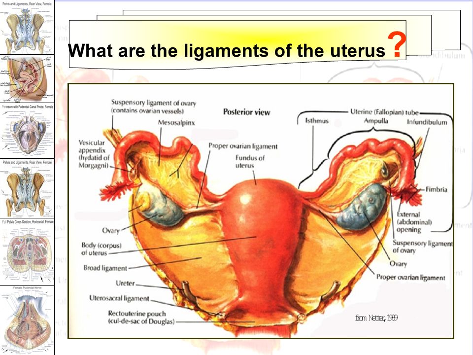
Fibroid symptoms can vary based on size and location in a normal size uterus. They can also be similar to other conditions that affect the reproductive system, so it is important to discuss symptoms with your doctor.
Typical symptoms of fibroids include:
- Heavy menstrual bleeding and longer periods
- Bleeding between periods
- Severe menstrual cramping
- Pain in the pelvis or lower back
- Frequent urination
- Stomach swelling
- Pain during sex
- Anemia
- Bloating and/or constipation
If you experience these symptoms, you should see your doctor. They may diagnose fibroids during a regular pelvic exam or use other tests to reach a diagnosis. They may recommend:
- Ultrasound
- MRI
- Hysterosonography
- Hysteroscopy
These tests can confirm a diagnosis of uterine fibroids.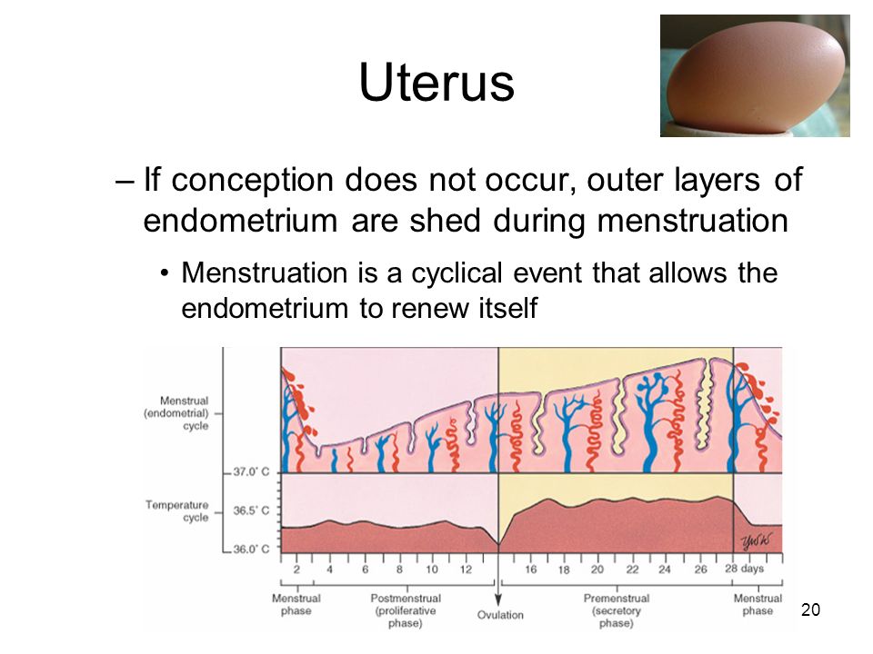
Fibroids can cause complications with pregnancy. They may grow in size during pregnancy because of the increase in progesterone. They can outgrow their blood supply, which may lead to severe pain and hospitalization to monitor the mother and baby’s conditions.
Fibroids can affect the baby’s position in the uterus, increasing the risk of miscarriage. They may also increase the risk for preterm delivery and the need for a cesarean section delivery.
Uterine Fibroid TreatmentUSA Fibroid Centers offers a minimally invasive treatment for uterine fibroids to help reduce their size and accompanying symptoms. UFE treats conditions to reduce or eliminate symptoms while keeping the uterus intact, unlike a hysterectomy.
When UFE is performed, a fibroid specialist will use ultrasound technology to locate the fibroid. They will insert a tiny catheter into the thigh or wrist and inject embolic materials into the artery feeding the fibroid.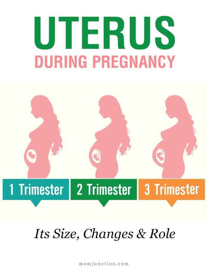 When the fibroid can no longer receive nutrients, it shrinks and dies.
If you are concerned about fibroids, how they can affect an average size uterus, or the symptoms that negatively impact your life, you can get help with USA Fibroid Centers. Our specialists will work with you to determine the right treatment plan. Schedule a free consultation online or call us at 855.615.2555 to visit one of our facilities.
Our Locations
Related Posts
Causes, Treatment, Outlook, and More
Enlarged Uterus: Causes, Treatment, Outlook, and MoreMedically reviewed by Alana Biggers, M.D., MPH — By Donna Christiano — Updated on September 18, 2018
Overview
The average uterus, which is also known as a woman’s womb, measures 3 to 4 inches by 2.5 inches. It has the shape and dimensions of an upside-down pear. A variety of medical conditions can cause the uterus to increase in size, including pregnancy or uterine fibroids.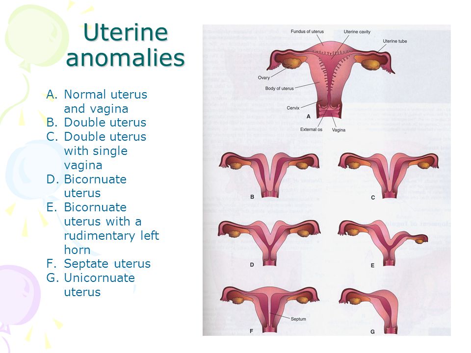
You may feel a heaviness in your lower abdomen or notice your abdomen protruding as your uterus enlarges. You may not have any noticeable symptoms, however.
Read on to learn more about the causes and symptoms of an enlarged uterus, and how to treat this condition.
A number of common conditions can cause a uterus to stretch beyond its normal size.
Pregnancy
The uterus normally fits into the pelvis. When you’re pregnant, your growing baby will cause your uterus to increase in size 1,000 times, from the size of a clenched fist to a watermelon or larger by the time you deliver.
Fibroids
Fibroids are tumors that can grow inside and outside the uterus. Experts aren’t sure what causes them. Hormonal fluctuations or genetics may contribute to the development of these growths. According to the Office on Women’s Health at the U.S. Department of Health and Human Services, up to 80 percent of women have experienced fibroids by the time they turn 50.
Fibroids are rarely cancerous, but they can cause:
- heavy menstrual bleeding
- painful periods
- discomfort during sex
- lower back pain
Some fibroids are small and may not cause any noticeable symptoms.
Others can grow so large that they weigh several pounds and can enlarge the uterus to such an extent that you may look several months pregnant. For example, in a case report published in 2016, a woman with fibroids was found to have a uterus weighing 6 pounds. For comparison’s sake, the average uterus is about 6 ounces, which is roughly the weight of a hockey puck.
Adenomyosis
Adenomyosis is a condition in which the uterine lining, called the endometrium, grows into the uterine wall. The exact cause of the condition is unknown, but adenomyosis is tied to estrogen levels.
Most women see a resolution of their symptoms after menopause. That’s when the body stops producing estrogen and periods cease. The symptoms are similar to those of fibroids and include:
- heavy menstrual bleeding
- painful cramping
- pain with sex
Women may also notice tenderness and swelling in their lower abdomen. Women with adenomyosis can have a uterus that is double or triple its normal size.
Reproductive cancers
Cancers of the uterus, endometrium, and cervix can all produce tumors. Depending on the size of the tumors, your uterus can swell.
Additional symptoms include:
- abnormal vaginal bleeding, such as bleeding not related to your menstrual cycle
- pain with sex
- pelvic pain
- pain while urinating or feeling like you can’t empty your bladder
An enlarged uterus is usually found incidentally. For example, your doctor may identify an enlarged uterus during a routine pelvic exam as part of a well-woman checkup. It may also be identified if your doctor is treating you for other symptoms, like abnormal menstruation.
If your uterus in enlarged because of pregnancy, it will naturally begin to shrink after you deliver. By one week postpartum, your uterus will be reduced to half its size. By four weeks, it’s pretty much back to its original dimensions.
Other conditions causing an enlarged uterus could need medical intervention.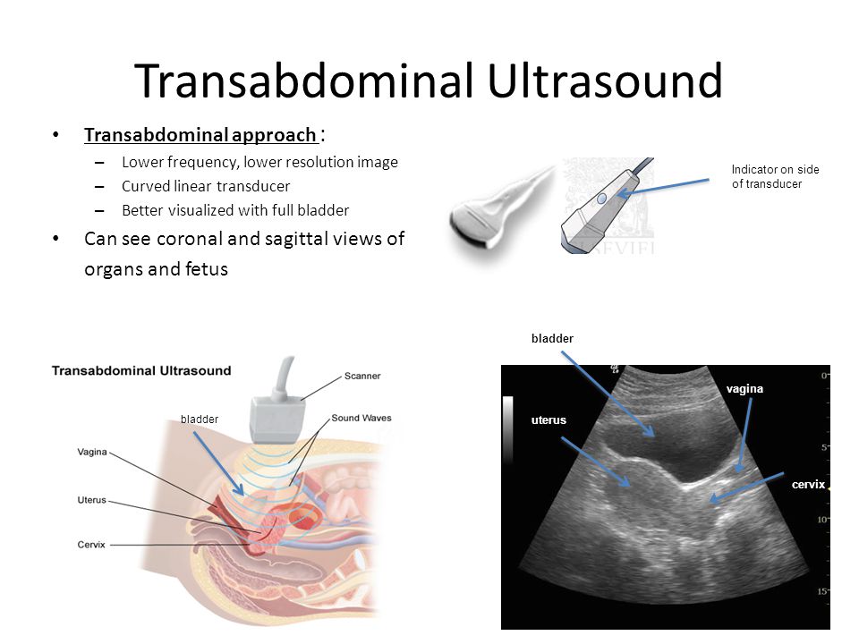
Fibroids
Fibroids that are large enough to stretch the uterus will probably need some kind of medical treatment.
Your doctor may prescribe birth control drugs, such as birth control pills that contain estrogen and progesterone or a progesterone-only device like an IUD. Birth control medication may halt the growth of the fibroids and limit menstrual bleeding.
Another treatment, known as uterine artery embolization,uses a thin tube inserted into the uterus to inject small particles into the arteries of the uterus. That cuts off the blood supply to the fibroids. Once the fibroids are deprived of blood, they will shrink and die.
In some cases, you may need surgery. Surgery to remove the fibroids is called a myomectomy. Depending on the size and location of the fibroids, this may be done with a laparoscope or through traditional surgery. A laparoscope is a thin surgical instrument with a camera on one end that’s inserted through a small incision or through traditional surgery.
Complete surgical removal of the uterus, called a hysterectomy, may also be advised. Fibroids are the No. 1 reason hysterectomies are performed. They’re generally done on women whose fibroids cause a lot of symptoms, or on women with fibroids who don’t want children or are near or past menopause.
A hysterectomy can be done laparoscopically, even on a very large uterus.
Adenomyosis
Anti-inflammatory medications, like ibuprofen (Advil, Motrin) and hormonal contraception such as the birth control pill can help relieve the pain and heavy bleeding associated with adenomyosis. These medications won’t help to decrease the size of an enlarged uterus, however. In severe cases, your doctor may recommend a hysterectomy.
Reproductive cancers
Like other cancers, cancers of the uterus and endometrium are typically treated with surgery, radiation, chemotherapy, or a combination of these treatments.
An enlarged uterus doesn’t produce any health complications, but the conditions that cause it can.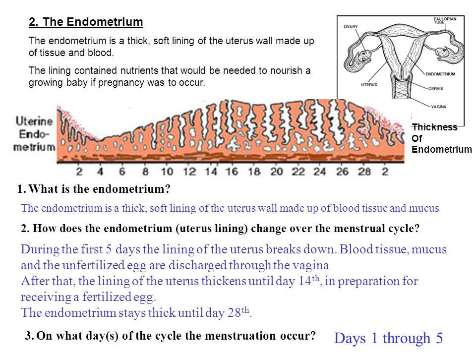 For example, besides the pain and discomfort associated with fibroids, these uterine tumors can reduce fertility, and cause pregnancy and childbirth complications.
In one study published in Obstetrics and Gynecology Clinics of North America, fibroids are present in up to 10 percent of infertile women. Additionally, up to 40 percent of pregnant women with fibroids will experience pregnancy complications, such as requiring a cesarean delivery, having premature labor, or experiencing excessive bleeding problems postdelivery.
Many of the conditions that cause an enlarged uterus aren’t serious, but they can be uncomfortable and should be evaluated. See your gynecologist if you experience abnormal, excessive, or prolonged:
- vaginal bleeding
- cramping
- pelvic pain
- fullness or bloating in your lower abdomen
You should also contact your doctor if you have a frequent need to urinate or pain during sex. There are successful treatments, especially when conditions are caught early.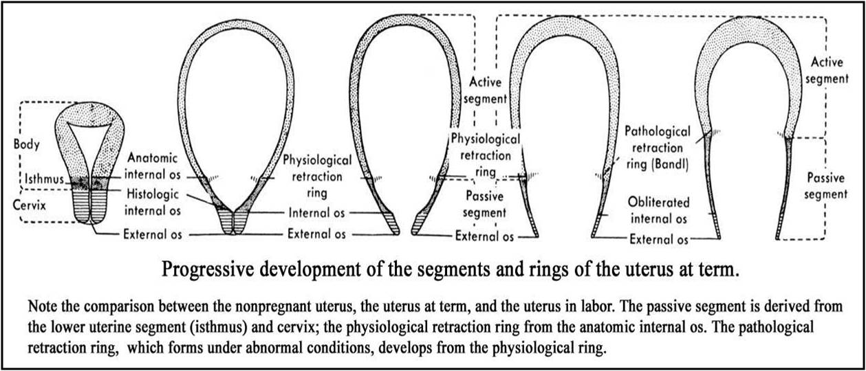
Last medically reviewed on September 19, 2017
How we reviewed this article:
Healthline has strict sourcing guidelines and relies on peer-reviewed studies, academic research institutions, and medical associations. We avoid using tertiary references. You can learn more about how we ensure our content is accurate and current by reading our editorial policy.
- Kehde BH, et al. (2016). Large uterus: What is the limit for a laparoscopic approach? [Abstract].DOI:
10.4322/acr.2016.025 - Uterine fibroids. (2017).
womenshealth.gov/a-z-topics/uterine-fibroids - Mayo Clinic Staff. (2015). Adenomyosis: Treatments and drugs.
mayoclinic.org/diseases-conditions/adenomyosis/basics/treatment/con-20024740 - Mayo Clinic Staff. (2015). Adenomyosis: Causes.
mayoclinic.org/diseases-conditions/adenomyosis/basics/causes/con-20024740 - Xiaoxiao CG, et al.
 (2012). The impact and management of fibroids for fertility: An evidence-based approach. DOI:
10.1016/j.ogc.2012.09.005
Our experts continually monitor the health and wellness space, and we update our articles when new information becomes available.
Share this article
Medically reviewed by Alana Biggers, M.D., MPH — By Donna Christiano — Updated on September 18, 2018
Read this next
What You Should Know About Retroverted Uterus
Medically reviewed by Debra Sullivan, Ph.D., MSN, R.N., CNE, COI
A retroverted uterus is a uterus that curves in a backwards position at the cervix instead of a forward position. Many women are either born with a…
READ MORE
10 Symptoms Women Shouldn’t Ignore
Medically reviewed by Alana Biggers, M.D., MPH
Some symptoms are easy to identify as potentially serious health problems.
 Chest pain, high fever, and bleeding are all typically signs that something…
READ MORE
8 Questions About Your Period You’ve Always Wanted to Ask
Medically reviewed by Stacy Sampson, D.O.
READ MORE
7 Potential Causes of Ovary Pain: How They’re Diagnosed and Treated
Medically reviewed by Valinda Riggins Nwadike, MD, MPH
Occasional ovary pain is likely related to your menstrual cycle, but it may be a sign of an underlying condition. Learn 7 potential causes.
READ MORE
Getting Pregnant With Endometriosis: Is It Possible?
Medically reviewed by Carolyn Kay, M.D.
Endometriosis can affect a person’s fertility, but it’s still possible to get pregnant. Here’s what you should know.
READ MORE
6 Questions Everyone Should Ask Themselves About Their Fertility, Right NowREAD MORE
The Best Women’s Health Blogs of 2020
From everyday essentials like food and fitness, to reproductive and sexual health, there’s no one-size-fits-all approach to women’s wellness.
 We’ve…
READ MORE
Can Stress Lead to Miscarriage? Prince Harry and Meghan Markle Discuss Their Experience in Netflix Doc
Meghan Markle and Prince Harry opened up about how the duchess experienced a miscarriage in July 2020. The couple believes that the high levels of…
READ MORE
Birth Control Access: Pharmacists Can Write Prescriptions in 20 States
In the past few years, many states have passed laws permitting pharmacists to prescribe hormonal birth control. By allowing pharmacists to prescribe…
READ MORE
1 in 10 Pregnant People At Risk for Hypertension Following Childbirth
New research shows that 1 in 10 pregnant people may develop hypertension within a year of childbirth despite no prior history of high blood pressure…
READ MORE
How to decipher the results of ultrasound of the uterus?
2017. 02.26 6:33
After receiving the ultrasound results, you may be interested in what exactly the doctor wrote. Let's find out in more detail what the main terms that sonographers write in their conclusions mean.
Position of the uterus.
The body of the uterus is in a certain position in the pelvis. Normally, the body of the uterus is tilted anteriorly, and the fold between the body of the uterus and the cervix forms an angle. In the conclusion of the ultrasound, this situation can be described by two Latin words: "anteversio" and "anteflexio". This is the usual (normal) position of the uterus. If the ultrasound report says that the body of the uterus is in the “retroversio” position, “retroflexio” means that the uterus is tilted backwards and there is a backward bending of the uterus. The backward bend of the uterus can indicate certain diseases, adhesions in the pelvis, and sometimes can cause infertility. nine0007
Size of the uterus.
Ultrasound can determine 3 dimensions of the uterus: transverse dimension, longitudinal dimension and anterior-posterior dimension. The longitudinal size (length of the uterus) is normally 45-50 mm (in women giving birth up to 70 mm), the transverse size (width of the uterus) is 45-50 mm (in women giving birth up to 60 mm), and the anterior-posterior size (thickness of the uterus) normally 40-45 mm. Slight deviations in the size of the uterus are found in many women and do not indicate illness. However, too large size of the uterus can talk about uterine fibroids, adenomyosis, pregnancy. nine0007
M-echo.
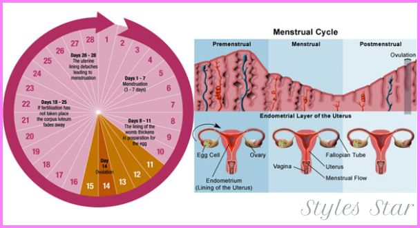
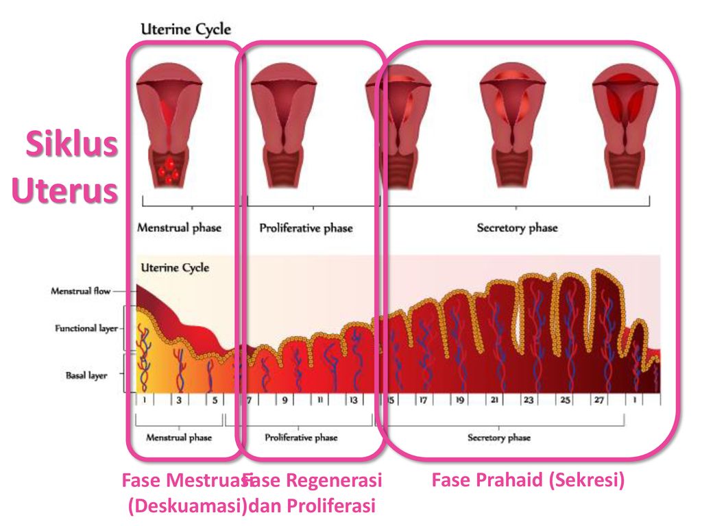
The thickness of the inner layer of the uterus (endometrium) is determined by ultrasound using M-echo. The thickness of the endometrium depends on the day of the menstrual cycle: the fewer days left until the next period, the thicker the endometrium. In the first half of the menstrual cycle, the M-echo ranges from 0. 3 to 1.0 cm; in the second half of the cycle, the thickness of the endometrium continues to grow, reaching 1.8-2.1 cm a few days before the onset of menstruation. If you have already entered menopause (menopause), then the thickness of the endometrium should not exceed 0.5 cm. If the thickness of the endometrium is too large, this may indicate endometrial hyperplasia. In this case, you need an additional examination in order to exclude uterine cancer. nine0007
Structure of the myometrium.
The myometrium is the muscular, thickest layer of the uterus. Normally, its structure should be homogeneous. The heterogeneous structure of the myometrium may indicate adenomyosis. But do not be afraid ahead of time, as you will need an additional examination to clarify the diagnosis.
Uterine fibroids on ultrasound
Uterine fibroids is a benign tumor that almost never develops into uterine cancer. With the help of ultrasound, the gynecologist determines the location of the fibroid and its size. nine0003 For fibroids, the size of the uterus is given in weeks of pregnancy. This does not mean that you are pregnant, but that the size of your uterus is the same as the size of the uterus at a certain stage of pregnancy.
Uterine fibroids can vary in size on different days of the menstrual cycle. So, in the second half of the cycle (especially shortly before menstruation), the fibroids increase slightly. Therefore, ultrasound with uterine fibroids is better to take place immediately after menstruation (on the 5-7th day of the menstrual cycle).
The location of uterine fibroids can be intramural (in the wall of the uterus), submucosal (under the inner lining of the uterus) and subserous (under the outer lining of the uterus). nine0007
Endometriosis of the uterus (adenomyosis) on ultrasound
Endometriosis of the uterus, or adenomyosis, is a disease in which endometrial-like cells grow into the muscle layer.
In case of adenomyosis on ultrasound of the uterus, the doctor finds that the myometrium (the muscular layer of the uterus) has a heterogeneous structure with heterogeneous hypoechoic inclusions.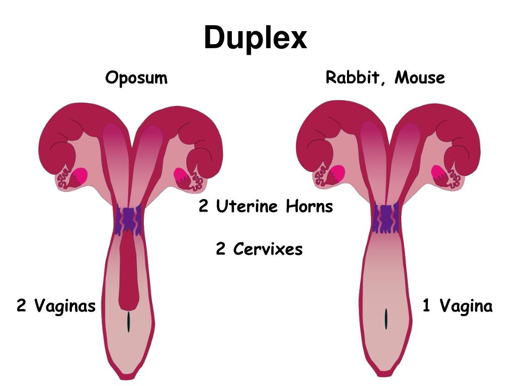 In "translation into Russian" this means that in the muscular layer of the uterus there are sections of the endometrium, which has formed vesicles (or cysts) in the myometrium. Very often, with adenomyosis, the uterus is enlarged in size. nine0007
Pregnancy on ultrasound
Ultrasound of the uterus during pregnancy is an extremely important diagnostic step. Here are just a few of the benefits of ultrasound during pregnancy:
-
- Helps determine gestational age and fetal size
- Helps to clarify the position of the fetus in the uterus
- Helps detect ectopic pregnancy
- Helps to follow the development of the fetus and detect any abnormalities in time nine0054 Helps determine the sex of the baby
- Used in first trimester screening
- Used for amniocentesis
How to interpret the results of ovarian ultrasound?
Ultrasound of the ovaries determines the size of the right and left ovaries, as well as the presence of follicles and cysts in the ovary. The normal size of the ovaries is on average 30x25x15 mm. A deviation of a few millimeters is not a sign of illness, since one or both ovaries may enlarge slightly during the menstrual cycle. nine0007
Ultrasound of an ovarian cyst
Ultrasound of an ovarian cyst looks like a round vesicle, the size of which can reach several centimeters. With the help of ultrasound, the doctor can not only determine the size of the ovarian cyst, but also suggest its type (follicular cyst, corpus luteum cyst, dermoid cyst, cystadenoma, and so on. , which is noticeable during ultrasound.The volume of the ovary also increases: if the normal volume of the ovary does not exceed 7-8 cm3, then with polycystic ovaries it increases to 10-12 cm3 or more.Another sign of polycystic ovaries is a thickening of the ovarian capsule, as well as the presence of many follicles in the ovary (usually, 12 More with a diameter of 2 to 9 folliclesmm).
In our center you can undergo the following ultrasound examinations:
-
- abdominal organs, kidneys, adrenal glands
- uterus and ovaries, folliculometry, mammary glands
- prostate, bladder with determination of residual urine, scrotum
- thyroid
- salivary glands
- lymph nodes, soft tissues
- hearts
- vessels of the neck and brain,
- veins and arteries of the lower extremities
- knee and shoulder joints
- Ultrasound of the internal organs for children from 3 years of age and older
See also:
How often and what ultrasound should be done to monitor health?
full name*
Your e-mail
Contact phone*
Comment
Ultrasound of the uterus - Ultrasound of the cervix and appendages in Nizhny Novgorod, transvaginal ultrasound
Ultrasound of the uterus is one of the most important and common methods of examining this organ.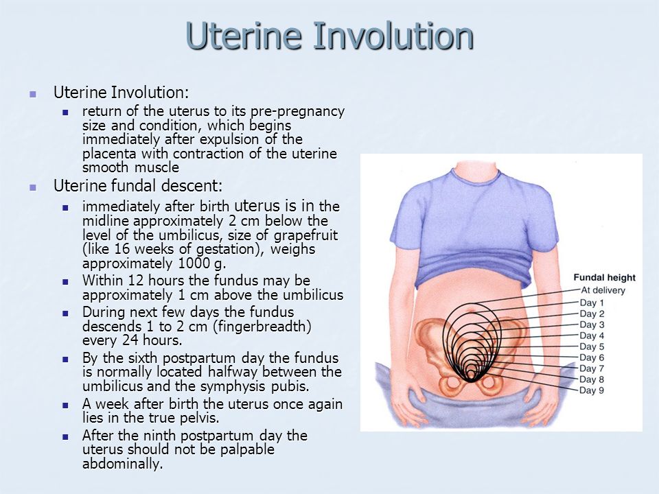 For a better understanding of the process of conducting ultrasound of the uterus and appendages, consider the uterus as an organ of the female reproductive system.
The uterus is a hollow muscular organ consisting of three layers: endometrium (inner), myometrium (middle), perimetrium (outer), and is located in the pelvic cavity, between the bladder and rectum. The normal anatomical position of the uterus in the small pelvis is provided by the supporting muscular pelvic floor and the suspension ligamentous apparatus of the uterus. The uterus has a fundus (upper part of the uterus), a body, and a cervix. The cervix connects the uterus to the vagina. Therefore, the vaginal and supravaginal parts of the uterus are isolated. nine0007
The function of the uterus is to provide a place and conditions for the implantation of the embryo and for the further development of the fetus.
Fallopian tubes and ovaries are the so-called appendages of the uterus. The ovaries are paired sex glands that work alternately. They contain many different degrees of development of follicles in which eggs mature. The fallopian tubes run from the uterus to the ovaries. They are where fertilization takes place.
Therefore, in order to enjoy the happiness of motherhood, it is necessary to undergo prophylactic Ultrasound of the uterus and appendages , and to conduct timely diagnosis and treatment of the uterus and other genital organs of a woman.
Ultrasound of the uterus and appendages a gynecologist can prescribe if there are any symptoms of a disease of the female genital organs:
- Various pains in the lower abdomen, pain during menstruation
- Irregular menses
- Uterine bleeding
- Anomalies in the development of the uterus
- Bleeding during menopause nine0070
- Clarification of the presence and location of the intrauterine contraceptive (spiral)
- Establishing pregnancy
- Neoplasm diagnostics
- Uterine length 30-40 mm, cervix 40-50 mm, anterior-posterior size 28-45 mm, width 35-50 mm.
- Position of the uterus in anteflexio - inclination towards the bladder. Bend of the uterus to the rectum (retroflexio) is a normal variant, but less common
- The thickness of the endometrium changes during the menstrual cycle
By the 5-7th day of the menstrual cycle, the endometrium is clearly defined, its thickness is up to 6-9 mm. - Myometrium homogeneous in structure and without formations
- Developmental anomalies (unicornuate, bicornuate, bifid, with septum)
- Endometriosis
- Adenomyosis
- Metroendometritis
- Endometritis
- Uterine polyps
- Tumors (benign and malignant)
As well as ultrasound of the uterus and appendages is used for:
For the study of the pelvic organs in a sexually active woman, transvaginal ultrasound of the uterus is performed. To prepare for a transvaginal ultrasound of the uterus, 2 days before the examination, do not eat foods that cause gas formation, and, if necessary, make an enema. nine0007
Transvaginal ultrasound of the uterus is not performed for girls before the onset of sexual activity. To do this, use the transabdominal method of conducting ultrasound of the uterus. It is necessary before the study to try to fill the bladder as much as possible - drink about 1 liter of water or tea. This is necessary in order to visualize the uterus well on ultrasound.
The most important condition for transvaginal ultrasound and transabdominal ultrasound of the uterus and appendages is its 5-7 day of the menstrual cycle or during the first 2 days after the end of menstruation. nine0007
With an ultrasound of the uterus, the doctor determines the position, size and structure of the uterus, and assesses the condition of the cervix. It is also taken into account when conducting an ultrasound of the uterus, that before childbirth, the size of the uterus in a girl is minimal, and in a woman who has given birth, it is maximum.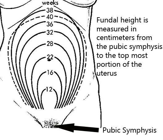
Normal ultrasound findings of the uterus in reproductive age (from 20 to 35 years):
Ultrasound of the uterus in the early reproductive period (from the onset of menstruation to 20 years) shows the smallest size of the uterus. With ultrasound of the uterus in postmenopause (begins at the age of 45 to 55 years), the uterus decreases and becomes homogeneous in echostructure. The endometrium may not be visualized. nine0007
Ultrasound of the uterus helps to detect the following diseases:
In addition to ultrasound of the uterus, ultrasound of the cervix is also performed:
When ultrasound of the cervix is evaluated, its shape, structure and thickness of the cervical canal, the structure of the muscular layer of the cervix.

