Vag ultrasound in early pregnancy
Early in Pregnancy - What Should Show • Vision Xray Group
Home » Blog » Early Weeks of Pregnancy: What Ultrasound Should Show
Early Weeks of Pregnancy: What Ultrasound Should Show
By Peter Kitchener 22 Jan, 2014 Ultrasound
Carrying life is an amazing experience for a woman, and many are anxious to see more of their future child via ultrasound scans. As technology has developed and improved greatly over the years, ultrasounds can now provide more information than ever. While later ultrasounds are much increased in detail, even the earliest ultrasounds can present technicians, doctors, and expectant parents with particulars regarding their baby’s growth and health.
5 weeks is the earliest point at which a pregnancy ultrasound can be completed. This ultrasound is typically a transvaginal ultrasound, as an abdominal ultrasound will not reveal many signs of pregnancy development yet. At approximately 5 weeks, the gestational sac can be seen via transvaginal ultrasound. This is the structure in which the embryo will grow, and can generally be seen before the embryo itself is visible.
6-7 Weeks
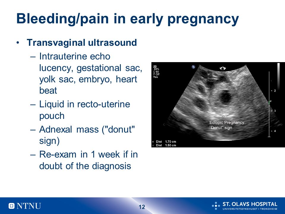
Around 6 or 7weeks, an abdominal ultrasound will show the gestational sac. A transvaginal ultrasound given at this time is likely to show images of an early developing embryo. At this point the ultrasound technician can see the location of the embryo in the uterus and if it is an the proper, healthy location. These scans can help determine if there are signs of an ectopic pregnancy. The transvaginal ultrasound at 6-7 weeks can also show signs of the fetal heartbeat! At this point you will likely know if you have a twin pregnancy.
8 Weeks
It is around the 8th week of pregnancy that ultrasound photos will begin to have increased detail. The heartbeat is clearly accessible, as well as arm and leg buds on the growing fetus.
First Trimester Pregnancy Ultrasound (12 weeks)
Although the earlier scans are possible (and typically done by transvaginal ultrasound), the most typical first trimester ultrasound is recommended around 10-14 weeks of gestation. This is generally performed by abdominal ultrasound and should provide images of the baby which look more “familiar” to the expectant parents. This as an exciting event as the parents finally get a real “look” at their baby for the first time!
This ultrasound shows greater detail and allows the technician to assess the baby’s size and growth. At this time, a more accurate due date can be given. The technician will measure the size of the fetus from its head to its bottom, called the Crown Rump length. This measurement helps determine the speed of the baby’s growth and is important in determining due date. During this pregnancy ultrasound, movement of the baby can be clearly seen, as well as recognizably developed arms and legs.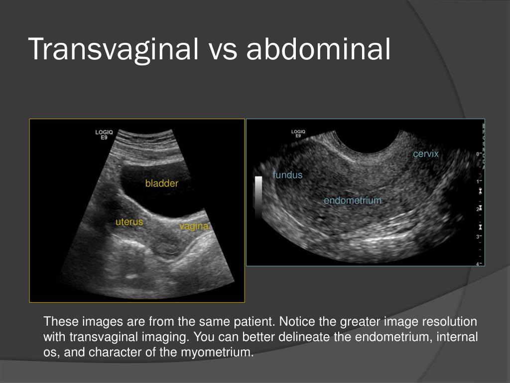 Although the ultrasound can detect movement of the baby, the mother will not yet be able to feel it inside her.
12 weeks is approximately the time when fairly accurate screening tests can be performed for Down Syndrome. This can be assessed using a test called nuchal translucency, which measures the amount of fluid in the base of the fetus’ neck. Those with a likelihood for Down Syndrome commonly have more fluid in this region. This is not always indicative of the presence of Down Syndrome, however, and further tests are needed to confirm.
What to Expect During Your First Trimester Ultrasound? – Sneak A Peek Ultrasound
Typically, an ultrasound is going to be done anytime within the first trimester of pregnancy. Frequently, ultrasounds during pregnancy will be done externally with a transabdominal approach; it could be required to complete the ultrasound. Most often in the first trimester a transvaginal ultrasound, a wand coated with gel and a condom is set in the vagina to permit for visualization of the baby. When a transabdominal ultrasound is done, the bladder usually must be full to obtain the best view of their uterus, ovaries along with the baby.
How the Test is Performed
You may experience some discomfort from pressure on a full bladder. The conducting gel may feel slightly cold and wet. You won’t feel the ultrasound waves.
You will have to have a full bladder to acquire the best ultrasound picture. You could be asked to drink 2 to 3 glasses of liquid an hour before the test.
How to Get Ready for the Test
The individual performing the test will spread a transparent, water-based gel or lotion onto your belly and pelvis area. A probe will then be transferred over the stomach. The gel helps transmit sound waves. These waves bounce off the entire body structures, including the developing baby, to create an image on the ultrasound device.
To have the process:
Sometimes, a pregnancy ultrasound may be done by placing the probe to the vagina; this is much more likely in early pregnancy.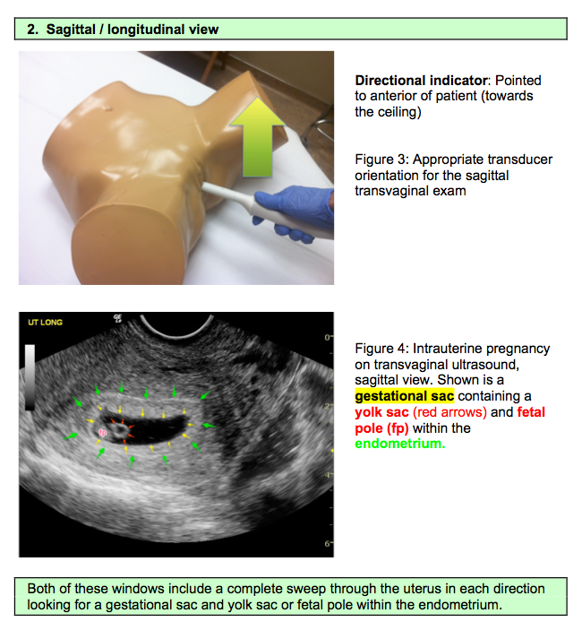 Many women will have the length of the cervix measured by ultrasonography about 20 to 24 weeks of pregnancy.
First trimester ultrasounds are done for various reasons including:
To accurately date your pregnancy to determine how far along you are, evaluate the size of the baby and qualities of the infant’s gestational sac, for example, dimensions and appearance. Occasionally, the gestational sac can be viewed as early as 5 weeks gestation. Detect the heartbeat, which may be early as 5-7 weeks gestation. Verify that the baby is growing within the uterus rather than in another structure such as the fallopian tube assess the presence of numerous infants, particularly in women who failed IVF or other assistive fertility remedies.
Make sure to speak with your physician or midwife about your ultrasound requirements.
Your doctor may recommend ultrasounds in some instances, primarily if your pregnancy is categorized as insecure, or whether you are undergoing processes such as chorionic villus sampling, an amniocentesis translucency screening, and biophysical profiles.
If you get an ultrasound during 8 to 11 weeks, it will show your growing baby, which during this time has recognizable features such as the body, arms, head, and thighs, and is moving relatively quickly within the gestational sac.
If your ultrasound is performed between 6- and five-weeks’ gestation, you will likely only be able to observe the gestational and yolk sacs. The yolk sac allows your doctor or midwife to confirm your pregnancy, although the infant may not be recognizable yet.
Ultrasound in early pregnancy
Modern high-tech equipment provides high accuracy and informativeness of the method even with the smallest size of the ovum. Ultrasound in the early stages helps to fully control the most vulnerable period of pregnancy.
- the latest Mindray DC-70 PRO device
- specialists with more than 15 years of experience
- detailed description of the examination and comments of the doctor during the diagnostic process
You can undergo an ultrasound scan to determine pregnancy in the early stages at the Tauras-Med Medical Center. The procedure is carried out by experienced obstetrician-gynecologists who are certified in the field of ultrasound diagnostics and understand all its intricacies. Our specialists are guided by international standards for the provision of medical care, regularly improve their professionalism and apply a personalized approach to each patient.
Indications
First trimester ultrasound screening from 11 to the end of 13 weeks. However, in special situations, the doctor may recommend an ultrasound at an earlier date, namely:
- From 4-5 weeks. The study is carried out to confirm the fact of pregnancy, with negative test results or the inability to conduct laboratory diagnostics (analysis for hCG). Ultrasound in the early stages is prescribed to confirm pregnancy resulting from IVF. The procedure allows you to clarify the location of the fetal egg and exclude ectopia, which is especially important in the presence of cases of ectopic pregnancy in history.
- From 6-7 weeks.
 Ultrasound of the fetus in the early stages may be relevant for assessing its viability. During the study, the location, size, area of attachment of the embryo, as well as the presence and speed of the heartbeat, are evaluated. The procedure is especially relevant in the presence of a history of miscarriages, in case of abdominal pain or spotting in a pregnant woman.
 - From the 9th week. In this period, with the help of ultrasound, it is possible to assess the anatomy of the embryo and diagnose the first signs of developmental anomalies. Early diagnosis is relevant in the presence of genetic diseases in one or both parents.
Ultrasound in early pregnancy does not replace first trimester screening. Even after conducting several studies in the first weeks of gestation, the procedure is repeated from the 11th to the 13th week, because. diagnostic capabilities of the method are different.
How early ultrasound is performed
In the first trimester, when the fetus is small, ultrasound is performed transvaginally (through the vagina).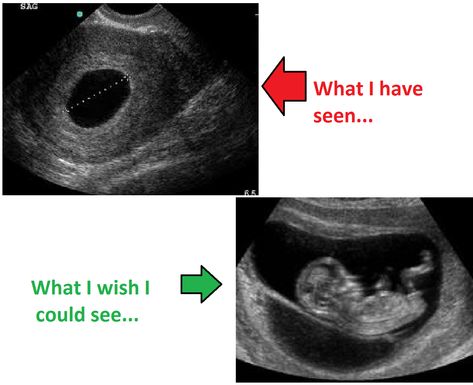 The study is carried out using a vaginal sensor using a disposable condom. It should be noted that the introduction of equipment into the pelvic cavity cannot harm the health of the mother and fetus, or provoke complications during pregnancy. The procedure is painless and takes no more than 10 minutes.
Make an appointment
Our administrator will call you back to set the time and answer your questions
Preparation for the procedure
No specific preparation is required. On the day of the study, a hygienic shower is shown. Immediately before entering the diagnostic room, you need to empty your bladder. Only with a transabdominal scan should the bladder be full. This method is used for heavy bleeding.
Early ultrasound contraindications
One of the main advantages of ultrasound diagnostics is the absolute absence of contraindications. Ultrasound has no effect on the condition of the uterus and fetus. The procedure has no restrictions regarding the multiplicity and frequency of its implementation.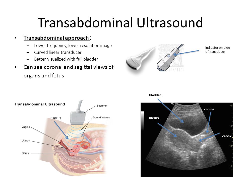 If necessary, ultrasound is done in dynamics until the threat of abortion disappears.
Where can I get an ultrasound to determine pregnancy? Residents of St. Petersburg can undergo an ultrasound to determine pregnancy, monitor the vital activity of the fetus and an initial assessment of its anatomy as prescribed by a doctor or on their own initiative. The clinic also performs screenings of the first, second and third trimesters, Doppler, ultrasound in 3D and 4D. The high professionalism of doctors and the availability of expert equipment guarantee high accuracy of the results.
The cost of ultrasound in the early stages
Prices for medical services are indicated in the appropriate section on the center's website. The cost of ultrasound during pregnancy depends on the goals and complexity of the procedure. To clarify the price, call by phone or fill out the feedback form on the website.
If you have any questions, call us at: 8 (812) 603-44-71
or write to WhatsApp or Telegram
Our specialists:
Reviews
Ultrasound in early pregnancy: 3D and 4D, when to do, is it harmful, what shows
Ultrasound in early pregnancy: 3D and 4D, when to do it, is it harmful, what shows - Junohome
Articles
ultrasound during pregnancy
An ultrasound is scheduled for every woman in an "interesting position". The expectant mother must undergo this procedure at least twice in 40 weeks. The study allows you to specify the gestational age, make measurements of the height and weight of the fetus. You will learn about the benefits and harms of ultrasound in our article.
Planned ultrasound during pregnancy
From 2021, the expectant mother will have to undergo at least two ultrasound examinations during pregnancy. The timing of the planned examination during pregnancy is determined by the Ministry of Health. These studies are called screening. Their task is to identify possible violations in the development of the fetus and the course of pregnancy and provide the woman with qualified medical care in time.
Until 2021, a pregnant woman underwent an ultrasound in trimesters - one in each specified period. But according to order N 1130n, now the expectant mother will be screened only twice - in the first and second trimester.
First trimester
The first screening ultrasound is performed at 11-14 weeks. At the same time, biochemical screening is done. The expectant mother donates blood for the determination of β-hCG and PAPP-A. The data obtained are evaluated together with the results of the first ultrasound screening. Together, these methods make it possible to identify disorders in the development of the fetus, such as Down syndrome and other chromosomal abnormalities.
In the first trimester, ultrasound can determine:
- Gestational age. If the expectant mother does not remember when her last menstruation was or she has an irregular menstrual cycle, an ultrasound examination will help. The doctor will specify the gestational age by ultrasound. But you need to remember: these calculations will not be too accurate, and therefore, if possible, gynecologists are guided by the date of the last menstruation and calculate the due date from it.
- Number of fruits. In a multiple pregnancy, the doctor examines the placenta (or chorion) and membranes in detail.
 The tactics of managing pregnancy and childbirth depend on their location and number.
 - Fetal malformations. For example, to diagnose Down syndrome, the doctor evaluates the thickness of the collar space and visualization and the length of the nasal bone, the length of the thigh. Ultrasound in early pregnancy can also reveal malformations of the internal organs and nervous system.
- An assessment of the condition of the cervix (cervicometry), uterine appendages and uterine wall is carried out.
Despite a thorough examination, it is impossible to completely exclude fetal malformations only by the results of ultrasound. If in doubt, your doctor may recommend an invasive test such as amniocentesis or cordocentesis. The second ultrasound screening also helps to clarify the diagnosis, in which it is possible to examine the organs and tissues of the fetus in more detail.
Second trimester
The second screening ultrasound is done at a period of 19-21 weeks. Here is what the doctor evaluates:
- Correspondence of the size of the fetus to the gestational age. If they are less than normal, they talk about fetal growth retardation.
- Structure of internal organs and nervous system. At this time, malformations of the heart, brain, digestive tract and other organs and systems can be detected.
- The state of the placenta and umbilical cord, features of blood flow in them. If the blood flow is disturbed, the fetus will suffer from a lack of oxygen.
- Volume of amniotic fluid. If there is too much amniotic fluid, they talk about polyhydramnios, a little - about oligohydramnios.
At the second ultrasound screening, the sex of the fetus can be determined. This is not necessary, and if the expectant mother wants a surprise, she can ask the doctor not to report the results.
The timing of the ultrasound during pregnancy and the interpretation of the results is done by a gynecologist observing a pregnant woman. The doctor will tell you when to do an ultrasound scan during pregnancy, and if necessary, he will prescribe an unscheduled examination.
Unscheduled ultrasound during pregnancy
Outside of screenings, ultrasound is prescribed in such situations:
- Confirm pregnancy. This is necessary in order to make a correct diagnosis - after all, tests are sometimes wrong, and a delay in menstruation is not always associated with an interesting situation. Such an ultrasound is done in the early stages - at 4-6 weeks.
- Determine the location of the gestational sac. This is necessary in order to exclude an ectopic pregnancy.
- When bloody discharge from the genital tract appears, ultrasound is done on an emergency basis at any stage of pregnancy - it is necessary to exclude the development of complications.
- In the later stages - if the fetus has stopped moving or, on the contrary, has become overly active. In addition to ultrasound, CTG (cardiotocography) is done from the 33rd week to assess the fetal heartbeat.

- Before childbirth - if there is a risk of complications. With ultrasound, you can clarify the weight and position and presentation of the fetus, the condition of the placenta, umbilical cord and amniotic fluid.
With multiple and complicated pregnancies, ultrasound can be done more often. The terms are set by the attending physician individually for each woman.
Features of ultrasound in early pregnancy
Many expectant mothers are wondering what week the ultrasound will show pregnancy. Modern devices allow this to be done at about 3-4 weeks if a vaginal sensor is used (transvaginal method). If a specialist conducts a study through the abdominal wall (transabdominal method), then he will be able to detect a fetal egg later, at 5-6 weeks.
Note
Knowing how long the ultrasound shows pregnancy, you can not run for an examination immediately after a missed period. For a very short time, the doctor may not see the fetal egg - and not because it is not there, but because the equipment is not perfect. There is no need to create a cause for alarm for yourself - it is better to wait until 5-6 weeks, when the fetal egg will be clearly visible.
In the early stages, ultrasound can detect serious problems such as ectopic or regressive (non-developing) pregnancy. The sooner the pathology is detected, the easier it will be to avoid complications.
Types of ultrasound during pregnancy
Modern equipment of ultrasound rooms allows for high-precision ultrasound examinations. In addition to the standard 2D ultrasound, three- and four-dimensional studies - 3D and 4D - have now become very popular. Let's consider them in detail.
- 2D is a study in which a black and white image is obtained in two dimensions - in height and length. This option is quite informative. The doctor can measure the growth and proportions of the fetus, as well as assess the condition of the placenta and amniotic fluid. 2d is the most common and "old" procedure among all ultrasound diagnostic formats.
- 3D is a more modern examination method. It gives a detailed and three-dimensional image of the object. 3D ultrasound during pregnancy allows not only to assess the condition of the fetus in detail, but also to take a photo of it. 3D ultrasound is not a mandatory procedure, it is carried out at the request of the baby's parents.
- 4D Pregnancy Ultrasound provides a video image of the fetus. Parents are given the opportunity to watch the child in real time: how he sleeps, eats or sucks his thumb. The video material, like the photo, is recorded on a disk and remains as a keepsake for mom and dad.
Experts say that all existing methods of ultrasound diagnostics are the same in terms of the impact on the fetus: the power of the ultrasound wave and its intensity are identical in all cases.
Many women are interested in seeing a pregnancy ultrasound photo by week. It is not necessary to do an ultrasound without indications so often, but you can find such photographs in scientific papers and see how a child develops in the mother's womb.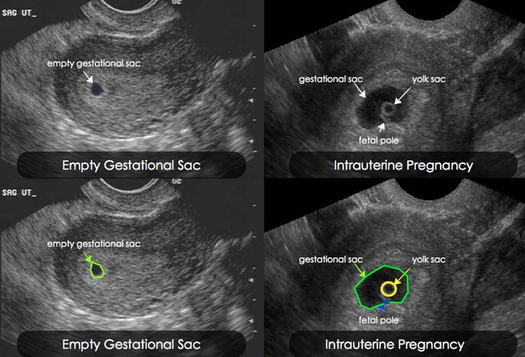
Is it harmful to do ultrasound for pregnant women
There is no consensus among experts: some believe that the study should be carried out without fail, others - that it is best to refuse the effect of ultrasound on the fetus. Russian and foreign specialists in the field of gynecology also do not find a compromise on this issue.
Meanwhile, according to statistics, not a single expectant mother or child in the womb suffered as a result of ultrasound diagnostics. So there are no scientific facts proving the harm of ultrasound to humans. In this regard, most experts who observe the pregnancy of their patients adhere to the principle of the "golden mean". They insist on carrying out two planned procedures, more - only according to indications.
Experts rightly believe that it is impossible to do without ultrasound. It allows you to control the development of the fetus and, if necessary, take timely measures to preserve the health of the baby.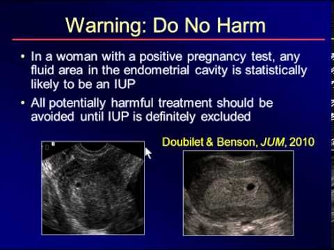
Other articles
01/31/2023
Management of multiple pregnancies
Multiple pregnancy is not only a great joy in the family, but also implies maximum attention to the expectant mother and babies, as it carries an increased risk for everyone. We will tell you more in our article.
01/05/2023
Ultrasound folliculometry: how and why is it performed?
Folliculometry is the observation of changes occurring in a woman's genitals during the menstrual cycle using ultrasound. Such an examination is necessary to identify the causes of menstrual irregularities, to monitor the growth and formation of the egg in the case of diagnosis and treatment of infertility.


