Pregnant inside your tubes
Ectopic Pregnancy (for Parents) - Nemours KidsHealth
What Is an Ectopic Pregnancy?
In a normal pregnancy, the fertilized egg implants and develops in the uterus. In an ectopic pregnancy, the egg implants somewhere other than the uterus — often, in the fallopian tubes. This is why ectopic pregnancies are commonly called "tubal pregnancies." The egg also can implant in the ovary, abdomen, or the cervix.
None of these areas has the right space or nurturing tissue for a pregnancy to develop. As the fetus grows, it will eventually burst the organ that contains it. This can cause severe bleeding and endanger the mother's life. A classical ectopic pregnancy does not develop into a live birth.
What Are the Signs & Symptoms of an Ectopic Pregnancy?
Ectopic pregnancy can be hard to diagnose because symptoms often are like those of a normal early pregnancy. These can include missed periods, breast tenderness, nausea, vomiting, tiredness, or frequent urination (peeing).
Often, the first warning signs of an ectopic pregnancy are pain or vaginal bleeding. There might be pain in the pelvis, abdomen, or even the shoulder or neck (if blood from a ruptured ectopic pregnancy builds up and irritates certain nerves). The pain can range from mild and dull to severe and sharp. It might be felt on just one side of the pelvis or all over.
These symptoms also might happen with an ectopic pregnancy:
- vaginal spotting
- dizziness or fainting (caused by blood loss)
- low blood pressure (also caused by blood loss)
- lower back pain
What Causes an Ectopic Pregnancy?
An ectopic pregnancy usually happens because a fertilized egg couldn’t quickly move down the fallopian tube into the uterus. The tube can get blocked from an infection or inflammation. The tube can get blocked from:
- pelvic inflammatory disease (PID)
- endometriosis, when cells from the lining of the uterus implant and grow elsewhere in the body
- scar tissue from previous abdominal or fallopian surgeries
- rarely, birth defects that changed the shape of the tube
How Is an Ectopic Pregnancy Diagnosed?
If a woman might have an ectopic pregnancy, her doctor may do an ultrasound to see where the developing fetus is. Often, pregnancies are too small to see on ultrasound until more than 5 or 6 weeks after a woman’s last menstrual period. If an external ultrasound can’t show the pregnancy, the doctor might do the test with a wand-like device in the vagina.
A woman might need testing every few days if the first tests can’t confirm or rule out an ectopic pregnancy.
How Is an Ectopic Pregnancy Treated?
How doctors treat an ectopic pregnancy depends on things like the size and location of the pregnancy.
Sometimes they can treat an early ectopic pregnancy with an injection of methotrexate, which stops the growth of the embryo. The tissue usually is then absorbed by the woman’s body.
If the pregnancy is farther along, doctors usually need to do surgery to remove the abnormal pregnancy.
Whatever treatment she gets, a woman will see her doctor regularly afterward to make sure her pregnancy hormone levels return to zero. This may take several weeks. An elevated level could mean that some ectopic tissue was missed. If so, she might need more methotrexate or surgery.
What About Future Pregnancies?
Most women who have had an ectopic pregnancy can have normal pregnancies in the future. Having had one ectopic pregnancy does increase a woman’s risk of having another one.
What Else Should I Know?
Any woman can have an ectopic pregnancy. But the risk is higher for women who are older than 35 and those who have had:
- PID
- a previous ectopic pregnancy
- surgery on a fallopian tube
- infertility problems or medicine to stimulate ovulation
Some birth control methods also can affect a woman's risk of ectopic pregnancy. Those who become pregnant while using an intrauterine device (IUD) might be more likely to have an ectopic pregnancy. Smoking and having multiple sexual partners also increase the risk of an ectopic pregnancy.
When Should I Call the Doctor?
If you believe you're at risk for an ectopic pregnancy, meet with your doctor to talk about your options before you become pregnant. If you are pregnant and have any concerns about the pregnancy being ectopic, talk to your doctor — it's important to find it early. Your doctor might want to check your hormone levels or schedule an early ultrasound to ensure that your pregnancy is developing normally.
Call your doctor right away if you're pregnant and having any pain, bleeding, or other symptoms of ectopic pregnancy.
What Is Ectopic Pregnancy? | Definition and Treatment
In This Section
- Ectopic Pregnancy
- How do I know if I have an ectopic pregnancy?
Ectopic pregnancy is when a pregnancy grows outside of your uterus, usually in your fallopian tube. Ectopic pregnancies are rare but serious, and they need to be treated.
What’s an ectopic pregnancy?
Normal pregnancies develop inside your uterus, after a fertilized egg travels through your fallopian tube and attaches to your uterine lining. Ectopic pregnancy is when a fertilized egg attaches somewhere else in your body, usually in your fallopian tube — that’s why it’s sometimes called “tubal pregnancy.”
Ectopic pregnancies can also happen on your ovary, or somewhere else in your belly.
Ectopic pregnancies are rare — it happens in about 2 out of every 100 pregnancies. But they’re very dangerous if not treated. Fallopian tubes can break if stretched too much by the growing pregnancy — this is sometimes called a ruptured ectopic pregnancy. This can cause internal bleeding, infection, and in some cases lead to death.
Am I at risk for an ectopic pregnancy?
We don’t always know the cause of ectopic pregnancy. But you may be more likely to have an ectopic pregnancy if you:
-
have had an STD, pelvic inflammatory disease or endometriosis
-
have already had an ectopic pregnancy
-
have had pelvic or abdominal surgery
-
are 35 or older
-
smoke cigarettes
If you get pregnant after you’ve been sterilized or while you have an IUD, it’s more likely to be ectopic. But this is very rare, because these types of birth control are super effective at preventing pregnancy.
Can I get pregnant again after an ectopic pregnancy?
Most people who have an ectopic pregnancy can have healthy pregnancies in the future, depending on the treatment you had and the condition of your fallopian tubes. If one of your fallopian tubes was removed or your tubes are scarred, it may be more difficult to get pregnant. If you have an ectopic pregnancy, you’re more likely to get another one in the future.
More questions from patients:
Can a doctor re-implant my ectopic pregnancy in my uterus? And is treating an ectopic pregnancy the same thing as having an abortion?
No, a doctor can’t re-implant or move your ectopic pregnancy into your uterus. Ectopic pregnancies can’t grow into fetuses: A pregnancy won’t survive if it’s ectopic, because a fertilized egg can’t grow or survive outside your uterus.
Untreated ectopic pregnancies can cause internal bleeding, infection, and in some cases lead to death. When you have an ectopic pregnancy, it’s extremely important to get treatment from a doctor as soon as possible.
Ectopic pregnancies are unsafely outside of your uterus (usually in the fallopian tubes), and are removed with a medicine called methotrexate or through a laparoscopic surgical procedure. The medical procedures for terminating a pregnancy in the uterus are usually different from the medical procedures for terminating an ectopic pregnancy.
Ectopic pregnancies are dangerous when left untreated and can’t lead to a baby. If you’re pregnant and have severe pain or bleeding, go to the emergency room right away. If you have any other symptoms of ectopic pregnancy, contact your doctor or nurse as soon as you can. The earlier an ectopic pregnancy is found and treated, the safer you’ll be.
Was this page helpful?- Yes
- No
Help us improve - how could this information be more helpful?
How did this information help you?
Please answer below.
Are you human? (Sorry, we have to ask!)
Please don't check this box if you are a human.
You’re the best! Thanks for your feedback.
Thanks for your feedback.
We couldn't access your location, please search for a location.
Zip, City, or State
Please enter a valid 5-digit zip code or city or state.
Please fill out this field.
Service All Services Abortion Abortion Referrals Birth Control COVID-19 Vaccine HIV Services Men's Health Care Mental Health Morning-After Pill (Emergency Contraception) Pregnancy Testing & Services Primary Care STD Testing, Treatment & Vaccines Transgender Hormone Therapy Women's Health Care
Filter By All Telehealth In-person
Please enter your age and the first day of your last period for more accurate abortion options. Your information is private and anonymous.
I'm not sure This field is required.
AGE This field is required.
Or call 1-800-230-7526
Physiological changes in the body during pregnancy
From the very first days of pregnancy, a woman's body undergoes profound transformations. These transformations are the result of the coordinated work of almost all body systems, as well as the result of the interaction of the mother's body with the child's body. During pregnancy, many internal organs undergo significant restructuring. These changes are adaptive in nature, and, in most cases, are short-lived and completely disappear after childbirth. Consider the changes in the basic systems of the vital activity of a woman's body during pregnancy.
The respiratory system during pregnancy works hard. The respiratory rate increases. This is due to an increase in the need of the mother and fetus for oxygen, as well as in the limitation of the respiratory movements of the diaphragm due to an increase in the size of the uterus, which occupies a significant space of the abdominal cavity.
The mother's circulatory system during pregnancy has to pump more blood to ensure an adequate supply of nutrients and oxygen to the fetus. In this regard, during pregnancy, the thickness and strength of the heart muscles increase, the pulse and the amount of blood pumped by the heart in one minute increase. In addition, the volume of circulating blood increases. In some cases, blood pressure increases. The tone of blood vessels during pregnancy decreases, which creates favorable conditions for increased supply of tissues with nutrients and oxygen. During pregnancy, the network of vessels of the uterus, vagina, and mammary glands decreases sharply. On the external genitalia, in the vagina, lower extremities, there is often an expansion of the veins, sometimes the formation of varicose veins. Heart rate decreases in the second half of pregnancy. It is generally accepted that the rise in blood pressure over 120-130 and a decrease to 100 mm Hg. signal the occurrence of pregnancy complications. But it is important to have data on the initial level of blood pressure.
And changes in the blood system. During pregnancy, blood formation increases, the number of red blood cells, hemoglobin, plasma and bcc increases. BCC by the end of pregnancy increases by 30-40%, and erythrocytes by 15-20%. Many healthy pregnant women have a slight leukocytosis. ESR during pregnancy increases to 30-40. Changes occur in the coagulation system that contribute to hemostasis and prevent significant blood loss during childbirth or placental abruption and in the early postpartum period.
Kidneys work hard during pregnancy. They secrete decay products of substances from the body of the mother and fetus (the waste products of the fetus pass through the placenta into the mother's blood).
Changes in the digestive system are represented by increased appetite (in most cases), cravings for salty and sour foods. In some cases, there is an aversion to certain foods or dishes that were well tolerated before the onset of pregnancy.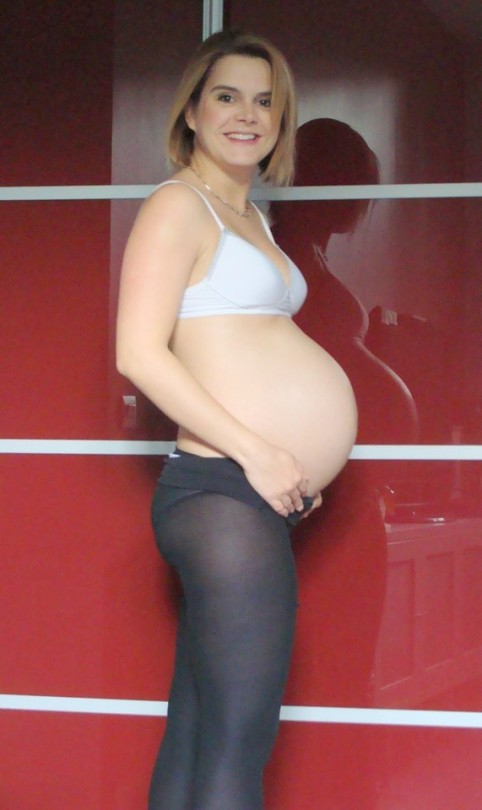 Due to the increased tone of the vagus nerve, constipation may occur.
The most significant changes, however, occur in the genitals of pregnant women. These changes prepare the woman's reproductive system for childbirth and breastfeeding.
The uterus of a pregnant woman increases significantly in size. Its mass increases from 50 g - at the beginning of pregnancy to 1200 g - at the end of pregnancy. The volume of the uterine cavity by the end of pregnancy increases by more than 500 times! The blood supply to the uterus is greatly increased. In the walls of the uterus, the number of muscle fibers increases. The cervix is filled with thick mucus that clogs the cavity of the cervical canal. The fallopian tubes and ovaries also increase in size. In one of the ovaries, there is a "corpus luteum of pregnancy" - a place for the synthesis of hormones that support pregnancy. Walls vaginas will loosen and become more elastic. External genitalia (labia minor and major), also increase in size and become more elastic. The tissues of the perineum are loosened. In addition, there is an increase in mobility in the joints of the pelvis and a divergence of the pubic bones. The changes in the genital tract described above are of extremely important physiological significance for childbirth. Loosening the walls, increasing the mobility and elasticity of the genital tract increases their throughput and facilitates the movement of the fetus through them during childbirth.
Skin in the genital area and in the midline of the abdomen usually becomes darker in color. Sometimes "stretch marks" form on the skin of the lateral parts of the abdomen, which turn into whitish stripes after childbirth.
Mammary glands increase in size, become more elastic, tense. When pressing on the nipple, colostrum (first milk) is released.
Changes in the bone skeleton and muscular system . An increase in the concentration of the hormones relaxin and progesterone in the blood contributes to the leaching of calcium from the skeletal system. This helps to reduce the rigidity of the joints between the bones of the pelvis and increase the elasticity of the pelvic ring. Increasing the elasticity of the pelvis is of great importance in increasing the diameter of the internal bone ring in the first stage of labor and further reducing the resistance of the birth tract to fetal movement in the second stage of labor. Also, calcium, washed out of the mother's skeletal system, is used to build the skeleton of the fetus.
It should be noted that calcium compounds are washed out of all bones of the maternal skeleton (including the bones of the foot and spine). As shown earlier, a woman's weight increases during pregnancy by 10 -12 kg. This additional load against the background of a decrease in bone stiffness can cause foot deformity and the development of flat feet. A shift in the center of gravity of the body of a pregnant woman due to an increase in the weight of the uterus can lead to a change in the curvature of the spine and the appearance of pain in the back and pelvic bones. Therefore, for the prevention of flat feet, pregnant women are advised to wear comfortable shoes with low heels. It is advisable to use insoles that support the arch of the foot. For the prevention of back pain, special physical exercises are recommended that can unload the spine and sacrum, as well as wearing a comfortable bandage. Despite an increase in calcium loss by the bones of the skeleton of a pregnant woman and an increase in their elasticity, structure and bone density (as is the case with osteoporosis in older women).
Changes in the nervous system . In the first months of pregnancy and at the end of it, there is a decrease in the excitability of the cerebral cortex, which reaches its greatest degree by the time of the onset of childbirth. By the same period, the excitability of the receptors of the pregnant uterus increases. At the beginning of pregnancy, there is an increase in the tone of the vagus nerve, in connection with which various phenomena often occur: changes in taste and smell, nausea, increased salivation, etc.
Active endocrine glands there are significant changes that contribute to the proper course of pregnancy and childbirth. Changes in body weight. By the end of pregnancy, a woman's weight increases by about 10-12 kg. This value is distributed as follows: fetus, placenta, membranes and amniotic fluid - approximately 4.0 - 4.5 kg, uterus and mammary glands -1.0 kg, blood - 1.5 kg, intercellular (tissue) fluid - 1 kg , an increase in the mass of adipose tissue of the mother's body - 4 kg.
Heart formation
So, a new life was born. Whether you wanted it or not, whether the fruit of your love is desirable or not, it doesn't matter. The egg, formed in the ovary, passed through the tubes, settled in the uterine mucosa, accepted and merged with the sperm. This is already a fertilized egg that will grow and eventually become your child.
This life, still only one cell, carries all the information contained in your genes, i.e. the smallest protein molecules, and in your partner's genes. We will return to this. But now, the cells have merged, and in the first two weeks after conception, the processes of formation of cell systems begin, which then turn into tissues and organs.
As the amazing poet Dmitry Kedrin once wrote:
“Still nausea and spots are not even in sight.
And your belt is just as narrow, even if you look in the mirror.
But you are elusive, according to secret female signs
Frightened guessed what you have inside ... "
In the beginning, new life takes the form of a disk. Sometimes such a small protein disk can be seen in the yolk of a broken chicken egg. It is called an embryo, and in the early days it is just a collection of wise cells that know exactly what they need to do. With each subsequent hour, the cells become more and more. They connect and fold into certain shapes, forming first two tubes, then, merging, one. This tube folds and descends from the primary disc to form a loop called the primary cardiac loop. The loop quickly lengthens, significantly outstripping the growth and increase in the number of cells surrounding it, lies to the right, in the form of such a ring as a mooring rope ring, which is thrown onto the bollard when a boat or vessel is moored. This loop normally lies only on the right, otherwise the future heart will lie not on the left, but on the right of the sternum. And on the 22nd day after conception, the first contraction occurs in the thickened lower part of the loop. The heart began to beat. You can try to remember what happened then with the future mother. What state was she in? What happened to her? And, if you, like the vast majority of married and single couples, did not pay attention to this, I can guarantee that you will not remember. You will say: “So what?”, - and you will be right. As a rule, nothing. But still, think about it. The first days may not solve anything. But the next will decide a lot.
The cardiovascular system of the fetus is the first of all its systems to form, because the fetus needs its own blood circulation for the full development of its other organs. The development and formation of the cardiovascular system begins in the third week and, in general, ends by the eighth week of the embryo's life, i.e. takes place over five weeks.
We will briefly describe these stages, but now we ask ourselves the question: “What is today 4-5 weeks of pregnancy?”. A woman is not yet sure if she is pregnant, especially if she does not expect this event too much. She does not change her lifestyle, habits, sometimes harmful. She can work in heavy and hazardous production or do hard physical work at home. She can carry a viral infection in the form of influenza on her feet. Usually a couple does not think yet, tries not to think about the future, but it - this future - is not only living, but also beating, shrinking, growing. But wait to execute yourself - there may be other reasons. About them - later. In the meantime, remember: today in the world they believe that the life of a child begins not from the moment of his birth, but from the moment of conception.
So, on the 22nd day, the future heart begins to pulsate, and on the 26th day in the body of the fetus, whose length is 3 millimeters, independent blood circulation begins. Thus, by the end of the fourth week, the fetus has a beating heart and circulation. So far, this is one stream, one curved tube, in the bend of which lies the "motor" - the heart. But every minute processes take place in it that lead to the final formation. It is very important to understand that these processes take place simultaneously in three-dimensional space and in order for “everything to fit together correctly and accurately”, their complete synchronization is needed. Moreover, if this did not happen, i.e. at some point, something did not connect where it was needed, the growth and development of the heart does not stop. Everything goes on. After all, when some musician suddenly plays a false note in the orchestra, the orchestra will still play the symphony. But the false sound will fly away and be forgotten, and few people will pay attention to it, but the forming heart will remember it..jpg) And now the growing partition has nowhere to attach, or the valve has nothing to hold on to. This is how birth defects are formed. In order for the heart to become four-, and not two-chambered (as in the third week), it is necessary that its septa grow (interatrial and interventricular), so that the common arterial trunk is divided into an aorta and a pulmonary artery, so that inside the common ventricle it is divided into right and left so that the aorta connects with the left ventricle so that the heart valves are fully formed. All this happens between the 4th and 8th weeks of pregnancy (at this time, the length of the fetus reaches only 3.5–4 cm). By the end of the second month of pregnancy, everything is already formed in the "inch" (3.5 cm) embryo. Obviously, the earlier in this process there was a violation of normal development, the more the heart becomes deformed, i.e. the more severe his congenital defect. The later this happened, the smaller the structural change will be and the easier it will be to correct the defect in the future.
And now the growing partition has nowhere to attach, or the valve has nothing to hold on to. This is how birth defects are formed. In order for the heart to become four-, and not two-chambered (as in the third week), it is necessary that its septa grow (interatrial and interventricular), so that the common arterial trunk is divided into an aorta and a pulmonary artery, so that inside the common ventricle it is divided into right and left so that the aorta connects with the left ventricle so that the heart valves are fully formed. All this happens between the 4th and 8th weeks of pregnancy (at this time, the length of the fetus reaches only 3.5–4 cm). By the end of the second month of pregnancy, everything is already formed in the "inch" (3.5 cm) embryo. Obviously, the earlier in this process there was a violation of normal development, the more the heart becomes deformed, i.e. the more severe his congenital defect. The later this happened, the smaller the structural change will be and the easier it will be to correct the defect in the future.


