Infant percentile chart
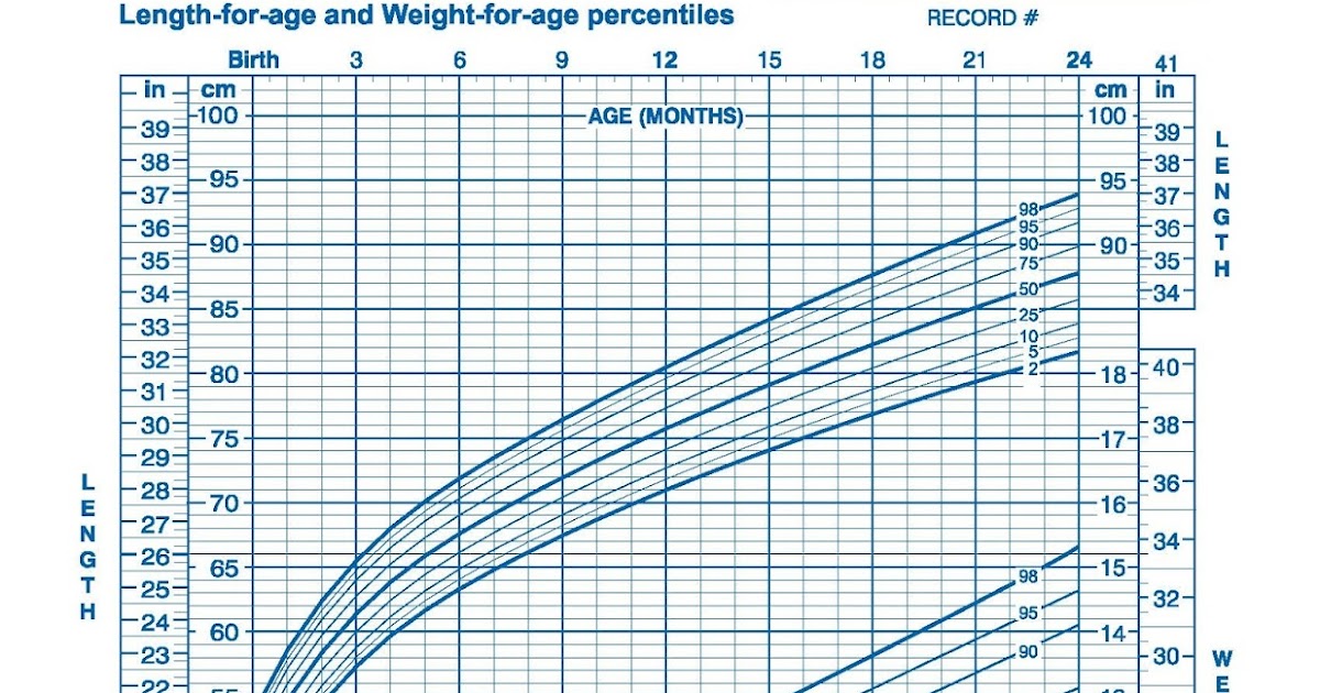
Baby weight and height tracker growth chart
- Baby
- Baby Development
Tip: Track your child's growth over time by logging into your account.
New to BabyCenter? Join now
Child's name
Date of birth
Enter birthday
Sex
Boy Girl
Date of measurement
Enter date
Weight
lb.kg. oz.
Height
in.cm.
Head circumference
cm.in.
By Rebekah Wahlberg
|
|
June 21, 2022
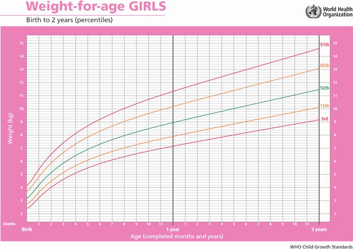
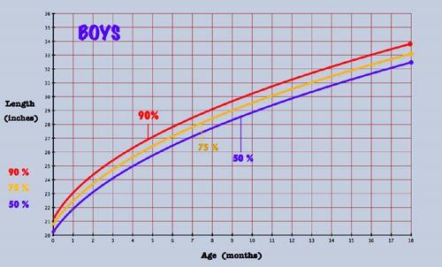
Our growth percentile calculator shows how your baby's size compares with other boys or girls the same age. Just enter your child's weight, height (aka length), and head circumference, and we'll calculate a percentile for each. The number reflects what percentage of kids is larger or smaller than your baby. So if your 3-month-old baby is in the 40th percentile for weight, that means 40 percent of 3-month-old babies weigh less than your baby, and 60 percent weigh more.
Using our baby growth chart
When your baby is first born, you'll have several visits at your pediatrician's office to make sure your baby's growth is on track. Your pediatrician will plot your baby's weight, height, and head circumference on a growth chart.
Don't be concerned if your baby doesn't fall right in the middle of the growth chart, though. There's a wide range of normal, and your baby's measurements now don't necessarily reflect what size they'll be as an adult. It's normal for some children to be in the 10th percentile for weight, while other children are in the 90th percentile. The most important thing is that your baby is growing at a healthy and consistent rate, no matter what percentile they're in.
Our growth percentile calculator is a general educational tool only.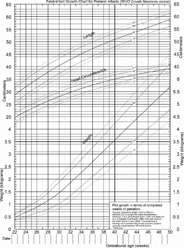 It's not the last word on how your child is growing, and it's not a substitute for having a healthcare provider monitor your child's growth at regularly scheduled exams. At these visits, your provider will determine whether your child is following a healthy growth pattern over time.
This tool is for children under age 2 and is based on World Health Organization data for kids in that age group. And if you'd like, you can find out how tall your older child is likely to be with our Child Height Predictor.
Read on to learn more about the measurements healthcare professionals take, and learn how to measure your baby at home.
Baby weight
If you want to see how your baby is growing between doctor visits, here's how to get a fairly accurate weight at home:
- Undress your baby completely (remove their diaper, too) and step on a scale while you're holding them. Record that weight.
- Then, set your baby down and weigh yourself. Subtract your own weight from your combined weight with your baby and record that number.
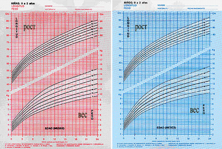
- This isn't as accurate as weighing your baby at the doctor's office, but it's a good estimate of how much your baby weighs.
- If you're worried about your baby's weight gain, call your baby's pediatrician. They may have you come in for a visit, since it's important to have your baby weighed on a consistent scale that can accurately measure weight in pounds and ounces.
Baby height
Before your baby can stand and walk on their own, their height is often recorded as their "length" – that is, how long they are from the crown of their head to the bottom of their heel while lying down. If you want to measure your baby's length at home between doctor visits, here's how:
- Lay your baby down on a flat surface and stretch a measuring tape from the top of their head to the bottom of their heel. Since babies' knees naturally bend a bit, you may need a second person to stretch out their legs while you take the measurement.
- Record their length to the nearest tenth of a centimeter or inch.
 (In the hospital, birth length is often recorded in centimeters, but your pediatrician's office will likely record their length in inches. What you do is up to you!) Your record might be a little different than the doctor's, but that's okay.
Baby head circumference
Your baby's healthcare provider will measure their head circumference for the first two years of their life to make sure your baby's brain is growing at a healthy rate.
You can measure your baby's head circumference at home, but it can be hard to do this accurately. Here's how to get a good estimate:
- Wrap a flexible measuring tape around the widest part of your baby's head, just above the eyebrows and ears and around the back where their head slopes out from their neck.
- Take the measurement three times (your baby may be wiggly, which could make getting an accurate measurement tough) and use the largest measurement to the nearest 0.1 centimeter.
Are you sure you want to delete this entry?
Yes Cancel
New entry
Edit entry
Your entry couldn't be saved. Please try again later.
advertisement | page continues below
advertisement
Featured video
All pregnancy, parenting, and birth videos >
Get the BabyCenter app
The #1 app for tracking pregnancy and baby growth.
Download now
Growth Charts - Clinical Growth Charts
The clinical growth charts reflect modifications in the format of the individual charts, whereby two individual charts appear on a single page, and data entry tables have been added. The clinical charts have the grids scaled to metric units (kg, cm), with English units (lb, in) as the secondary scale. Clinical charts are available for boys and for girls. The available clinical charts include the following:
Infants, birth to 36 months:
- Length-for-age and Weight-for-age
- Head circumference-for-age and Weight-for-length
Children and adolescents, 2 to 20 years
- Stature-for-age and Weight-for-age
- BMI-for-age
Preschoolers, 2 to 5 years
- Weight-for-stature
The clinical charts for infants and older children were published in two sets.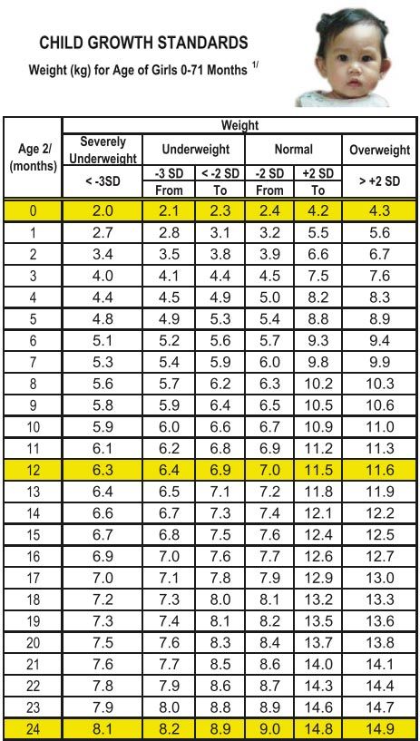
- Set 1 contains 10 charts (5 for boys and 5 for girls), with the 5th, 10th, 25th, 50th, 75th, 90th, and 95th smoothed percentile lines for all charts, and the 85th percentile for BMI-for-age and weight-for-stature.
- Set 2 contains 10 charts (5 for boys and 5 for girls), with the 3rd, 10th, 25th, 50th, 75th, 90th, and 97th smoothed percentile lines for all charts, and the 85th percentile for BMI-for-age and weight-for-stature.
Set 1 has the outer limits of the curves at the 5th and 95th percentiles. These are the charts that most users in the United States will find useful for the majority of routine clinical assessments. Set 2 has the outer limits of the curves at the 3rd and 97th percentiles for selected applications. Pediatric endocrinologists and others who assess the growth of children with special health care requirements may wish to use the format in set 2 for selected applications.
Two summary files, each with all 10 clinical charts in set 1 or set 2 are also available.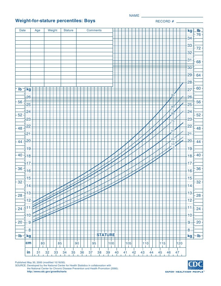 These summary files contain the clinical charts from either set 1 or set 2 as described above.
Infants (birth to 24 months) must be measured for length, and the sex appropriate length-for-age or weight-for-length charts for infants, birth to 36 months must be used to plot the measurements. At age 24 months and older, if children can stand unassisted and follow directions, stature should be measured and plotted on the stature-for-age chart for children (2 to 20 years). Otherwise, between 24 and 36 months, length can be used in place of stature.
BMI-for-age charts are recommended to assess weight in relation to stature for children ages 2 to 20 years. The weight-for-stature charts are available as an alternative to accommodate children ages 2-5 years who are not evaluated beyond the preschool years. However, all health care providers should consider using the BMI-for-age charts to be consistent with current recommendations.
All individual 2000 CDC growth charts have an initial publication date of May 30, 2000.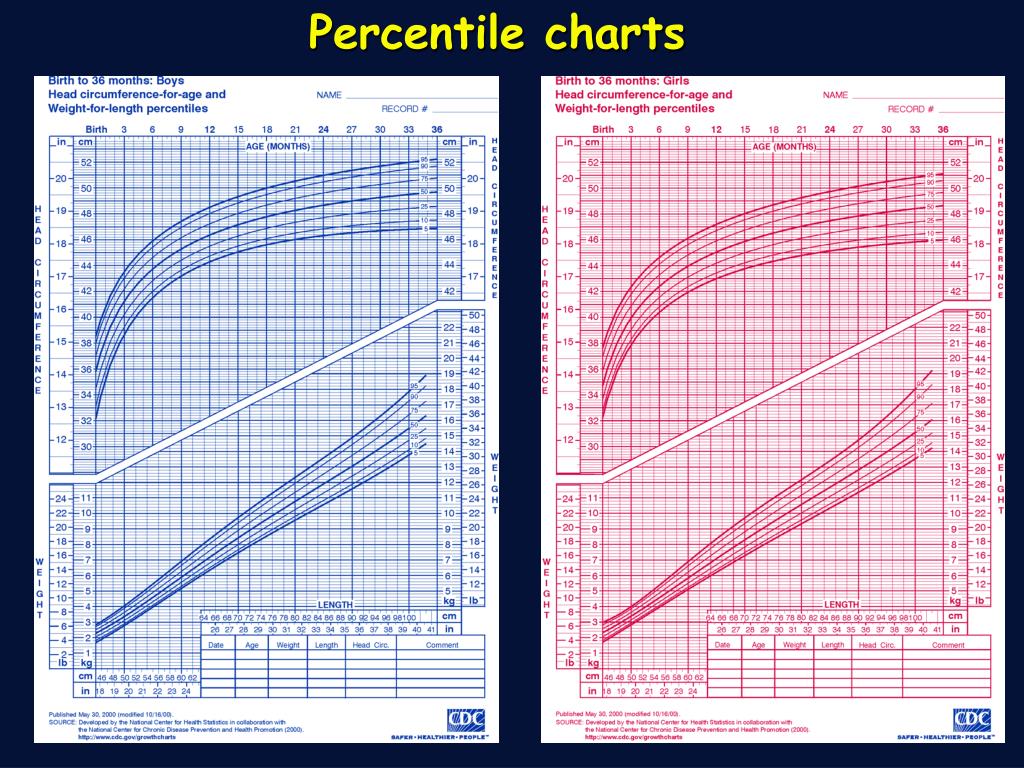 For various reasons, modifications were made to charts after the initial publication date. For example, the individual charts were modified to create the clinical charts, which were made available on October 16, 2000. Subsequent modifications were made to selected clinical charts to correct or enhance particular aspects of the scales on the graphs. In all cases, the data points in the corresponding data file for each modified chart remain unchanged from the initial release on May 30, 2000. Where applicable, when selected clinical charts were further modified, the date is indicated on each chart. The clinical growth charts for stature-for-age were modified because the scale for inches was not correctly aligned with the metric scale. The clinical growth charts for infant length-for-age and infant weight-for-age were revised to improve the appearance of the scale for inches on the length charts by extending the indicators at ½ inch increments, and enhancing alignment of the English with the metric scales on both the length and weight scales.
Users should use the most recent version of each chart, as available on the growth charts web site.
To view, print, and reproduce clinical growth charts
All clinical growth charts may be viewed, downloaded, and printed in Adobe Acrobat. For routine viewing on a computer monitor and printing on a laser printer, the individual charts are available as PDF files (Black and White). All clinical charts have been colorized for viewing and printing. When routed to a color printer, the clinical charts for boys will print in blue and the clinical charts for girls will print in red. Otherwise, these same charts can be routed to a black-and-white printer, and will print in black-and-white. Higher resolution PDF files (Color) are available to provide the highest resolution and are intended to be used as a high quality print master for quantity production when using the services of a commercial printing facility. The recommended ink colors for printing are Pantone 206 red (for girls) and Pantone 286 blue (for boys).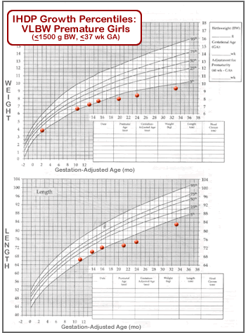 The recommended paper weight is 80#. Charts should be printed as two-sided copies, in the following combinations for each sex:
Infants, birth to 36 months:
- Side 1: Length for age + Weight-for-age
- Side 2: Head circumference-for-age + Weight-for-length
Children and adolescents, 2 to 20 years:
- Side 1: Stature-for-age + Weight-for-length
- Side 2: BMI-for-age or
- Weight-for-stature (age 2 to 5 years only)
Set 1: Clinical charts with 5th and 95th percentiles
Birth to 36 months (5th-95th percentile)
Boys Length-for-age and Weight-for-age
B&W [PDF – 41 KB] Modified 4/20/01
Color [PDF – 44 KB] Modified 4/20/01
Spanish Version, Color [PDF – 44 KB] Modified 4/20/01
French Version, Color [PDF – 44 KB] Modified 4/20/01
B&W Press-Ready [PDF – 186 KB] Modified 9/15/05
Color Press-Ready [PDF – 200 KB] Modified 9/15/05
Data table of Length-for-age charts
Data table of Weight-for-age charts
Boys Head circumference-for-age and Weight-for-length
B&W [PDF – 48 KB] Modified 10/16/00
Color [PDF – 48 KB] Modified 10/16/00
Spanish Version Color [PDF – 52 KB] Modified 10/16/00
French Version Color [PDF – 56 KB] Modified 10/16/00
B&W Press-Ready [PDF – 206 KB] Modified 9/15/05
Color Press-Ready [PDF – 236 KB] Modified 9/15/05
Data table of Head circumference-for-age charts
Data table of Weight-for-length charts
Girls Length-for-age and Weight-for-age
B&W [PDF – 40 KB] Modified 4/20/01
Color [PDF – 40 KB] Modified 4/20/01
Spanish Version, Color [PDF – 50 KB] Modified 4/20/01
French Version, Color [PDF – 44 KB] Modified 4/20/01
B&W Press-Ready [PDF – 186 KB] Modified 9/15/05
Color Press-Ready [PDF – 401 KB] Modified 9/15/05
Data table of Length-for-age charts
Data table of Weight-for-age charts
Girls Head circumference-for-age and Weight-for-length
B&W [PDF – 48 KB] Modified 10/16/00
Color [PDF – 55 KB] Modified 10/16/00
Spanish Version, Color [PDF – 60 KB] Modified 10/16/00
French Version, Color [PDF – 55 KB] Modified 10/16/00
B&W Press-Ready [PDF – 206 KB] Modified 9/15/05
Color Press-Ready [PDF – 440 KB] Modified 9/15/05
Data table of Head circumference-for-age charts
Data table of Weight-for-length charts
Children 2 to 20 years (5th-95th percentile)
Boys Stature-for-age and Weight-for-age
B&W [PDF – 77 KB] Modified 11/21/00
Color [PDF – 77 KB] Modified 11/21/00
Spanish Version, Color [PDF – 63 KB] Modified 11/21/00
French Version, Color [PDF – 78 KB] Modified 11/21/00
B&W Press-Ready [PDF – 208 KB] Modified 9/15/05
Color Press-Ready [PDF – 260 KB] Modified 9/15/05
Data table of Stature-for-age charts
Data table of Weight-for-age charts
Boys BMI-for-age
B&W [PDF – 61 KB] Modified 10/16/00
Color [PDF – 61 KB] Modified 10/16/00
Spanish Version, Color [PDF – 50 KB] Modified 10/16/00
French Version, Color [PDF – 64 KB] Modified 10/16/00
B&W Press-Ready [PDF – 141 KB] Modified 9/15/05
Color Press-Ready [PDF – 183 KB] Modified 9/15/05
Data table of BMI-for-age charts
Girls Stature-for-age and Weight-for-age
B&W [PDF – 77 KB] Modified 11/21/00
Color [PDF – 77 KB] Modified 11/21/00
Spanish Version, Color [PDF – 62 KB] Modified 11/21/00
French Version, Color [PDF – 79 KB] Modified 11/21/00
B&W Press-Ready [PDF – 208 KB] Modified 9/15/05
Color Press-Ready [PDF – 499 KB] Modified 9/15/05
Data table of Stature-for-age charts
Data table of Weight-for-age charts
Girls BMI-for-age
B&W [PDF – 67 KB] Modified 10/16/00
Color [PDF – 61 KB] Modified 10/16/00
Spanish Version, Color [PDF – 51 KB] Modified 10/16/00
French Version, Color [PDF – 64 KB] Modified 10/16/00
B&W Press-Ready [PDF – 142 KB] Modified 9/15/05
Color Press-Ready [PDF – 339 KB] Modified 9/15/05
Data table of BMI-for-age charts
Optional Chart (5th-95th percentile)
Boys Weight-for-stature
B&W [PDF – 40 KB] Modified 10/16/00
Color [PDF – 41 KB] Modified 10/16/00
B&W Press-Ready [PDF – 148 KB] Modified 9/15/05
Color Press-Ready [PDF – 180 KB] Modified 9/15/05
Data table of Weight-for-stature charts
Girls Weight-for-stature
B&W [PDF – 40 KB] Modified 10/16/00
Color [PDF – 41 KB] Modified 10/16/00
B&W Press-Ready [PDF – 147 KB] Modified 9/15/05
Color Press-Ready [PDF – 334 KB] Modified 9/15/05
Data table of Weight-for-stature charts
Set 2: Clinical charts with 3rd and 97th percentiles
Birth to 36 months (3rd -97th percentile)
Boys Length-for-age and Weight-for-age
B&W [PDF – 41 KB] Modified 4/20/01
Color [PDF – 47 KB] Modified 4/20/01
Spanish Version, Color [PDF – 47 KB] Modified 4/20/01
French Version, Color [PDF – 44 KB] Modified 4/20/01
B&W Press-Ready [PDF – 186 KB] Modified 9/15/05
Color Press-Ready [PDF – 201 KB] Modified 9/15/05
Data table of Length-for-age charts
Data table of Weight-for-age charts
Boys Head circumference-for-age and Weight-for-length
B&W [PDF – 48 KB] Modified 10/16/00
Color [PDF – 52 KB] Modified 10/16/00
Spanish Version, Color [PDF – 53 KB] Modified 10/16/00
French Version, Color [PDF – 51 KB] Modified 10/16/00
B&W Press-Ready [PDF – 207 KB] Modified 9/15/05
Color Press-Ready [PDF – 223 KB] Modified 9/15/05
Data table of Head circumference-for-age charts
Data table of Weight-for-length charts
Girls Length-for-age and Weight-for-age
B&W [PDF – 41 KB] Modified 4/20/01
Color [PDF – 48 KB] Modified 4/20/01
Spanish Version, Color [PDF – 48 KB] Modified 4/20/01
French Version, Color [PDF – 45 KB] Modified 4/20/01
B&W Press-Ready [PDF – 186 KB] Modified 9/15/05
Color Press-Ready [PDF – 390 KB] Modified 9/15/05
Data table of Length-for-age charts
Data table of Weight-for-age charts
Girls Head circumference-for-age and Weight-for-length
B&W [PDF – 48 KB] Modified 10/16/00
Color [PDF – 53 KB] Modified 10/16/00
Spanish Version, Color [PDF – 54 KB] Modified 10/16/00
French Version, Color [PDF – 52 KB] Modified 10/16/00
B&W Press-Ready [PDF – 206 KB] Modified 9/15/05
Color Press-Ready [PDF – 434 KB] Modified 9/15/05
Data table of Head circumference-for-age charts
Data table of Weight-for-length charts
Children 2 to 20 years (3rd-97th percentile)
Boys Stature-for-age and Weight-for-age
B&W [PDF – 79 KB] Modified 11/21/00
Color [PDF – 83 KB] Modified 11/21/00
Spanish Version, Color [PDF – 61 KB] Modified 11/21/00
French Version, Color [PDF – 79 KB] Modified 11/21/00
B&W Press-Ready [PDF – 208 KB] Modified 9/15/05
Color Press-ready [PDF – 250 KB] Modified 9/15/05
Data table of Stature-for-age charts
Data table of Weight-for-age charts
Boys BMI-for-age
B&W [PDF – 64 KB] Modified 10/16/00
Color [PDF – 68 KB] Modified 10/16/00
Spanish Version, Color [PDF – 52 KB] Modified 10/16/00
French Version, Color [PDF – 66 KB] Modified 10/16/00
B&W Press-Ready [PDF – 153 KB] Modified 9/15/05
Color Press-Ready [PDF – 173 KB] Modified 9/15/05
Data table of BMI-for-age charts
Girls Stature-for-age and Weight-for-age
B&W [PDF – 79 KB] Modified 11/21/00
Color [PDF – 84 KB] Modified 11/21/00
Spanish Version, Color [PDF – 62 KB] Modified 11/21/00
French Version, Color [PDF – 81 KB] Modified 11/21/00
B&W Press-Ready [PDF – 218 KB] Modified 9/15/05
Color Press-Ready [PDF – 488 KB] Modified 9/15/05
Data table of Stature-for-age charts
Data table of Weight-for-age charts
Girls BMI-for-age
B&W [PDF – 54 KB] Modified 10/16/00
Color [PDF – 58 KB] Modified 10/16/00
Spanish Version, Color [PDF – 52 KB] Modified 10/16/00
French Version, Color [PDF – 67 KB] Modified 10/16/00
B&W Press-Ready [PDF – 152 KB] Modified 9/15/05
Color Press-Ready [PDF – 334 KB] Modified 9/15/05
Data table of BMI-for-age charts
Optional Chart (3rd-97th percentile)
Boys Weight-for-stature
B&W [PDF – 31 KB] Modified 10/16/00
Color [PDF – 34 KB] Modified 10/16/00
B&W Press-Ready [PDF – 158 KB] Modified 9/15/05
Color Press-Ready [PDF – 168 KB] Modified 9/15/05
Data table of Weight-for-stature charts
Girls Weight-for-stature
B&W [PDF – 40 KB] Modified 10/16/00
Color [PDF – 46 KB] Modified 10/16/00
B&W Press-Ready [PDF – 157 KB] Modified 9/15/05
Color Press-Ready [PDF – 323 KB] Modified 9/15/05
Data table of Weight-for-stature charts
Summary files
Set 1 summary file; 5th, 10th, 25th, 50th, 75th, 90th, 95th percentiles
Download all 10 charts in Set 1
B&W [PDF – 409K]
Color [PDF – 418 KB]
Set 2 summary file; 3rd, 10th, 25th, 50th, 75th, 90th, 97th percentiles
Download all 10 charts in Set 2
B&W [PDF – 408K]
Color [PDF – 423 KB]
Use of percentile charts in myopia control
Axial length (AL) is perhaps the most important parameter on which to judge the progression of myopia.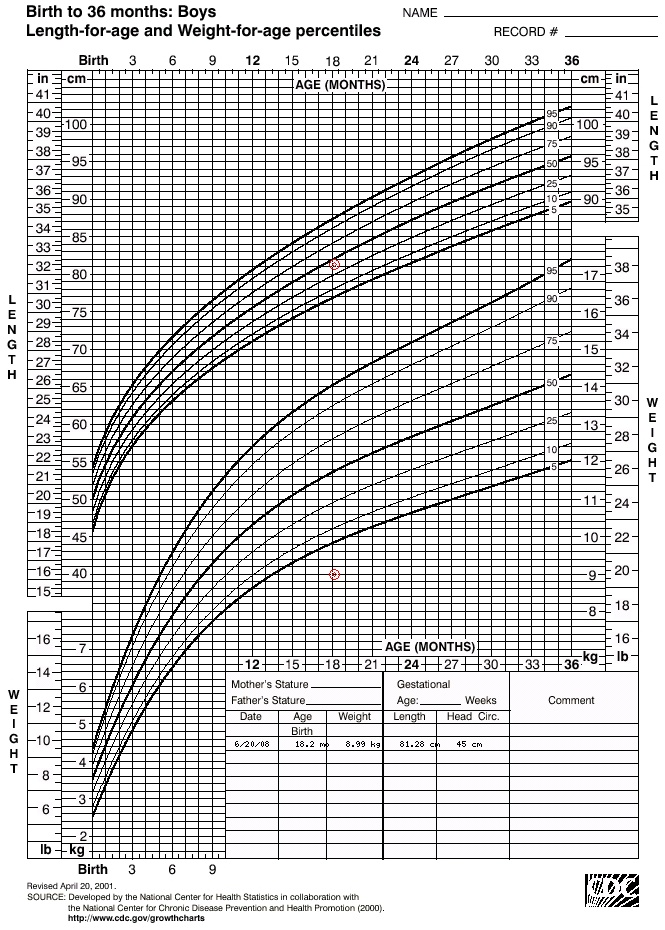 In the control of myopia, the AL value measured for a particular child and its change over the years are compared with the standard values obtained as average values for the population.
For this purpose, percentile charts are used, which present a set of curves for changing axial length with age for different percentiles (from 5th to 9th5th, step may be 5 or more). Percentiles reflect the prevalence of age-AL combination in a given population. So, if the AL value for a given age lies on the 10th percentile curve (on the 10th percentile), then this means that 10% of the population of this age have an axial length less than this AL value, and 90% more.
Axial length-age percentile charts (and corresponding tables) for European children can be taken, for example, from the article (https://bjo.bmj.com/content/early/2021/11/09/bjophthalmol-2021-319431), published in 2021 in the British Journal of Ophthalmology. Percentile curves are also used in the Myopia module of Haag-Streit's LENSTAR biometer.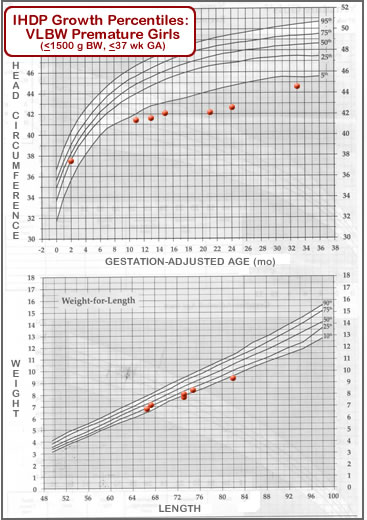
Consider 2 examples of the use of percentiles in myopia control. For a 7-year-old Chinese girl, the measured AL was 22.40 mm (ref. error +1.50 D). This AL value for 7 years on the percentile chart (for the Chinese population) is at the 35th percentile. This means that 35 out of 100 girls at the age of 7 years have eyes less than 22.40 mm, and 65 out of 100 girls at the age of 7 have eyes longer than 22.40 mm. By the age of 8, the girl's eye grew to 22.64 mm (ref. error +0.75 D), the percentile remained the same (35th), which indicates a stable growth pattern. However, at 9years, AL increased to 23.15 mm (ref. error -0.50 D), which already corresponds to the 45th percentile. This indicates that the child's AL has shifted by 10 percentiles. A 10 percent increase may indicate an unusual increase in eye length between 8 and 9 years. An upward shift in percentiles indicates a higher risk of developing myopia, which warrants further evaluation and treatment if needed.
Consider the second example. A 9 year old European boy has a measured AL of 23.5 mm (ref. error -1.00 D). These values are in the 65th percentile, indicating that the boy's AL is greater than 65% of boys 9years old. Considering this percentile, refractive status, and other risk factors, the use of myopia control methods was recommended. The AL values measured at 10 and 11 years old were 23.6 mm and 23.65 mm (ref. error in both cases -1.25 D). Although the AL increased, the percentile decreased to the 60th, indicating an improvement over both the baseline percentile and the population mean.
As these examples show, measured AL values and their percentiles indicate the location of a child's AL relative to population averages and can be used to control myopia. Measurement of AL and percentiles during follow-up of the child can help to observe the growth pattern of the eye and evaluate the effectiveness of the method of myopia control. nine0004
Adapted from publication (https://reviewofmm.com/listening-to-patients-needs-led-me-to-myopia-management/).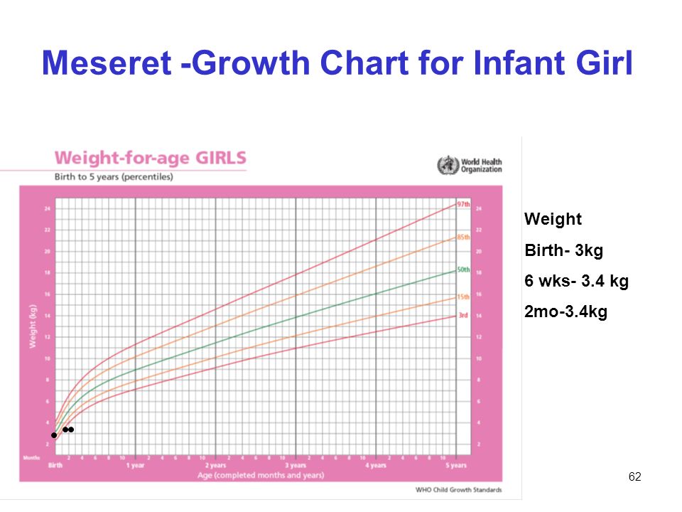
Vol 22, No 5 (2020)
ORIGINAL ARTICLE
Dynamics of the epidemic process and the course of the COVID-19 in pregnant women of the Far Eastern and Siberian Federal Districts
Belokrinitskaya T.E., Artymuk N.V., Filippov O.S., Shifman E.M.
Abstract
The objective of the study was to evaluate an epidemiological characteristic and the course of COVID-19in pregnant women from the Far East and Siberian Federal Districts of Russia.
Materials and methods. The analysis of operational information on the incidence of COVID-19 and the its features in pregnant women and after childbirth provided by the chief obstetrician-gynecologists of the Far Eastern Federal District and Siberian Federal District on May 25, 2020 and August 25, 2020 is carried out. Mathematical analysis included descriptive statistics methods, analysis of contingency tables, where the χ2 value, the achieved significance level (p)
Results. On August 25, 2020, 2010 cases of COVID-19 in pregnant women were detected in two subjects, including 134 active cases. In the population, the proportion of pregnant women among cases of COVID-19 was 1.45%. Pneumonia in pregnant women was registered in 44.8% of cases, severe course of the disease - in 3.7%, hospitalization in RAW - in 1.9% of cases, mechanical ventilation - 0. In the population, pneumonia was registered in 24.4% of cases, hospitalized in RAW - 2.1%, on mechanical ventilation - 0.77%. The incidence in pregnant women was 1464.7 per 100,000 (in the population - 591.8 per 100,000). 23 (1.76%) COVID-19 (+) newborns were identified, despite preventive measures and separation of mother and child immediately after childbirth, which does not exclude vertical transmission of the disease. Maternal mortality was 2 cases (0.1%) vs population mortality 1.49%. Perinatal mortality was 7 cases (0.35%). There were 5 (0.25%) stillbirths. Early neonatal mortality was 2 (0.1%) cases.
Conclusion. The incidence of COVID-19 in pregnant women in the Far East and Siberia is higher than in the general population with a higher incidence of pneumonia, but a milder course: less need for ICU, mechanical ventilation and mortality. 23 cases of SARSCoV-2-positive newborns, despite the separation of mother and child, do not exclude the possibility of vertical transmission of infection. nine0004
Keywords: COVID-19, pregnancy, epidemiology, incidence, pneumonia, mortality
Show
Hide
Gynecology . 2020;22(5):6-11
(Rus)
(JATS XML)
The result of 10 years of experience in monitoring indicators of infant and perinatal mortality in Khabarovsk Krai
Pestrikova T.Y.
Abstract
Aim. To evaluate the effectiveness of the obstetric care service in Khabarovsk Krai, based on a comparison of the dynamics of infant and perinatal mortality rates for the period 2009–2018.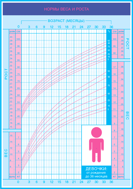
Materials and methods. A comparative analysis of official statistics of the Ministry of Health of Khabarovsk Krai and other territories of the Far Eastern Federal District was carried out using the Rosstat database for the period 2009–2018, taking into account the indicators of infant, perinatal mortality, and preterm birth. The trend of the dynamics was determined in the process of graphical analysis of the chart and by modeling trends. The results of the studies were subjected to methods of statistical processing of information. nine0004
Results. The results revealed in our study indicate that over the 10-year interval, the infant and perinatal mortality rates in Khabarovsk Krai decreased by 2 times, the rate of decrease was higher than similar processes in the Far Eastern Federal District and the Russian Federation. A graphical representation of infant and perinatal mortality rates underlines the instability of these values in Khabarovsk Krai.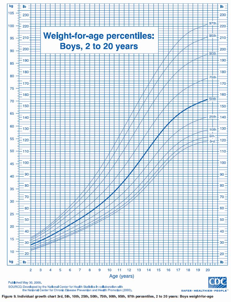 The decrease in infant and perinatal mortality in Khabarovsk Krai is mainly due to a decrease in early neonatal mortality. The ratio of stillbirth to early neonatal mortality indicates an excess of stillbirth by 7.88 and 6.95 times in 2017–2018. The relatively high stillbirth rate in Khabarovsk Krai indicates the instability of the results in providing assistance to pregnant women and women in childbirth.
Conclusion. The infant mortality rate is the main quality indicator recommended by WHO for assessing the level of care. This indicator must be considered in conjunction with the indicator of perinatal mortality, especially its constituent components of stillbirth and early neonatal mortality, namely the ratio of stillbirth to early neonatal mortality. The deformation of this proportion towards a multiple increase in stillbirth, against the background of a progressive decrease in early neonatal mortality, indicates the presence of an inadequate assessment of the organization of medical care in the obstetric care service.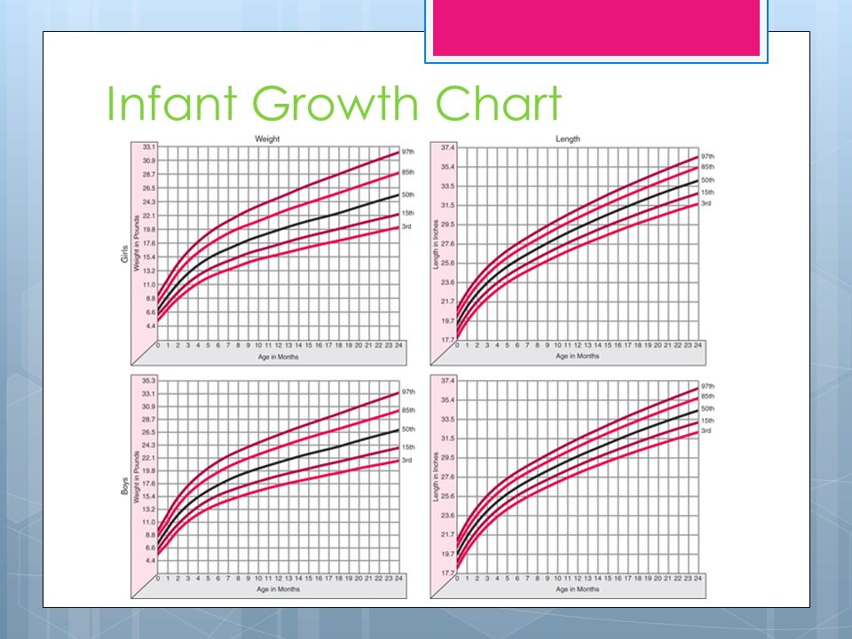 nine0004
Show
Hide
Gynecology . 2020;22(5):12-16
(Rus)
(JATS XML)
A prognostic model to calculate the probability of severe preeclampsia in a healthy young reproductive age woman
Frolova N.I., Belokrinitskaya T.E., Kolmakova K.A.
Abstract
Aim. To develop the prognostic model to calculate the probability of severe preeclampsia in a healthy young reproductive age woman. nine0004
Materials and methods. This study included two groups of healthy women aged 18–35 years with spontaneous singleton pregnancy: 100 patients with severe preeclampsia (patient group), and 100 women with non-complicated pregnancy (control group). All women had not a risk factors of preeclampsia such as a history of hypertension, autoimmune, metabolic, renal, or cardiac diseases, and family or individual history of preeclampsia or thromboembolism before this pregnancy.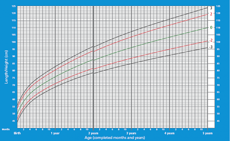 Their body mass index in the 1st trimester of gestation was nine0004
Results. Some prognostic models to calculate the probability of severe preeclampsia were build using an anamnestic, clinical and genotypic characteristics and multifactorial analysis. Combination of genotypes AGTR2 -1675AA/ eNOS3 -786CC; tobacco smoking; bacteriuria; acute respiratory infections and/or acute vulvovaginitis during 2–3 trimester of gestation were determinate as the most informative predictors of severe preeclampsia. Logistic model included three predictors: bacteriuria; acute respiratory infections and/or acute vulvovaginitis during 2–3 trimester of gestation had higher prognostic value. ROC analysis identified a high specificity (89.58%) and sensitivity (76.47%) of the model, and the integral index of the effectiveness of predictive markers (AUC=0.885), according to the expert scale of values which is indicative of a very high quality model.
Conclusion. It is recommended to use this elaborated predictive model for the purpose of individual risk assessment of severe preeclampsia in healthy young reproductive age woman.
Show
Hide
Gynecology . 2020;22(5):17-21
(Rus)
(JATS XML)
The efficacy and safety of using the topical medicine with combined action in patients with opportunistic vaginal infections
Prilepskaya V.N., Bairamova G.R., Mezhevitinova E.A., Donnikov A.E., Trofimov D.Y., Ivanets T.Y., Kukes I.V.
Abstract
Relevance. Infections of the lower genital tract are one of the most common reasons for women to visit a gynecologist. According to various authors, among all vulvovaginitis, the highest percentage of patients visiting a doctor is bacterial vaginosis (20–50%), vulvovaginal candidiasis (17–39%), trichomoniasis (10%) and other genital infections (10%).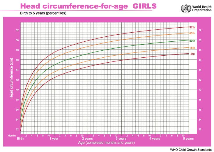 In the management of patients with infectious processes of the genitals, the choice of the method of therapy is of great importance, which allows not only to influence the microbes-associates, detected in high concentrations in the woman's vagina during the initial episode of the disease , but also to prevent the development of relapses.
To evaluate the clinical and laboratory efficacy, adherence and long-term results of benzydamine use in patients with vulvovaginal candidiasis, aerobic vaginitis and bacterial vaginosis
Materials and methods. The study included 31 women aged 18 to 49 years, who complained of abundant discharge from the genital tract, itching, burning, dyspareunia. A comprehensive clinical and laboratory examination was carried out, including the collection of anamnestic data, general and special gynecological examination, microscopy of vaginal smears, KOH test, determination of the pH of the vaginal environment, assessment of vaginal microbiocenosis by quantitative PCR in real time . All patients were prescribed benzydamine hydrochloride, which was used as monotherapy in the form of a vaginal solution according to the recommended treatment regimen specified in the instructions for medical use of the drug Tantum ® Rosa 1 (irrigation 2 times a day for 10 days).
Results. In 12–19 days after beginning treatment, 25 (80.6%) of 31 patients with laboratory signs of vaginal microflora alterations showed restoration of the normal vaginal biocenosis laboratory parameters. Of them, 8 patients initially had dysbiosis, 12 patients had yeast-like fungi, 4 patients had aerobic vaginitis and 1 patient had a combination of dysbiosis and fungi. Herewith, all patients also noted clinical signs of improvement. In 12–19days after beginning treatment, 6 (1.4%) of 31 patients with laboratory signs of vaginal biocenosis alterations had unsatisfactory laboratory parameters; that is vaginal biocenosis alterations (a decrease in Lactobacillus spp. percentage).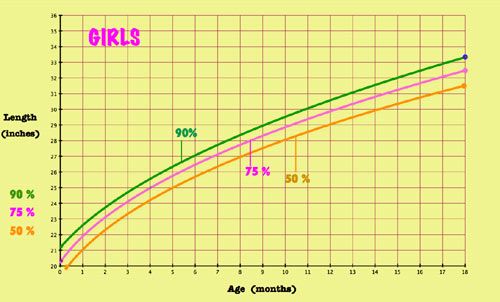 At the same time, there were no clinical signs of pronounced inflammation such as pathological discharge, hyperemia, vaginal swelling.
Conclusion. Benzydamine hydrochloride can be widely used in gynecological practice due to its combined effects on inflammation of the vagina, good clinical and microbiological results. It is important to note that additional therapy with drugs which contain lactobacilli is required only in 19.4% of cases. The clinical and laboratory effectiveness of benzydamine hydrochloride (Tantum ® Rosa) was 96.8%, since the relapse rate after 30–60 days of the study did not exceed 3.2%.
Show
Hide
Gynecology . 2020;22(5):22-26
(Rus)
(JATS XML)
Features of the ovarian reserve of women of reproductive age suffering from autoimmune diseases
Andreeva E.N., Grigoryan O.R., Absatarova Y.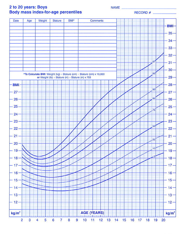 S., Yarovaya I.S., Mikheev R.K. nine0004
Abstract
The reproductive potential of a woman depends on indicators of the ovarian reserve, such as the anti-Muller hormone (AMH) and the number of antral follicles (NAF). Autoimmune diseases have a significant effect on fertility and contribute to the development of premature ovarian failure.
To evaluate the parameters of the ovarian reserve in patients with type 1 diabetes mellitus, carriers of antibodies to the thyroid gland in a state of euthyroidism and compare them with similar parameters in healthy women. nine0004
Materials and methods. In the first block of the study, the level of AMH, follicle-stimulating hormone, luteinizing hormone, NAF was studied among 224 women with diabetes and 230 healthy women in the control group. In block II, the level of the above hormonal indices was studied in 35 carriers of antithyroid antibodies in the state of euthyroidism and 35 healthy women.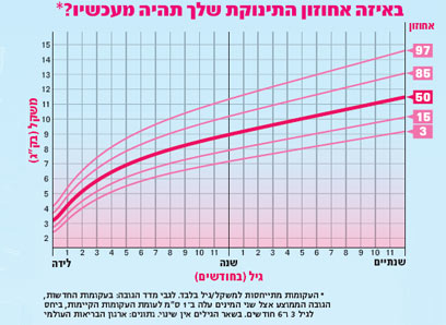
Results. In patients with type 1 diabetes, the level of AMH, NAF was statistically significantly lower when compared with the control group. Among carriers of antithyroid antibodies and healthy women, no difference in AMH and NAF was found. nine0004
Conclusion. The autoimmune processes accompanying diabetes are more influenced by the ovarian reserve indices than autoimmune aggression to the tissues of the thyroid gland.
Show
Hide
Gynecology . 2020;22(5):27-30
(Rus)
(JATS XML)
Promising directions of medical technologies for the correction of sleep disorders in menopausal women of various ethnic groups nine0024
Semenova N.V., Madaeva I.M., Kolesnikova L.I.
Abstract
ethnic groups.
Materials and methods. The objects of the study were menopausal women of Russian and Buryat ethnic groups (n=466), who were divided into a control group (without sleep disturbances) and main subgroups (with insomnia and comorbidity of insomnia with obstructive sleep apnea syndrome).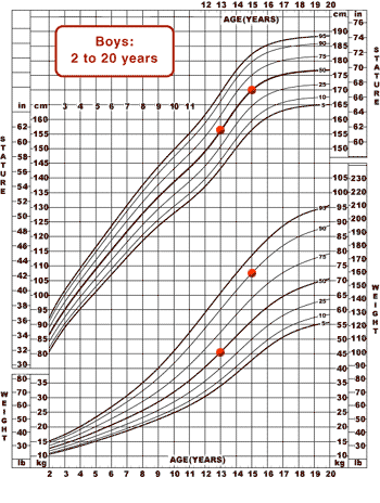 All women underwent clinical and anamnestic examination, genotyping for 3111T /C Clock gene polymorphism, determination of the circadian profile of melatonin secretion, parameters of lipid metabolism and the system "lipid peroxidation - antioxidant protection".
Results. 3111T /T genotype and the Clock gene 3111T allele was revealed as compared to the control level in Mongoloids; personalized prescription of antioxidant therapy in all patients; specific therapy to eliminate breathing disorders during sleep in those who have obstructive sleep apnea syndrome.
Conclusions. Presence of insomnia, chronobiological rhythms of melatonin secretion, lipid peroxidation processes in sleep disorders are ethnospecific.
Show
Hide
Gynecology . 2020;22(5):31-36
(Rus)
(JATS XML)
REVIEW
Vaginal bleeding during menopause hormone therapy.
 Is it always endometrial cancer? How to choose the appropriate tactics for examination and management of patients? nine0024
 Protasova A.E., Solntseva I.A., Vandeeva E.N.
Abstract
The article provides an overview of the possible causes of bleeding from the genital tract in peri- and postmenopausal women. The correct approach to patients with abnormal uterine bleeding during menopausal hormone therapy is described. A modern analysis of data on the diagnosis and treatment of postmenopausal vaginal bleeding against the background of menopausal hormone therapy was carried out. The modern possibilities of endometrial diagnostics and treatment are shown. nine0004
Show
Hide
Gynecology . 2020;22(5):37-43
(Rus)
(JATS XML)
Ovarian endometriosis associated infertility: a modern view to the problem
Orazov M.R., Khamoshina M.B., Abitova M.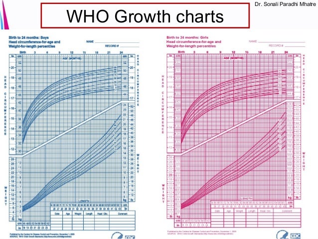 Z., Mikhaleva L.M., Volkova S.V., Aryutin D.G., Shustova V.B.
Abstract
This review summarizes the current understanding of the pathogenesis of one of the most common forms of external genital endometriosis – ovarian endometrioma’s. Due to their frequent occurrence in young women of reproductive age and extremely negative impact on the morphophysiological state of the ovaries, this disease makes a significant contribution to the structure of endometriosis-associated infertility. The main determinant of the negative effect of ovarian endometrioma's on reproductive function is a decrease in ovarian reserve, which can occur either due to the direct gonadotoxic effect of the endometriod cyst itself, or due to the unintentional removal of healthy ovarian tissue during surgery or the use of aggressive methods of electrosurgery. Hence, the question of methods for achieving hemostasis during surgery in terms of iatrogenic effects on healthy ovarian tissue is debatable. The management strategy for patients with infertility associated with ovarian endometriosis consists of two components: surgical treatment and/or the use of assisted reproductive technologies.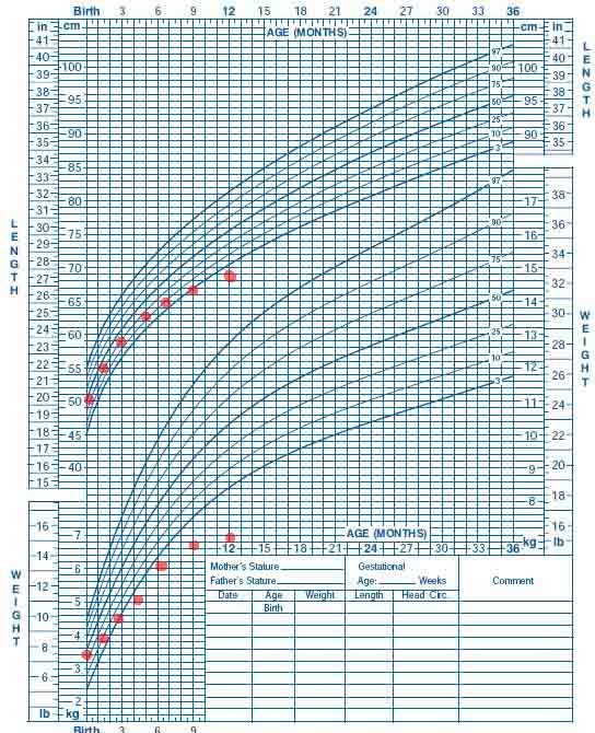 Laparoscopic cystectomy is indicated for cysts larger than 3 cm. Repeated surgical interventions in case of endometriosis do not improve fertility outcomes. Assisted reproductive technologies methods should be considered as a priority tactic in patients of older reproductive age with low ovarian reserve indicators or infertility duration of more than 2 years, as well as in cases of recurrent ovarian endometrioma’s. Management of such patients must be personalized and take into account the age, state of the ovarian reserve, duration of infertility, stage and number of surgical interventions for this disease. nine0004
Show
Hide
Gynecology . 2020;22(5):44-49
(Rus)
(JATS XML)
Deep infiltrative endometriosis. Contentious issues: pros and cons
Rusina E.I., Yarmolinskaya M.I., Piankova V.O.
Abstract
Relevance. Deep infiltrative endometriosis (DIE) is a particular form of endometriosis with a more severe symptoms and dysfunction of the neighboring organs.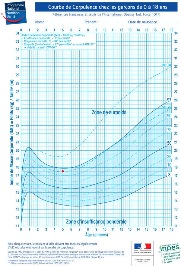 Inspite of the fact that in recent years, much had been done to understand the pathogenesis of the disease and its associated symptoms; there are still several unresolved issues. nine0004
To describe debated issues of pathogenesis and management of patients with DIE using current data.
Materials and methods. Foreign and domestic scientific articles on this topic that is available in PubMed database and on Internet resources have been examined over the past 5 years.
Results. The review consolidated of a system analysis of data on the pathogenesis of pain, infertility, the choice of conservative or surgical treatment in dealing with specific problems of managing patients with DIE. nine0004
Conclusion. Despite there are different options of managing and treating, most specialists agree that the treatment strategy should be based on the assumption that it is a chronic recurrent disease. The treatment choice should be personal depending on the severity of symptoms, dysfunction of the neighboring organs affected by endometriosis, such as the intestines, bladder, ureters, as well as the woman’s age and reproductive life plans.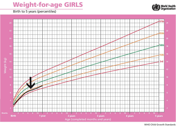 Depending on the clinical case, it is possible to use various regimens of conservative management. Surgical management planning should be highly balanced. A radical surgery with removing foci of endometriosis performed by an experienced surgeon in a specialized hospital in coupled with pharmacotherapy and assisted reproductive technologies increases the patient’s chances of pregnancy, a healthy childbearing. Subsequent combination therapy provides a long-term remission. Pregnancy and childbirth in patients with deep infiltrative endometriosis should be considered as a high risk condition for the severe complications. nine0004
Show
Hide
Gynecology . 2020;22(5):50-56
(Rus)
(JATS XML)
Current views on the molecular mechanisms of the initial stages of folliculogenesis
Marchenko L.A., Mashaeva R.I., Chernukha G.E.
Abstract
The ovary is a unique structure of the female body, which simultaneously presents various morphohistological units—from primordial to dominant follicles.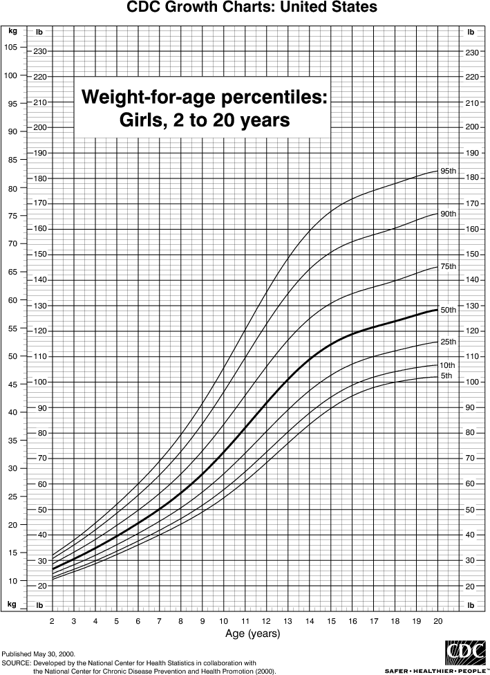 Over the past decades, scientists have focused on studying the mechanisms of folliculogenesis at the gonadotropin-dependent stage. While more complex and lengthy processes that determine the fate of the follicle occur from the moment of their recruitment to the preantral stage of maturity (about 290 days), another 60 days pass before the dominant maturity. Currently, it has been proven that intercellular communication is established within the follicle, which involves a bidirectional exchange of information between the oocyte and its “companions” – granulose and Teka cells through auto-and paracrine interactions using various genes, growth factors and cytokines. The purpose of this review was to study intrafollicular factors that control the early stages of folliculogenesis and other disorders that may ultimately lead to the development of premature ovarian failure. nine0004
Show
Hide
Gynecology . 2020;22(5):57-60
(Rus)
(JATS XML)
Omega-3 polyunsaturated fatty acids in supporting pregnancy and fetal development: dosing considerations
Gromova O.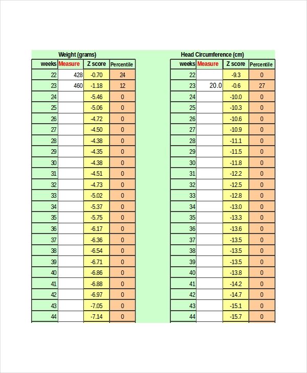 A., Torshin I.I., Grishina T.R., Maliavskaia S.I.
Abstract
Being complex regulators of inflammation processes omega-3-polyunsaturated fatty acids (PUFAs) are vitally important for supporting pregnancy and fetal development. Docosahexaenoic acid (DHA) and eicosapentaenoic acid (EPA) are the most important omega-3-PUFAs. The article presents the results of a systematic analysis of 1973 studies of dose-dependent effects of DHA and EPA in nutritional support of pregnancy. The results analysis suggest that DHA/EPA donations for pregnant women are effective only in doses of at least 200 mg/day. According to the analysis results, the optimal doses of DHA/EPA are in the 400- to 800-mg/day range. The dose of omega-3-PUFA for a particular patient can be assessed by the measurement of the DHA and EPA levels in the erythrocyte membrane. To achieve specific therapeutic effects (for example, lowering triglyceride levels, therapy for postpartum depression, etc.), higher doses of omega-3-PUFAs (3–5 g/day) may be required. With an adequate choice of dosing regimen, DHA / EPA donations have a beneficial effect on the somatic health of women of reproductive age, help to prevent pregnancy complications and determine the child development program in the subsequent years of life. DHA/EPA drugs are effective in the adjuvant therapy of various chronic disorders. nine0004
Show
Hide
Gynecology . 2020;22(5):61-69
(Rus)
(JATS XML)
Cesarean scar defect: terminological aspects
Martynov S.A., Adamyan L.V.
Abstract
The review aim was to summarize information and to provide terminological analysis of the foreign and Russian literature on assessing the status of the uterine scar after a caesarean section, as well as to identify terms that are most appropriate to use out and during pregnancy, in childbirth and in the postpartum period. nine0004
Show
Hide
Gynecology . 2020;22(5):70-75
(Rus)
(JATS XML)
IN AID OF THE CLINICIAN
Recurrent vulvovaginitis and vaginosis. Possibilities of effective therapy
Prilepskaya V.N., Uruimagova A.T., Ivanova A.A.
Abstract
Infectious and inflammatory diseases of the genitals caused by various opportunistic and pathogenic microorganisms are found in 70% of all gynecological patients who seek help from medical institutions. Currently, scientists and clinicians in their practice are increasingly faced with insufficient effectiveness of treatment of vulvovaginitis and vaginosis, a tendency to a long and protracted course, recurrence of the process. One of the reasons for the longer and more severe course, the recurrence of the process is the common increasingly mixed infection with the possible simultaneous predominance of aerobes, anaerobes, and Candida fungi, which suggests the appointment of appropriate drugs. One of these drugs is Tergynan, which demonstrates a good clinical effect and tolerability of the drug in the treatment of recurrent vulvovaginitis and dysbiotic conditions of the lower genital tract.
Show
Hide
Gynecology . 2020;22(5):76-80
(Rus)
(JATS XML)
CLINICAL CASE
Experience of pyomyoma treatment
Gabidullina R.I., Mingazetdinov M.A., Druzhkowa E.B., Sirmatova L.I.
Abstract
Uterine pyomyoma is a rare and dangerous complication of uterine fibroids, which develops as a result of myomatous node infarction with the addition of an infectious component. In the literature there are descriptions of 50 cases of pyomyomas, half of which are associated with pregnancy. Incorrect diagnosis, lack of antibacterial and surgical treatment can lead to death of the patient. This article describes a clinical case of uterine piomioma development after the first birth by Caesarean section. Method of uterine artery embolization in combination with antibiotic therapy has helped keep the uterus of a young woman. On subsequent observation, menstrual function has fully recovered. nine0004
Show
Hide
Gynecology . 2020;22(5):81-83
(Rus)
(JATS XML)
Pelvic schwannoma
Sinchikhin S.P., Magakyan S.G., Magakyan O.G.
Abstract
Relevance. A neoplasm originated from the myelonic sheath of the nerve trunk is called neurinoma or neurilemmoma, neurinoma, schwannoglioma, schwannoma. This tumor can cause compression and dysfunction of adjacent tissues and organs. The most common are the auditory nerve neurinomas (1 case per 100,000 population per year), the brain and spinal cord neurinomas are rare. In the world literature, there is no information on the occurrences of this tumor in the pelvic region. nine0004
Description. Presented below is a clinical observation of a 30-year-old patient who was scheduled for myomectomy. During laparoscopy, an unusual tumor of the small pelvis was found and radically removed. A morphological study allowed to identify the remote neoplasm as a neuroma.
Conclusion. The presented practical case shows that any tumor can hide under a clinical "mask" of another disease. The qualification of the doctor performing laparoscopic myomectomy should be sufficient to carry out, if necessary, another surgical volume. nine0004
Show
Hide
Gynecology . 2020;22(5):84-86
(Rus)
(JATS XML)
Vitamin D deficiency and iron deficiency anemia in women in menopausal transition period. clinical case
Tatarova N.A., Airapetian M.S.
Abstract
Vitamin D is a regulator of immune response, and plays a role in the development of cardiovascular diseases, insulin resistance and diabetes mellitus, obesity, autoimmune disorders, iron binding, anemia, respiratory viral and other diseases.