How long does it take to get pelvic ultrasound results
Ultrasound scan for ovarian cancer
You might have an external ultrasound of your lower tummy (pelvis) or a vaginal ultrasound to help diagnose ovarian cancer.
Ultrasound scans use high frequency sound waves to create a picture of a part of the body. It can show the ovaries, womb and surrounding structures.
During an external ultrasound of your pelvis, the doctor or radiographer moves a probe over the lower part of your tummy. For a vaginal ultrasound, they insert the probe into your vagina. This is called a transvaginal ultrasound.
Why you have it
Pelvic ultrasound and vaginal ultrasound scans can show whether:
- your ovaries are the right size
- your ovaries look normal in texture
- there are any cysts in your ovaries
Vaginal ultrasound can help to show whether any cysts on your ovaries contain cancer or not. If a cyst has any solid areas it is more likely to be cancer.
Sometimes, in women who are past their menopause, the ovaries do not show up on an ultrasound. This means that the ovaries are small and not likely to be cancerous.
If you have a suspicious looking cyst, your specialist will recommend that you have surgery to remove it. The cyst will be looked at closely in the laboratory.
Risk of malignancy index (RMI)
Doctors can use a tool called the risk of malignancy index (RMI) to decide if an abnormality is more likely to be cancer or not. This index combines the results of the ultrasound, CA125 blood levels and menopausal status (whether or not you are past the menopause).
This gives doctors a final score. Women with a high score are referred to a specialist multidisciplinary team (MDT). They decide on which further tests and surgery may be necessary.
Your specialist may ask you to have a CT scan to show the ovaries more clearly. Sometimes though, it is not possible to diagnose ovarian cancer for certain without an operation.
If your specialist thinks it unlikely that you have cancer, but cannot completely rule it out, they may ask you to come back for a repeat ultrasound scan in 3 months time, to see if anything has changed.
External ultrasound scans
Preparing for your scan
Check your appointment letter for any instructions about how to prepare for your scan.
You might need to stop eating for 6 hours beforehand. Let the scan team know if this will be a problem for any reason, for example if you are diabetic.
They might ask you to drink plenty before your scan so that you have a comfortably full bladder.
Take your medicines as normal unless your doctor tells you otherwise.
Before the scan
Before the test you might be asked to remove your clothing and put on a hospital gown. It will depend on what part of the body you're having scanned as to whether you have to undress or not.
During the scan
You lie on the couch next to the ultrasound machine. You might be able to sit up depending on which part of your body is being scanned.
The sonographer will spread a clear gel onto your skin over the area they are checking. The gel feels cold. It helps to transmit the sound waves to the probe. The scan appears on a screen next to you.
You might feel a little pressure as the sonographer presses the probe against your skin and moves it around the area they are scanning. Tell them if this is uncomfortable.
An ultrasound scan can take up to 45 minutes depending on what's being scanned.
Vaginal ultrasound
You might have a vaginal ultrasound to look at the ovaries, womb and surrounding structures. It is called transvaginal ultrasound or TVS.
Preparation for the scan
There is no special preparation needed for this scan. So you can eat and drink normally. And you can take your medications as normal.
The doctor or sonographer will ask you to empty your bladder before you have the scan. They may ask you to change into a hospital gown or undress from the waist down. You will have a sheet to cover you.
Before the scan
The doctor or sonographer asks you to lie on your back with your bottom at the end of a short scanning couch. There are supports for your feet so you can bend your knees and have your legs apart. If this position is difficult for you, you may be able to lie on your side with your knees drawn up to your chest.
During the scan
The doctor inserts a small thin ultrasound probe into your vagina. This looks like the same shape and size as a tampon. The probe is covered with a protective sheath like a condom and has some lubricating gel on it. The test may be uncomfortable and a little cool from the gel, but shouldn't hurt. This type of scan does not take long.
The sonographer will gently move the probe to get the pictures they need. At times the sonographer may place their hand on your tummy and press to move some of your organs to get a clear view on the screen.
What happens afterwards
You can eat and drink normally after the test. You can go straight home or back to work afterwards.
Possible risks
An ultrasound scan is a very safe procedure. It doesn’t involve radiation and there are usually no side effects.
Getting your results
You should get your results within 1 or 2 weeks. The doctor may be able to let you know if they have seen any abnormal areas that have been sent to the laboratory.
Waiting for results can make you anxious. Ask your doctor or nurse how long it will take to get them.
Contact the doctor who arranged the test if you haven’t heard anything after a couple of weeks.
You might have contact details for a specialist nurse who you can contact for information if you need to. It can help to talk to a close friend or relative about how you feel.
For information and support, you can also call the Cancer Research UK nurses on freephone 0808 800 4040. The lines are open from 9am to 5pm, Monday to Friday.
Last reviewed:
04 Jan 2022
Next review due:
04 Jan 2025
Print page
-
The recognition and initial management of ovarian cancer
National Institute for Health and Care Excellence (NICE), April 2011 -
Newly diagnosed and relapsed epithelial ovarian carcinoma: ESMO Clinical Practice Guidelines for diagnosis, treatment and follow-up
J Ledermann and others; ESMO Guidelines Working Group
Annals of Oncology.
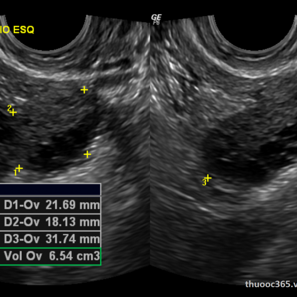 2013 Oct;24 Suppl 6:vi24-32.
 -
The Royal Marsden Manual of Clinical Nursing Procedures, 9th edition
L Dougherty and S Lister (Editors)
Wiley-Blackwell, 2015
Transvaginal Ultrasound: Purpose, Procedure, and Results
What is a transvaginal ultrasound?
An ultrasound test uses high-frequency sound waves to create images of your internal organs. Imaging tests can identify abnormalities and help doctors diagnose conditions.
A transvaginal ultrasound, also called an endovaginal ultrasound, is a type of pelvic ultrasound used by doctors to examine female reproductive organs. This includes the uterus, fallopian tubes, ovaries, cervix, and vagina.
“Transvaginal” means “through the vagina.” This is an internal examination.
Unlike a regular abdominal or pelvic ultrasound, where the ultrasound wand (transducer) rests on the outside of the pelvis, this procedure involves your doctor or a technician inserting an ultrasound probe about 2 or 3 inches into your vaginal canal.
There are many reasons why a transvaginal ultrasound might be necessary, including:
- an abnormal pelvic or abdominal exam
- unexplained vaginal bleeding
- pelvic pain
- an ectopic pregnancy (which occurs when the fetus implants outside of the uterus, usually in the fallopian tubes)
- infertility
- a check for cysts or uterine fibroids
- verification that an IUD is placed properly
Your doctor might also recommend a transvaginal ultrasound during pregnancy to:
- monitor the heartbeat of the fetus
- look at the cervix for any changes that could lead to complications such as miscarriage or premature delivery
- examine the placenta for abnormalities
- identify the source of any abnormal bleeding
- diagnose a possible miscarriage
- confirm an early pregnancy
In most cases, a transvaginal ultrasound requires little preparation on your part.
Once you’ve arrived at your doctor’s office or the hospital and you’re in the examination room, you have to remove your clothes from the waist down and put on a gown.
Depending on your doctor’s instructions and the reasons for the ultrasound, your bladder might need to be empty or partially full. A full bladder helps lift the intestines and allows for a clearer picture of your pelvic organs.
If your bladder needs to be full, you have to drink about 32 ounces of water or any other liquid about one hour before the procedure begins.
If you’re on your menstrual cycle or if you’re spotting, you have to remove any tampon you’re using before the ultrasound.
When it’s time to begin the procedure, you lie down on your back on the examination table and bend your knees. There may or may not be stirrups.
Your doctor covers the ultrasound wand with a condom and lubricating gel, and then inserts it into your vagina. Make sure your provider is aware of any latex allergies you have so that a latex-free probe cover is used if necessary.
You might feel some pressure as your doctor inserts the transducer. This feeling is similar to the pressure felt during a Pap smear when your doctor inserts a speculum into your vagina.
Once the transducer is inside of you, sound waves bounce off your internal organs and transmit pictures of the inside of your pelvis onto a monitor.
The technician or doctor then slowly turns the transducer while it’s still inside of your body. This provides a comprehensive picture of your organs.
Your doctor may order a saline infusion sonography (SIS). This is a special kind of transvaginal ultrasound that involves inserting sterile salt water into the uterus before the ultrasound to help identify any possible abnormalities inside the uterus.
The saline solution stretches the uterus slightly, providing a more detailed picture of the inside of the uterus than a conventional ultrasound.
Although a transvaginal ultrasound can be done on a pregnant woman or a woman with an infection, SIS cannot.
There are no known risk factors associated with transvaginal ultrasound.
Performing transvaginal ultrasounds on pregnant women is also safe, for both mother and fetus. This is because no radiation is used in this imaging technique.
When the transducer is inserted into your vagina, you’ll feel pressure and in some cases discomfort. The discomfort should be minimal and should go away once the procedure is complete.
If something is extremely uncomfortable during the exam, be sure to let the doctor or technician know.
You might get your results immediately if your doctor performs the ultrasound. If a technician performs the procedure, the images are saved and then analyzed by a radiologist. The radiologist will send the results to your doctor.
A transvaginal ultrasound helps diagnose multiple conditions, including:
- cancer of the reproductive organs
- routine pregnancy
- cysts
- fibroids
- pelvic infection
- ectopic pregnancy
- miscarriage
- placenta previa (a low-lying placenta during pregnancy that may warrant medical intervention)
Talk with your doctor about your results and what type of treatment, if any, is necessary.
There are virtually no risks associated with a transvaginal ultrasound, although you might experience some discomfort. The entire test takes about 30 to 60 minutes, and the results are typically ready in about 24 hours.
If your doctor is unable to get a clear picture, you might be called back to repeat the test. A pelvic or abdominal ultrasound is sometimes done before a transvaginal ultrasound depending on your symptoms.
If you experience too much discomfort from a transvaginal ultrasound and can’t tolerate the procedure, your doctor may perform a transabdominal ultrasound. This involves your doctor applying gel to your stomach and then using a handheld device to view your pelvic organs.
This approach is also an option for children when pelvic images are needed.
stages of preparation for the procedure - MEDSI
When is pelvic ultrasound required?
Ultrasound is prescribed to detect diseases and pathologies of the organs and structures of the genitourinary system. This analysis is considered one of the most informative, safe and painless. Preparation for ultrasound of the pelvic organs is not complicated. Therefore, it is recommended for use in examining patients of any age and gender.
The doctor refers the patient to this test when there is a suspicion of such diseases:
- The appearance of neoplasms (tumors, adenomas, fibroids)
- Cyst and polyp formation
- Pathological change in the structure of organs
- Presence of stones (stones, sand) in the bladder
- Cystitis
- Inflammations
- Infertility
- Salpingoophoritis
- Endometriosis
- Problems in the development of the genital organs in children
- Fetal development disorders in pregnancy
- Prostatitis
Similar ailments may be accompanied by the following symptoms:
- Pain in the lower abdomen and groin
- Bloody, mucus or purulent discharge in urine
- In women, bleeding outside of menses or after menopause, cycle disorders
- Problems with urination (soreness, too frequent urge, inability to empty the bladder)
- Pain in the lumbar region
If you experience any of these sensations and discomfort, you should immediately contact a qualified doctor.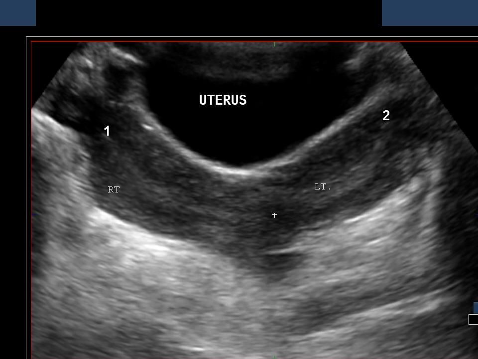
Ultrasound is also prescribed for women in such cases as:
- Determination of pregnancy and its timing
- Organ monitoring:
- Before and after abortion
- Before and after surgery
- If breast diseases are diagnosed
- For choosing a contraceptive, in some cases for its installation and control
It is recommended for both men and women to undergo a preventive examination of the genitourinary system every six months or at least once a year.
Types of pelvic ultrasound
Ultrasound examination methods differ depending on a number of factors:
- Which pelvic organs should be examined
- Suspicion of what disease to confirm or disprove
- Patient gender
- Is a woman sexually active
- Does the person have severe pain when trying to assume the position required for the examination
Depending on the above factors, one or more ultrasound methods are used:
- Abdominal
- Transvaginal
- Transrectal
Abdominal examination allows to determine the presence of inflammation, neoplasms or diseases in the bladder, uterus, prostate, ovaries, other organs and tissues of the small pelvis. It is non-invasive or endoscopic: it does not require abdominal incisions or insertion of sensors into the body. Such an inspection can also be carried out in cases where it is impossible to use the second and third methods.
Transvaginal ultrasound is a test in which a transducer is placed inside the pelvic organs through the vagina. It helps to examine in more detail minor violations or formations in the uterus and other structures. It also allows you to take a tissue sample for analysis. But it is not used if the patient is not yet sexually active, so as not to violate the integrity of the hymen. For obvious reasons, it cannot be assigned to a man.
Transrectal examination is used to clarify abnormalities and tumors in the prostate or testicles and is most often indicated in male patients if an abdominal ultrasound is not available or the results are inaccurate. With this method, the transducer is inserted into the rectum. It can also be assigned to a woman or girl if the first type of study is not enough.
Preparing for your procedure
Preparing for a pelvic ultrasound is simple but necessary. Most of the preliminary actions are recommended before the abdominal examination:
- In two or three days (better - a week), you need to start adhering to a certain diet
- The last meal before analysis should be no later than six o'clock in the evening on the eve of
- It is recommended to use preparations to remove food residues from the intestines or to give an enema
All of these steps are necessary to avoid the accumulation of gas in the intestines, flatulence and bloating, which will prevent you from getting the clearest image. If the patient has disturbances in flatulence or constipation, then he should consult a doctor about which drugs are better to use before analysis.
When adjusting nutrition before performing abdominal ultrasound of the small pelvis, you must follow the rules:
- Easy-to-digest foods can be used as food:
- Kashi
- Low-fat fish, meat dishes
- Hard cheeses
- Boiled eggs or scrambled eggs
- Weak tea
- It is necessary to exclude from the diet that which causes excessive gas formation, requires a long processing by the body:
- Vegetables (cabbage, potatoes and others)
- Legumes (beans, corn, peas)
- Milk and dairy products (cottage cheese, kefir)
- Fatty foods, including meat and fish
- Coffee
- Alcohol
- Carbonated drinks
- Fast food and sweets
- Brown bread, pastries
Immediately before the examination (one hour before), it is necessary to drink half a liter or a liter of water, since a filled bladder provides the best echolocation.
Transvaginal examination does not require special preparation. It should be remembered that before it is necessary to empty the bladder. It is also important to pay attention to personal hygiene.
This method is not used during monthly bleeding, but in case of emergency it can be done during this period.
Transrectal ultrasound requires an empty bowel, for which an enema should be given several hours before the test. On the eve, you can use cleansing preparations (this should be consulted with a doctor).
It is usually performed with an empty bladder, but in some cases it may be necessary to fill this reservoir with urine (investigation of the causes of infertility, erectile dysfunction). Then, before the analysis, you need to drink about 4 glasses of water or other unsweetened and non-carbonated liquid.
It is not recommended to smoke, take alcohol or other toxic substances before any of the examinations.
How a Pelvic Ultrasound is Performed
Once the preparations for the pelvic ultrasound have been completed, it's time for the exam itself.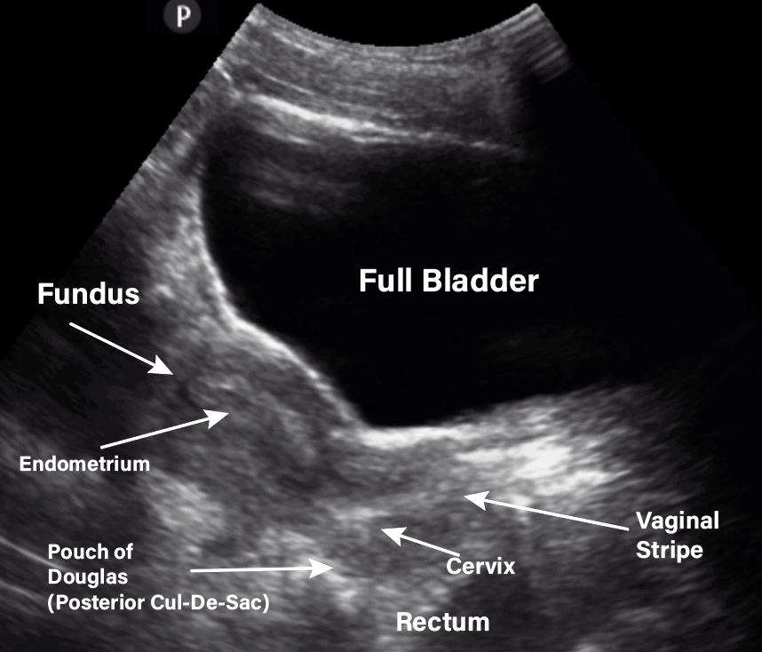 Its the procedure depends on the method by which it will be carried out.
During abdominal examination:
- The patient lies on the couch on his back and releases the abdomen and its lower part from clothing
- The doctor applies a gel to the patient's body, which is necessary for the signal to pass through the tissues in the best possible way
- Then the sonologist moves the probe over the area under study, and at this time the image is fed directly to the monitor
- Depending on which particular organ (or organs) needs to be examined, the procedure takes from ten to twenty minutes
During the transvaginal examination:
- The patient lies on her back, bends her knees and spreads her hips
- The doctor puts a condom on the transducer and lubricates it with gel to facilitate the procedure and improve the signal
- The probe is inserted into the patient's vagina a few centimeters, if necessary, it can move slightly from side to side
- During this examination, a tissue sample can be taken for examination
- Examination lasts about five minutes
This type of ultrasound is not used to diagnose virgin girls.
For transrectal analysis:
- Patient lies on side, knees drawn to chest
- Medic uses condom and probe lubricant
- The device is inserted into the rectum
- During this examination, it is also possible to take a sample for analysis
- Duration of the procedure - from five to twenty minutes
Ultrasound examination of the female body is usually performed in the first week of the cycle, after menstruation. But if there is a need to track the dynamics of the development of any formation or disease, then the examination can be done several times in different phases of the cycle.
Control examination during pregnancy is carried out several times: from the twelfth to the fourteenth, from the twentieth to the twenty-fourth and from the thirtieth to the thirty-second week.
Complications of Pelvic Ultrasound
Since ultrasound is one of the safest examinations, there are usually no complications when using it.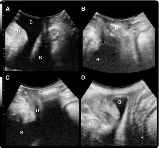 But when performing a pelvic ultrasound, there are rules that entail several nuances:
- Transvaginal ultrasound:
- Do not use in late pregnancy as insertion of the transducer may induce preterm labor or uterine contractions
- If used to diagnose a non-sexual girl, the sensor will damage the hymen (so it is not used in such cases)
- Transrectal ultrasound
- It should not be used if the patient has an anal fissure or rectal injury as the transducer may aggravate the situation
In all other cases, no problems or complications after ultrasound were recorded.
Advantages of the procedure at MEDSI
- MEDSI clinics use modern ultrasound equipment for all types of ultrasound examinations (more than thirty types)
- All devices are certified and suitable for patients of any age and gender
- Doctors of the highest qualification categories will decipher the results and make the most accurate diagnosis, as well as help to implement the preparation for ultrasound of the pelvic organs
- Clinics are conveniently located in all districts of Moscow - more than twenty diagnostic centers
- Emergency examination possible if necessary
If you experience sudden bleeding or abdominal pain, do not hesitate and contact your doctor - phone 8 (495) 7-800-500.
Do not delay treatment, see a doctor right now:
- Ultrasound
- Ultrasound at home
- Laboratory diagnostics
How is pelvic ultrasound performed in women and how to prepare for it
The body of a woman is a fragile mechanism that requires constant care. To determine what changes occur in the female body, you can check the condition of internal organs using ultrasound. One of the most common types of such diagnostics is the study of the pelvic organs.
Reasons for the study
Ultrasound diagnostics is a highly accurate and safe study for humans. The signals that the sensor sends are reflected from the internal organs of the person and returned. Thus, a picture is created on the monitor, which the doctor explains.
For examination of the pelvic organs, this type of study is prescribed. With the help of ultrasound, the condition of the internal organs is assessed, the presence of diseases and pregnancy is determined. Ultrasound of the small pelvis in Kaluga you can do at the Elite Medical Center. A referral for examination is acquired from a therapist; also, in case of violation of menstruation, a gynecologist can refer to the procedure. An ultrasound of the pelvis should be done according to the following indications:
- Frequent pain in the lower abdomen;
- For heavy bleeding;
- For unusual discharge;
- When education is suspected.
Ultrasound diagnostics can detect cysts, kidney stones, tumors, fibroids and other diseases. Preparation for the study of the small pelvis is carried out in the following ways:
- Transabdominal;
- Transvaginal;
- For pregnant women - obstetric.
Before the first type of examination, you need to fill the bladder, you will need to drink about a liter of liquid. It is necessary to empty the bladder during transvaginal diagnosis. There are no recommendations for screening and obstetric ultrasound.
Preparation for each type of examination is different, but there is a general rule: do not eat food that can cause gas a few days before the examination. The procedure will have to be postponed if you had an x-ray two or three days ago. This may affect the results. On the day when you will be examined, you need to clean the intestines.
When is the best time to have the procedure?
When examining in the direction of a gynecologist, it is important to know on which day of the cycle to do a pelvic ultrasound. It should be carried out in the first week after the end of menstruation. If you have an allergy, you need to tell your doctor about it. There are no contraindications to the procedure, it is impossible to conduct an ultrasound of the small pelvis only in the presence of menstruation.
Indicative will be the study in the first seven to ten days of the menstrual cycle. If the doctor suspects that you have fibroids, then a pelvic ultrasound should be performed immediately after your period has ended. When planning a pregnancy to detect folliculogenesis, a study is carried out for 5, 9days and after two weeks of the menstrual cycle. The timing may vary, depending on the specific case, on the duration of menstruation.
An examination by a female doctor should be carried out at least once a year, including an ultrasound examination. If you experience discomfort or changes in your menstrual cycle, contact your doctor immediately.
Survey results
Based on the image that is displayed on the screen, the doctor explains the results, diagnoses and assesses the size of the organs, their echogenicity. A complete conclusion is made only by a specialist who considers compliance with the size, location of the female organs, determines whether there are follicles, formations, stones. After the procedure, the doctor explains in writing or orally all the information about the compliance of the size of the internal organs with the norms, their condition and the presence of formations.


