Fundus of uterus function
Uterus (Anatomy): Definition, Function, Location
Uterus Definition
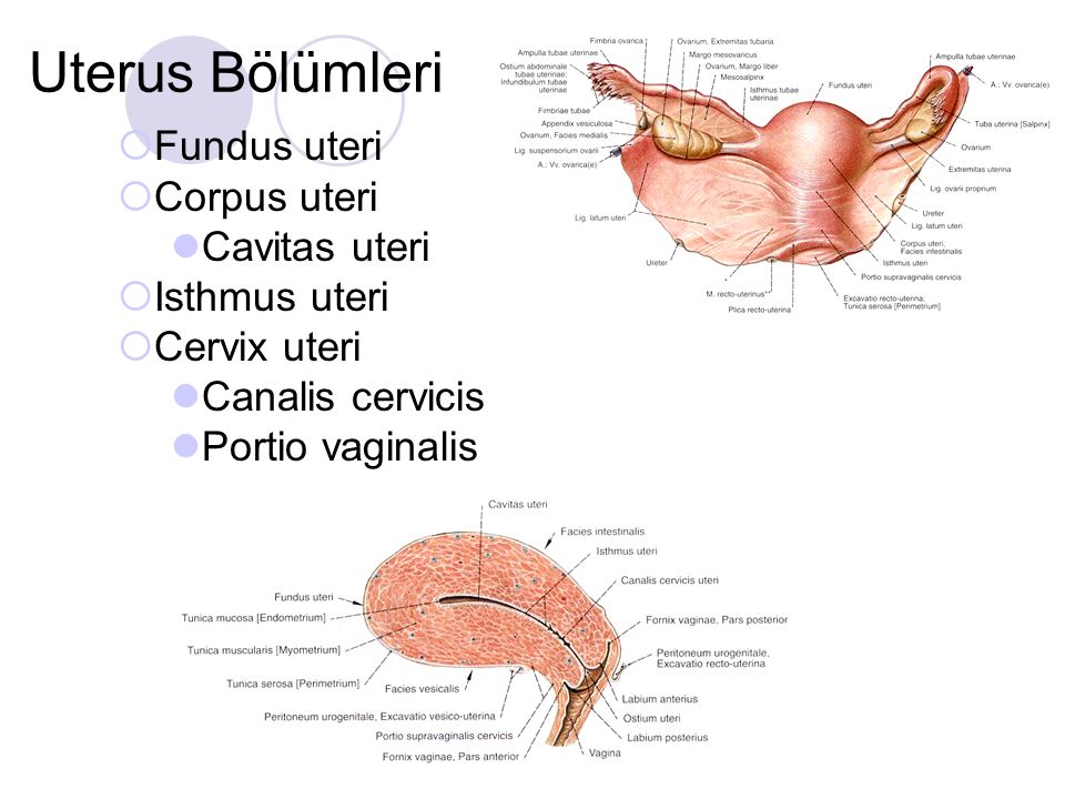
The uterus, otherwise known as the womb, is the female sex organ that carries a huge significance in many species’ survival – ours included. The uterus itself is a hollow organ that is shaped in the form of a pear, and interestingly enough measures about that size. It is neatly tucked into the pelvic area of most mammals and, of course, in humans. It is important to dissect the anatomy of the human uterus. In the female body, the upper end of the uterus, called the fundus, will join the fallopian tubes at either side while the lower end will open into the vagina. The wide portion at the top of the uterus is called the fundus, and will be the superior-most region that will host a fertilized embryo as it grows into a baby. A little below the fundus lies the muscular corpus region.
The corpus, in turn, is composed of three tissue layers. Post-pubescent women will have an innermost endometrium, which is the layer of muscle that is shed when the menstrual cycle commences in non-pregnant women. The endometrial tissue will thicken as the month’s cycle goes by in preparation for a fertilized egg to implant itself there. But in the absence of a fertilized egg, this layer will simply be shed away in what we know as menstruation. The middle muscle layer is called the myometrium, and is the layer that will expand during pregnancy and contract during childbirth. The outermost layer, the parametrium, will likewise expand and contract at these stages. Expanding will allow the uterus to house a growing baby, while the contractions will facilitate the newborn’s exit from the womb.
The image above depicts a diagram of the Uterus, with labeled tissue and arterial landmarks.
Function of the Uterus
Perhaps the principal, albeit lofty function of the uterus is to preserve life. It is the site of nourishment for the growing baby, making it one of the most important reproductive organs in the female body. This all begins when an egg, or ovum, is fertilized by a sperm and will make its downward trek in search of a better home.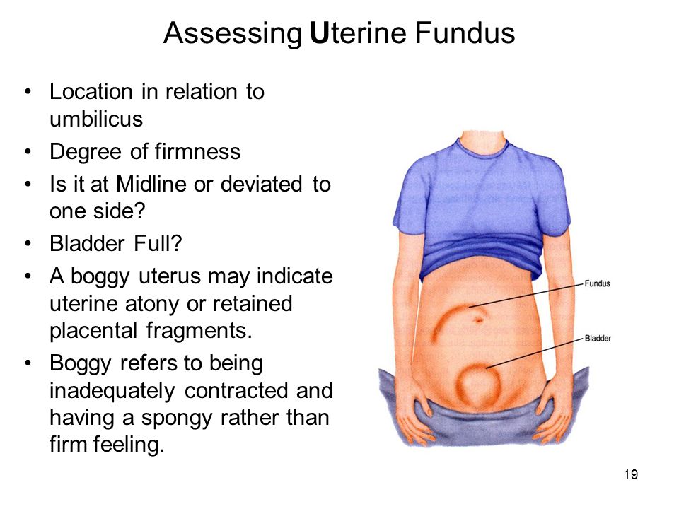 The tight fallopian tubes will not provide enough space to house the growing embryo! This is where the uterus meets all of these requirements, and more! The uterus’s thick, muscular nature will allow it to contract and expand to make room for the developing baby. The uterus is also rich in vasculature. There are many blood vessels supplying the muscle layers at any given time. This especially applies to the endometrium which is highly vascular and will come to nourish the embryo. In fact, many of the endometrial vessels that will come to supply the embryo will form just for this purpose. All of this explains why the fertilized ovum will choose to implant itself in the uterine lining – coined, the “site of implantation.” Thus, the uterus is the site that allows ours, and many species, to continue reproducing!
Location of the Uterus
The uterus measures about three inches long and two inches wide, and has a thick muscular lining within its walls. The lowest tip of the uterus will dip into the vagina in the area of the cervix, while the top most part will connect with the fallopian tubes through which the eggs travel.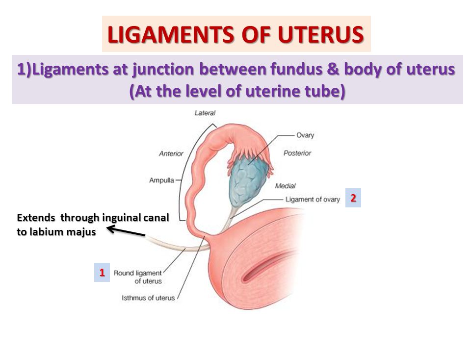 But a better way to pin its location is by describing its region as the area that lies between the belly button and the hip bones.
Abnormalities of the Uterus in Pregnancy
Nothing quite demonstrates the reproductive role of the uterus as the difficulties that arise from having an abnormal uterus. While the normal uterus will roughly measure three by two by one inches, some women will have uteruses that differ in shape and size. Many species’ evolution, including our own, has depended on having these precise dimensions to best support the growing embryo and to bring it to full term. But women with uterine abnormalities may realize they have this only once they have attempted and failed to conceive. These complications will surface either while trying to become pregnant, or after experiencing miscarriage. Examples of physical deformations of the uterus may include having a uterus with two inner cavities or vaginas (affecting roughly one in 350 women), having only one fallopian tube that will connect to the uterus, or having a heart shaped uterus instead of a pear-shaped one that is evolutionarily optimized to bear a child.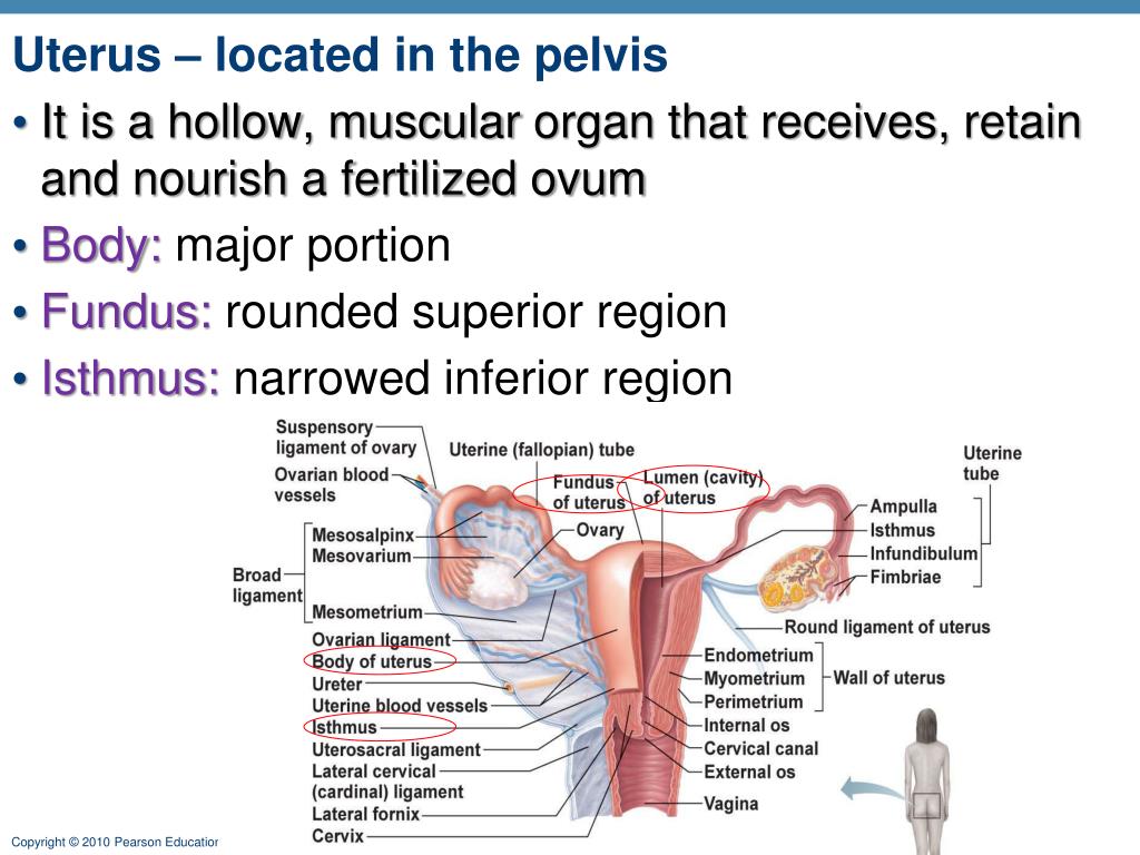 Moreover, some afflicted women will have a septum that parts the uterus, or a slight indentation at the top of the uterus that will likewise compromise the ability for these women to have a baby. These abnormalities, however, are not all an absolute guarantee of infertility or miscarriage, but instead may lessen the probability of carrying a child to full-term.
Quiz
1. Which of the following is the outermost muscle layer of the corpus?
A. Endometrium
B. Parametrium
C. Myometrium
D. None of the above
Answer to Question #1
B is correct. The parametrium, like the myometrium, will expand and contract during the crucial phases of pregnancy and childbirth. But the difference here is that the parametrium is the outermost layer of the uterine’s corpus region.
2. Which uterine layer nourishes the growing embryo?
A.:watermark(/images/watermark_5000_10percent.png,0,0,0):watermark(/images/logo_url.png,-10,-10,0):format(jpeg)/images/atlas_overview_image/1747/sBJ05xT2kyBAOyn8SVqaBg_uterus-and-ovaries_portuguese.jpg) Endometrium
Endometrium
B. Parametrium
C. Myometrium
D. None of the above
Answer to Question #2
A is correct. The endometrial lining is highly vascular (blood vessel rich) and is actually the layer that sheds off during the menstrual cycle. Its access to vessels will provide crucial nourishment to the growing fertilized embryo, as it will obtain its nutrients here.
3. Which of the following provides the best explanation for why the uterus is the main female reproductive organ?
A. It is the site of fertilization of the ovum
B. It is muscular and able to contract and expand
C. Its myometrial layer supplies the growing embryo
D. Both A and B
Answer to Question #3
B is correct. Of the answer choices, answer B is the only accurate choice by process of elimination. The uterus is not the actual site of fertilization, but instead the site of implantation but not at the myometrium but instead the endometrium.
References
- MedicineNet (2017). “Uterus.” Medicine Net. Retrieved on 2017-08-26 from http://www.medicinenet.com/script/main/art.asp?articlekey=5918
- BabyCentre Medical Advisory Board (2016). “Abnormalities of the uterus in pregnancy.” Retrieved on 2017-08-26 https://www.babycentre.co.uk/a551934/abnormalities-of-the-uterus-in-pregnancy
- Danielsson, K. “Abnormal Uterus Shapes and iscarriage Risk.” Very Well. Retrieved on 2017-08-27 from https://www.verywell.com/abnormal-uterus-and-miscarriage-risk-2371694
Uterus | Definition, Function, & Anatomy
uterus
See all media
- Related Topics:
- fallopian tube cervix puerperium endometrium bicornate uterus
See all related content →
uterus, also called womb, an inverted pear-shaped muscular organ of the female reproductive system, located between the bladder and the rectum.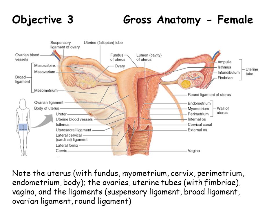 It functions to nourish and house a fertilized egg until the fetus, or offspring, is ready to be delivered.
The uterus has four major regions: the fundus is the broad curved upper area in which the fallopian tubes connect to the uterus; the body, the main part of the uterus, starts directly below the level of the fallopian tubes and continues downward until the uterine walls and cavity begin to narrow; the isthmus is the lower, narrow neck region; and the lowest section, the cervix, extends downward from the isthmus until it opens into the vagina. The uterus is 6 to 8 cm (2.4 to 3.1 inches) long; its wall thickness is approximately 2 to 3 cm (0.8 to 1.2 inches). The width of the organ varies; it is generally about 6 cm wide at the fundus and only half this distance at the isthmus. The uterine cavity opens into the vaginal cavity, and the two make up what is commonly known as the birth canal.
Lining the uterine cavity is a moist mucous membrane known as the endometrium. The lining changes in thickness during the menstrual cycle, being thickest during the period of egg release from the ovaries (see ovulation).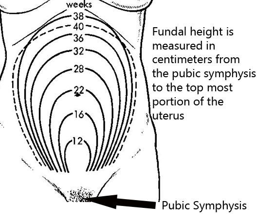 If the egg is fertilized, it attaches to the thick endometrial wall of the uterus and begins developing. If the egg is unfertilized, the endometrial wall sheds its outer layer of cells; the egg and excess tissue are then passed from the body during menstrual bleeding. The endometrium also produces secretions that help keep both the egg and the sperm cells alive. The components of the endometrial fluid include water, iron, potassium, sodium, chloride, glucose (a sugar), and proteins. Glucose is a nutrient to the reproductive cells, while proteins aid with implantation of the fertilized egg. The other constituents provide a suitable environment for the egg and sperm cells.
The uterine wall is made up of three layers of muscle tissue. The muscle fibres run longitudinally, circularly, and obliquely, entwined between connective tissue of blood vessels, elastic fibres, and collagen fibres. This strong muscle wall expands and becomes thinner as a child develops inside the uterus. After birth, the expanded uterus returns to its normal size in about six to eight weeks; its dimensions, however, are about 1 cm (0.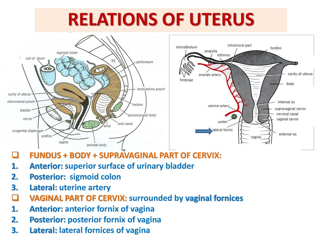 4 inch) larger in all directions than before childbearing. The uterus is also slightly heavier and the uterine cavity remains larger.
The uterus of a female child is small until puberty, when it rapidly grows to its adult size and shape. After menopause, when the female is no longer capable of having children, the uterus becomes smaller, more fibrous, and paler. Some afflictions that may affect the uterus include infections; benign and malignant tumours; malformations, such as a double uterus; and prolapse, in which part of the uterus becomes displaced and protrudes from the vaginal opening. Uterus transplantation, in which a uterus from a healthy female is transplanted into the affected woman, has been considered a potential form of treatment in extreme cases of uterine disease or absence of the uterus; the first birth of a healthy infant to a uterus transplant recipient occurred in 2014.
The Editors of Encyclopaedia Britannica This article was most recently revised and updated by Kara Rogers.
51. Structure and functions of the uterus
The structure and functions of the uterus.
Uterus, uterus , — unpaired muscular pear-shaped organ for the development of the embryo in the case fertilization of the egg, and removal of the fetus during childbirth. In the uterus emit: bottom, reversed up and forward; body - middle part triangular shape and lower - neck. Cervix lower the end is connected to the vagina. Place transition of the uterine body to the cervix narrow and is called isthmus uterus. Part of neck uterus that enters the vagina is called vaginal part, and outside it supravaginal part. B The uterus is divided into two surfaces: anterior - cystic, and back - intestinal, a also two edges: right and left.
Uterine cavity , small, triangular forms. Top from the sides open into it the fallopian tubes.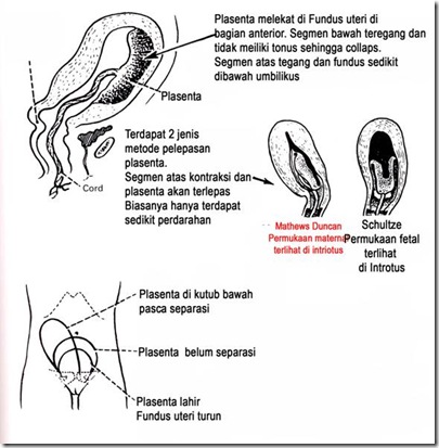 Below the uterine cavity goes to channel necks, which opens into the vagina with a hole uterus. It is limited two lips: front, and back. Neonatal the uterus is cylindrical, and the cervix longer than in adult women. Body uterus develops rapidly the onset of puberty. Topography. The uterus is located in the pelvis between bladder anteriorly and straight gut - behind. The fundus of the uterus is turned forward, its front surface is down and forward, and the back - up and back. The body of the uterus forms with the cervix open anterior bend. Large part of the uterus up to the level of the cervix is in the peritoneal cavity of the small pelvis, so loops of thin or sigmoid colon.
The structure of the uterus. The wall of the uterus consists of three shells: 1) inner mucous, containing many uterine glands; 2) medium, most thick, muscular,), having three layers: longitudinal, circular, longitudinal; 3) outdoor serous, Mucous lining of the uterus in mature women changes cyclically due to menstruation.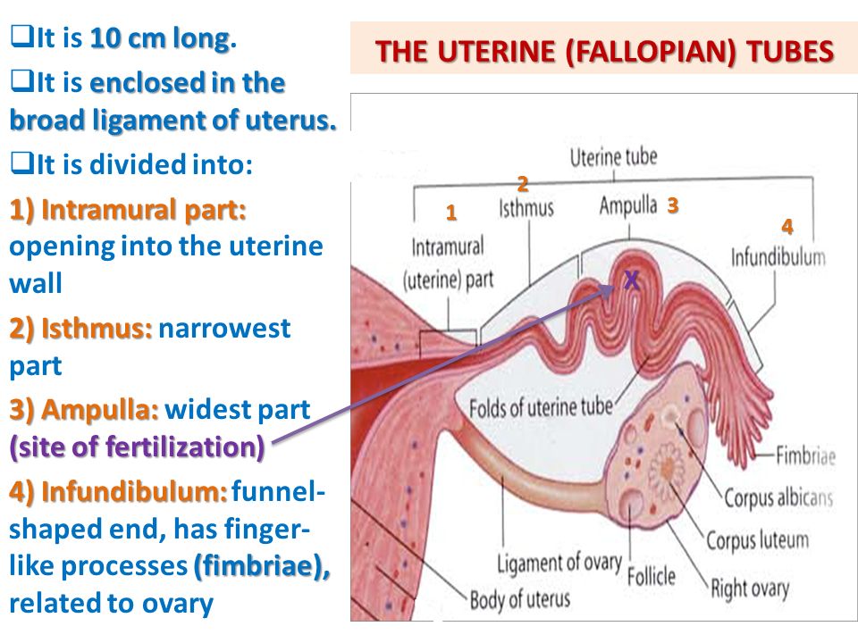 . After the end menstruation mucosa quickly is being restored. Serous membrane - peritoneum - covers the fundus of the uterus, as well as its anterior and posterior surfaces. nine0007 front she covers the uterus to the level of the cervix and passes to the bladder vesicouterine deepening. Rear peritoneum lines the posterior surface uterus and posterior vaginal fornix and passes nor the rectum, forming recto-uterine deepening. On the edges the peritoneum passes to the pelvic wall, forming wide uterine ligament, between the leaves of which are periuterine fiber, and fallopian tube, uterine artery, powerful uterine venous plexus and nervous plexus. From the upper corners of the uterus go to the deep inguinal ring nine0007 round bundles uterus. Through the inguinal channel they reach the symphysis, where and are running out.
The size of the uterus varies depending on on the age of the woman, the number of births and pregnancies.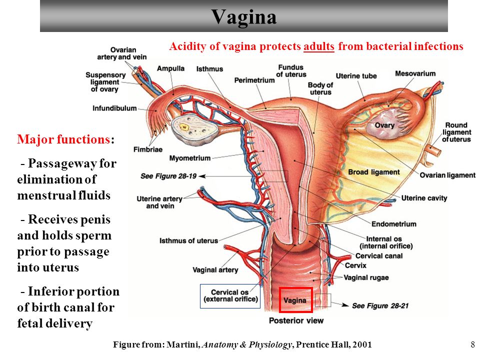 So, in a nulliparous women its length is 7-8 cm, width - 5 cm, weight does not exceed 50 g. Thanks to structural features, during pregnancy, the uterus is able to stretch up to 32 cm long and up to 20 cm wide.
Uterine menstruation occurs in the uterus cycle - a cycle of changes in the endometrium. nine0005
1 phase - desquamation. There is a rejection functional layer of the endometrium due to a sharp spasm of the spiral arteries mucous membranes that have muscle pulp at a level between basal and functional layers of the endometrium. Spasm due to low levels of progesterone and an increase in estrogen levels. Goes enzymatic degradation by proteolytic enzymes secreted leukocytes that migrate to mucosa at the end of the secretion phase. Phase lasts 3-5 days and is clinically expressed secretion of dark blood with mucus, without pieces of tissue from the female genital tract. nine0005
Phase 2 - regeneration. Regeneration starts from 2-3 days of the cycle.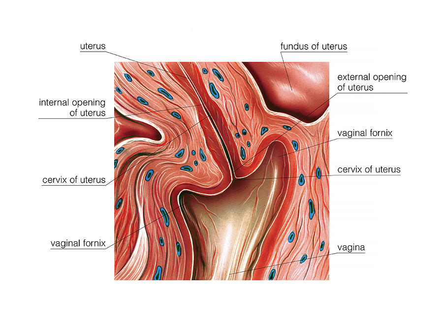 Gradually discharge from the genital tract stops. After rejection, a wound is formed surface that is epithelialized due to the donets tubular glands of the basal layer.
3 phase - proliferation, Complete restoration of the functional layer, glands, blood vessels, nerves. This phase ends by the 12th day of the cycle and lasts 8 days. Phases 2 and 3 are under the influence estrogen.
4 phase - secretions. Under the influence of the growing concentration of progesterone in epithelial accumulates in endometrial glands glycogen, glycoprotein lipids and all necessary for nutrition and implantation blastocysts. Getting ready to receive fertilized egg. When pregnancy occurs decidual transformation occurs endometrium. By the 20-21st day of the cycle, the endometrium ready to receive the blastocyst. nine0005
Ultrasound of the uterus - Ultrasound of the cervix and appendages in Nizhny Novgorod, transvaginal ultrasound
Ultrasound of the uterus is one of the most important and common methods for examining this organ.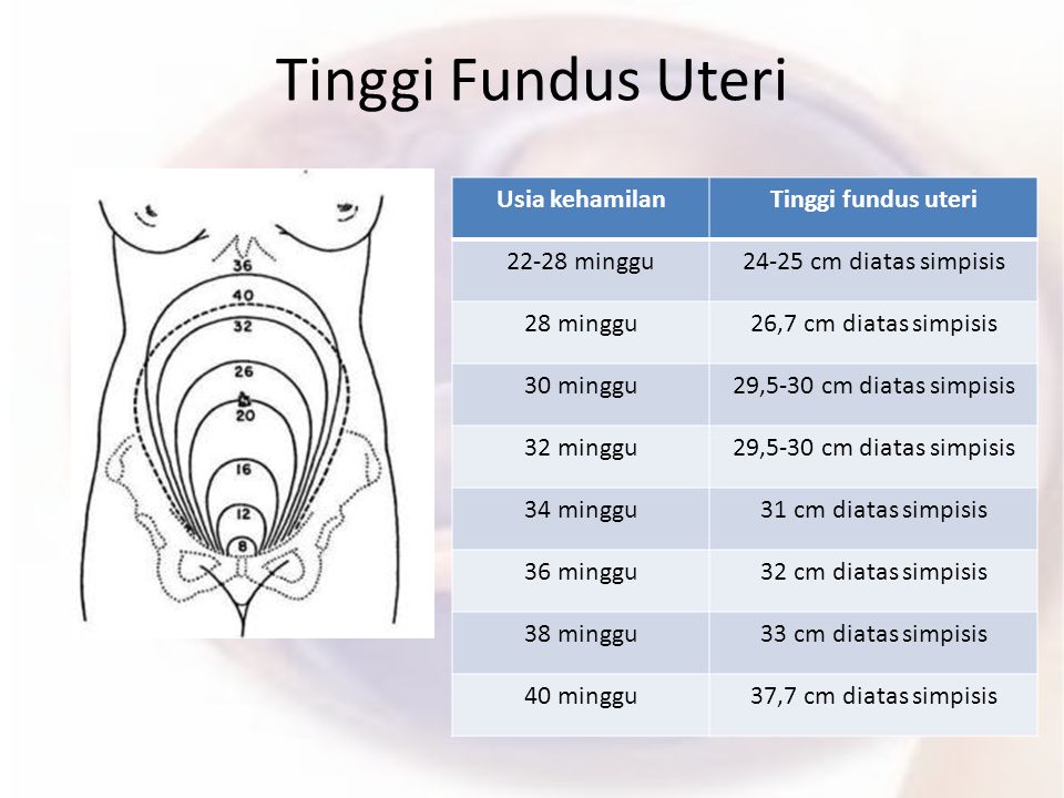 For a better understanding of the process of conducting ultrasound of the uterus and appendages, consider the uterus as an organ of the female reproductive system.
The uterus is a hollow muscular organ, consisting of three layers: endometrium (inner), myometrium (middle), perimetrium (outer), and is located in the cavity of the small pelvis, between the bladder and rectum. The normal anatomical position of the uterus in the small pelvis is provided by the supporting muscular pelvic floor and the suspension ligamentous apparatus of the uterus. The uterus has a fundus (upper part of the uterus), a body, and a cervix. The cervix connects the uterus to the vagina. Therefore, the vaginal and supravaginal parts of the uterus are isolated. nine0005
The function of the uterus is to provide a place and conditions for the implantation of the embryo and for the further development of the fetus.
Fallopian tubes and ovaries are the so-called appendages of the uterus.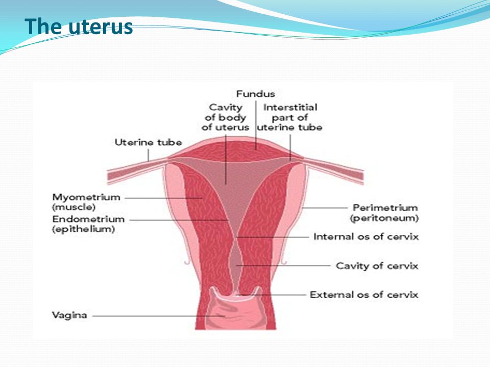 The ovaries are paired sex glands that work alternately. They contain many different degrees of development of follicles in which eggs mature. The fallopian tubes run from the uterus to the ovaries. They are where fertilization takes place.
Therefore, in order to enjoy the happiness of motherhood, it is necessary to undergo prophylactic Ultrasound of the uterus and appendages , and to conduct timely diagnosis and treatment of the uterus and other genital organs of a woman.
Ultrasound of the uterus and appendages a gynecologist may prescribe if there are any symptoms of a disease of the female genital organs:
- Various pains in the lower abdomen, pain during menstruation
- Irregular menses
- Uterine bleeding
- Anomalies in the development of the uterus
- Bleeding during menopause nine0113
- Clarification of the presence and location of the intrauterine contraceptive (spiral)
- Establishing pregnancy
- Neoplasm diagnostics
- Uterine length 30-40 mm, cervix 40-50 mm, anterior-posterior size 28-45 mm, width 35-50 mm.
- Position of the uterus in anteflexio - inclination towards the bladder. Bend of the uterus to the rectum (retroflexio) is a normal variant, but less common
- The thickness of the endometrium changes during the menstrual cycle
By the 5-7th day of the menstrual cycle, the endometrium is clearly defined, its thickness is up to 6-9 mm. - Myometrium homogeneous in structure and without formations
- Developmental anomalies (unicornuate, bicornuate, bifid, with septum)
- Endometriosis
- Adenomyosis
- Metroendometritis
- Endometritis
- Uterine polyps
- Tumors (benign and malignant)
As well as ultrasound of the uterus and appendages is used for:
For examination of the pelvic organs in a sexually active woman, transvaginal ultrasound of uterus is performed. To prepare for a transvaginal ultrasound of the uterus, 2 days before the examination, do not eat foods that cause gas formation, and, if necessary, make an enema. nine0005
Transvaginal ultrasound of the uterus is not performed for girls before the onset of sexual activity. To do this, use the transabdominal method of conducting ultrasound of the uterus. It is necessary before the study to try to fill the bladder as much as possible - drink about 1 liter of water or tea. This is necessary in order to visualize the uterus well on ultrasound.
The most important condition for transvaginal ultrasound and transabdominal ultrasound of the uterus and appendages is its 5-7 day of the menstrual cycle or during the first 2 days after the end of menstruation. nine0005
With an ultrasound of the uterus, the doctor determines the position, size and structure of the uterus, and evaluates the condition of the cervix. It is also taken into account when conducting an ultrasound of the uterus, that before childbirth, the size of the uterus in a girl is minimal, and in a woman who has given birth, it is maximum.
Normal ultrasound data of the uterus in reproductive age (from 20 to 35 years):
Ultrasound of the uterus in the early reproductive period (from the onset of menstruation to 20 years) shows the smallest size of the uterus. With ultrasound of the uterus in postmenopause (begins at the age of 45 to 55 years), the uterus decreases and becomes homogeneous in echostructure. The endometrium may not be visualized. nine0005
Ultrasound of the uterus helps to detect the following diseases:
In addition to ultrasound of the uterus, ultrasound of the cervix is also performed:
When ultrasound of the cervix is evaluated, its shape, structure and thickness of the cervical canal, the structure of the muscular layer of the cervix.