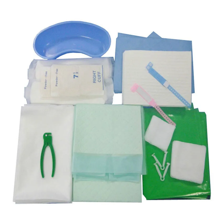
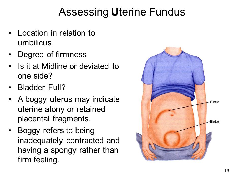
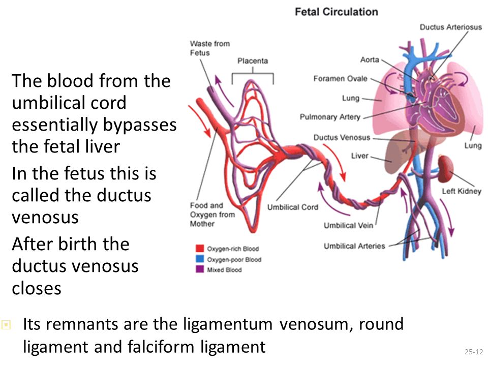
Baby delivery tools
EQUIPMENT, SUPPLIES, DRUGS AND LABORATORY TESTS - Pregnancy, Childbirth, Postpartum and Newborn Care
L2. EQUIPMENT, SUPPLIES, DRUGS AND TESTS FOR ROUTINE AND EMERGENCY PREGNANCY AND POSTPARTUM CARE
Warm and clean room
Hand washing
Clean water supply
Soap
Nail brush or stick
Clean towels
Waste
Bucket for soiled pads and swabs
Receptacle for soiled linens
Container for sharps disposal
Sterilization
Miscellaneous
Equipment
Supplies
Gloves:
- →
utility
- →
sterile or highly disinfected
- →
long sterile for manual removal of placenta
Urinary catheter
Syringes and needles
IV tubing
Suture material for tear or episiotomy repair
Antiseptic solution (iodophors or chlorhexidine)
Spirit (70% alcohol)
Swabs
Bleach (chlorine base compound)
Impregnated bednet
Condoms
Alcohol-based handrub
Tests
Syphilis testing (e.
g. RPR)
Proteinuria dip sticks
Container for catching urine
HIV testing kit (2 types)
Haemoglobin testing kit
Rapid diagnostic tests or Light microscopy
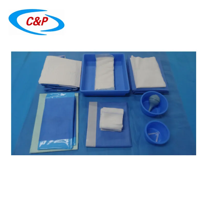
Disposable delivery kit
Drugs
Oxytocin
Ergometrine
Misoprostol
Magnesium sulphate
Calcium gluconate
Diazepam
Hydralazine
Ampicillin
Gentamicin
Metronidazole
Benzathine penicillin
Cloxacillin
Amoxycillin
Ceftriaxone
Trimethoprim + sulfamethoxazole
Clotrimazole vaginal pessary
Erythromycin
Ciprofloxacin
Tetracycline or doxycycline
Artesunate/Artemether
Quinine
Lignocaine 2% or 1%
Adrenaline
Ringer lactate
Normal saline 0.
 9%
9%Glucose 50% solution
Water for injection
Paracetamol
Gentian violet
Iron/folic acid tablet
Low-dose aspirin
Calcium tablets
Mebendazole
Sulphadoxine-pyrimethamine
Nevirapine (infant)
Zidovudine (AZT) (infant)
Once-daily fixed-dose combination of ARVs recommended as first-line ART according to national guidelines
Betamethasone or Dexamethasone
Vaccine
Tetanus toxoid
L3. EQUIPMENT, SUPPLIES AND DRUGS FOR CHILDBIRTH CARE
Warm and clean room
Delivery bed: a bed that supports the woman in a semi-sitting or lying in a lateral position, with removable stirrups (only for repairing the perineum or instrumental delivery)
Clean bed linen
Curtains if more than one bed
Clean surface (for alternative delivery position)
Work surface for resuscitation of newborn near delivery beds
Light source
Heat source
Room thermometer
Hand washing
Clean water supply
Soap
Nail brush or stick
Clean towels
Waste
Container for sharps disposal
Receptacle for soiled linens
Bucket for soiled pads and swabs
Bowl and plastic bag for placenta
Sterilization
Miscellaneous
Equipment
Blood pressure machine and stethoscope
Body thermometer
Fetal stethoscope
Baby scale
Self inflating bag and mask - neonatal size
Suction apparatus with suction tube
Infant stethoscope
Delivery instruments (sterile)
Supplies
Gloves:
- →
utility
- →
sterile or highly disinfected
- →
long sterile for manual removal of placenta
- →
Long plastic apron
Urinary catheter
Syringes and needles
IV tubing
Suture material for tear or episiotomy repair
Antiseptic solution (iodophors orchlorhexidine)
Spirit (70% alcohol)
Swabs
Bleach (chlorine-base compound)
Clean (plastic) sheet to place under mother
Sanitary pads
Clean towels for drying and wrapping the baby
Cord ties (sterile)
Blanket for the baby
Baby feeding cup
Impregnated bednet
Alcohol-based handrub
2 ml and 1 ml syringes (for giving ARV to babies)
Drugs
Oxytocin
Ergometrine
Misoprostol
Magnesium sulphate
Calcium gluconate
Diazepam
Hydralazine
Ampicillin
Gentamicin
Metronidazole
Benzathine penicillin
Lignocaine
Adrenaline
Ringer lactate
Normal saline 0.
 9%
9%Water for injection
Eye antimicrobial (1% silver nitrate or 2.5% povidone iodine)
Tetracycline 1% eye ointment
Vitamin A
Izoniazid
Nevirapine (infant)
Zidovudine (AZT) (infant)
Once-daily fixed-dose combination of ARVs recommended as first-line ART according to national guidelines
Vaccines
BCG
OPV
Hepatitis B
Contraceptives
(see Decision-making tool for family planning providers and clients)
Test
Syphilis testing (e.g. RPR)
Proteinuria dip sticks
Container for catching urine
HIV testing kits (2 types)
Haemoglobin testing kit
L4. LABORATORY TESTS
Check urine for protein
Label a clean container.
Give woman the clean container and explain where she can urinate.
Teach woman how to collect a clean-catch urine sample. Ask her to:
- →
Clean vulva with water
- →
Spread labia with fingers
- →
Urinate freely (urine should not dribble over vulva; this will ruin sample)
- →
Catch the middle part of the stream of urine in the cup. Remove container before urine stops.
Analyse urine for protein using either dipstick or boiling method.
DIPSTICK METHOD
Dip coated end of paper dipstick in urine sample.
Shake off excess by tapping against side of container.
Wait specified time (see dipstick instructions).
Compare with colour chart on label. Colours range from yellow (negative) through yellow-green and green-blue for positive.
BOILING METHOD
Put urine in test tube and boil top half. Boiled part may become cloudy. After boiling allow the test tube to stand.
 A thick precipitate at the bottom of the tube indicates protein.
Add 2-3 drops of 2-3% acetic acid after boiling the urine (even if urine is not cloudy)
- →
If the urine remains cloudy, protein is present in the urine.
- →
If cloudy urine becomes clear, protein is not present.
- →
If boiled urine was not cloudy to begin with, but becomes cloudy when acetic acid is added, protein is present.
Check haemoglobin
Draw blood with syringe and needle or a sterile lancet.
Insert below instructions for method used locally.
Check blood for malaria parasites
Blood for the test is commonly obtained from a finger-prick.
The two methods in routine use for parasitological diagnosis are light microscopy and rapid diagnostic tests (RDTs).
The choice between RDTs and microscopy depends on local context, including the skills available, patient case-load, epidemiology of malaria and the possible use of microscopy for the diagnosis of other diseases.
L5. PERFORM RAPID PLASMAREAGIN (RPR) TEST FOR SYPHILIS
Perform rapid plasmareagin (RPR) test for syphilis
Seek consent.
Explain procedure.
Use a sterile needle and syringe. Draw up 5 ml blood from a vein. Put in a clear test tube.
Let test tube sit 20 minutes to allow serum to separate (or centrifuge 3-5 minutes at 2000-3000-rpm). In the separated sample, serum will be on top.
Use sampling pipette to withdraw some of the serum.
Take care not to include any red blood cells from the lower part of the separated sample.
Hold the pipette vertically over a test card circle. Squeeze teat to allow one drop (50-μl) of serum to fall onto a circle. Spread the drop to fill the circle using a toothpick or other clean spreader.
Important: Several samples may be tested on one card. Be careful not to contaminate the remaining test circles. Use a clean spreader for every sample. Carefully label each sample with a patient's name or number.
Attach dispensing needle to a syringe. Shake antigen.1
Draw up enough antigen for the number of tests to be done (one drop per test).
Holding the syringe vertically, allow exactly one drop of antigen (20-μl) to fall onto each test sample.
DO NOT stir.
Rotate the test card smoothly on the palm of the hand for 8 minutes.2
(Or rotate on a mechanical rotator.)
Interpreting results
After 8 minutes rotation, inspect the card in good light. Turn or lift the card to see whether there is clumping (reactive result). Most test cards include negative and positive control circles for comparison.
Non-reactive (no clumping or only slight roughness) – Negative for syphilis
Reactive (highly visible clumping) – Positive for syphilis
Weakly reactive (minimal clumping) – Positive for syphilis
NOTE: Weakly reactive can also be more finely granulated and difficult to see than in this illsutration.
EXAMPLE OF A TEST CARD
L6. PERFORM RAPID HIV TEST (TYPE OF TEST USED DEPENDS ON THE NATIONAL POLICY)
Use rapid HIV testing with same-day results using rapid diagnostic tests (RDTs) in antenatal care. If the laboratory testing is the policy for antenatal care you may use RDTs for the pregnant woman who comes to ANC late in pregnancy, the woman who only comes in labour or has not received her HIV results prior to labour.
Explain the procedure and seek consent according to the national policy.
Use test kits recommended by the national and/or international bodies and follow the instructions of the HIV rapid test selected.
Prepare your worksheet, label the test, and indicate the test batch number and expiry date. Check that expiry time has not lapsed.
Wear gloves when drawing blood and follow standard safety precautions for waste disposal.
Inform the woman for how long to wait at the clinic for her test result (same day or they will have to come again).
Draw blood for all tests at the same time (tests for Hb, syphilis and HIV can often be coupled at the same time).
- →
Use a sterile needle and syringe when drawing blood from a vein.
- →
Use a lancet when doing a finger prick.
Perform the test following manufacturer's instructions.
Interpret the results as per the instructions of the HIV rapid test selected.
- →
If the first test result is negative, no further testing is done. Record the result as – HIV-negative.
- →
If the first test result is positive, perform a second HIV rapid test using a different test kit.
- →
If the second test is also positive, record the result as - HIV-positive.
- →
If the first test result is positive and second test result is negative, repeat the testing.
Do a finger prick and repeat both tests.
- →
If both tests are positive or both are negative, record accordingly.

- →
If tests show different results, use another test, or record the results as inconclusive. Repeat the tests after 2 weeks or refer the woman to hospital for a confirmatory test.
- →
Send the results to the health worker. Respect confidentiality A2.
Record all results in the logbook.
Footnotes
- 1
Make sure antigen was refrigerated (not frozen) and has not expired.
- 2
Room temperature should be 73°-85°F (22.8°-29.3°C).
Forceps or vacuum delivery - NHS
Assisted delivery
An assisted birth (also known as an instrumental delivery) is when forceps or a ventouse suction cup are used to help deliver the baby.
Ventouse and forceps are safe and only used when necessary for you and your baby. Assisted delivery is less common in women who've had a spontaneous vaginal birth before.
What happens during a ventouse or forceps delivery?
Your obstetrician or midwife should discuss with you the reasons for having an assisted birth, the choice of instrument and how it will be carried out.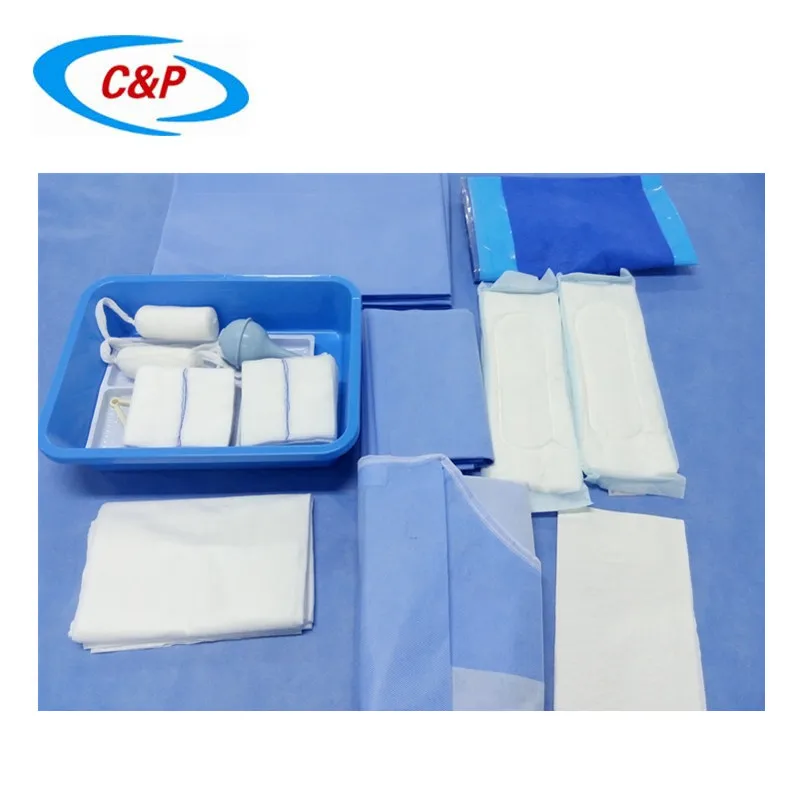 Your consent will be needed before the procedure can be carried out.
Find out more about consent to treatment.
You'll usually have a local anaesthetic to numb your vagina and the skin between your vagina and anus (perineum) if you have not already had an epidural.
If your obstetrician has any concerns, you may be moved to an operating theatre so a caesarean section can be carried out if needed.
It is likely a cut (episiotomy) will be needed to make the vaginal opening bigger. Any tear or cut will be repaired with stitches. Depending on the circumstances, your baby can be delivered and placed on your tummy, and your birth partner may still be able to cut the cord if they want to.
Ventouse
A ventouse (vacuum cup) is attached to the baby's head by suction. A soft or hard plastic or metal cup is attached by a tube to a suction device. The cup fits firmly on to your baby's head.
During a contraction and with the help of your pushing, the obstetrician or midwife gently pulls to help deliver your baby.
If you need an assisted birth and you are giving birth at less than 36 weeks pregnant, then forceps may be recommended over ventouse. This is because forceps are less likely to cause damage to your baby's head, which is softer at this point in your pregnancy.
Forceps
Forceps are smooth metal instruments that look like large spoons or tongs. They're curved to fit around the baby's head. The forceps are carefully positioned around your baby's head and joined together at the handles.
With a contraction and your pushing, an obstetrician gently pulls to help deliver your baby.
There are different types of forceps. Some are specifically designed to turn the baby to the right position to be born, such as if your baby is lying facing upwards (occipito-posterior position) or to one side (occipito-lateral position).
Why might I need ventouse or forceps?
An assisted delivery is used in about 1 in 8 births, and may be needed if:
- you have been advised not to try to push out your baby because of an underlying health condition (such as having very high blood pressure)
- there are concerns about your baby's heart rate
- your baby is in an awkward position
- your baby is getting tired and there are concerns that they may be in distress
- you're having a vaginal delivery of a premature baby – forceps can help protect your baby's head from your perineum
A children's doctor (paediatrician) is usually present to check your baby's condition after the birth. After the birth you may be given antibiotics through a drip to reduce your risk of getting an infection.
What are the risks of a ventouse or forceps birth?
Ventouse and forceps are safe ways to deliver a baby, but there are some risks that should be discussed with you.
Vaginal tearing or episiotomy
This will be repaired with dissolvable stitches.
3rd or 4th degree vaginal tear
There's a higher chance of having a vaginal tear that involves the muscle or wall of the anus or rectum, known as a 3rd- or 4th-degree tear.
This kind of tear affects:
- 3 in every 100 women having a vaginal birth
- 4 in every 100 women having a ventouse delivery
- 8 to 12 in every 100 women having a forceps delivery
Higher risk of blood clots
After an assisted birth, there's a higher chance of blood clots forming in the veins in your legs or pelvis. You can help prevent this by moving around as much as you can after the birth.
You may also be advised to wear special anti-clot stockings and have injections of heparin, which makes the blood less likely to clot.
Urinary incontinence
Urinary incontinence (leaking pee) is not unusual after childbirth. It's more common after a ventouse or forceps delivery. You should be offered physiotherapy to help prevent this happening, including advice on pelvic floor exercises.
Anal incontinence
Anal incontinence (involuntary farting or leaking poo) can happen after birth, particularly if there's been a 3rd or 4th degree tear. Because there's a higher risk of these tears happening with an assisted delivery, anal incontinence is more likely.
Are there any risks to the baby?
The risks to your baby include:
- a mark on your baby's head (chignon) being made by the ventouse cup – this usually disappears within 48 hours
- a bruise on your baby's head (cephalohaematoma) – this happens to around 1 to 12 of all 100 babies during a ventouse assisted delivery – the bruise is usually nothing to worry about and should disappear with time
- marks from forceps on your baby's face – these usually disappear within 48 hours
- small cuts on your baby's face or scalp – these affect 1 in 10 babies born using assisted delivery and heal quickly
- yellowing of your baby's skin and eyes – this is known as jaundice, and should pass in a few days
Afterwards
You'll sometimes need a small tube that drains your bladder (a catheter) for up to 24 hours.
You're more likely to need this if you've had an epidural as you may not have fully regained sensation in your bladder and therefore do not know when it's full.
The Royal College of Obstretricians and Gynaecologists (RCOG) has further information about assisted delivery.
healthtalk.org has videos and written interviews of women talking about their experiences of vaginal birth, including forceps and ventouse.
Find out more about what happens in labour and pain relief in labour.
Video: what is involved in an assisted birth?
In this video, a midwife explains what an assisted birth is and what is involved.
Media last reviewed: 20 March 2020
Media review due: 20 March 2023
Page last reviewed: 9 June 2020
Next review due: 9 June 2023
Obstetric instruments
main work obstetrician in animal care performs by hand and obstetric ropes. If necessary, the doctor uses special tools. depending tools are distinguished from the purpose auxiliary, for repulsion and extraction of the fetus and for fetotomy (Fig. thirty.).
Obstetrics rope and braid used to fix, correct position and extraction of the fetus. They are not allowed to be used in other purposes. They must be smooth and strong. Obstetric ropes are better to have a length 1.5-3 m, thick 0.5-0.7 cm. One end of a rope or ribbon has eyelet through which the free the end of the rope to form a loop. A loop of rope is put on the middle and ring fingers of the hand, injected into the birth paths and impose on parts of the body of the fetus, to be fixed. On limbs loops are applied above the carpal and hock, can be higher putovyh. The fetal head is fixed imposing loops and halters. Loop on the head of the fetus must be securely fortified. For a warning slipping off her mouth loop shifted to the back of the head, capturing both or one ear.
Obstetrics extractor, proposed by A. I. Varganov and A. D. Yumakin, used to remove fruits in cows.
Ophthalmic hooks are available with a sharp or more blunt point, larger and smaller sizes. They are used to correct the position of the head and fruit extraction. On living fruit single or double hooks with ropes strengthened in the inner corner eye orbit, and on the dead - enter into strong tissues of the fetus (skin, tendons, bony holes). Insert the hook into uterus with hand between middle and index fingers, its sharp part is closed thumb. To avoid breakdown hook and trauma to the uterus, after strengthening in tissues, fingers keep its sharp part deep fabrics, and the eye of the hook is pressed against the palm of your hand.
Auxiliary tools . This group of tools includes loop guides and handles for obstetric ropes and ribbons. Loop conductor Lindhorst is an iron elliptical ring length 14 cm wide 4 cm Loop conductor Afanasiev similar to the previous one, but narrower and most convenient in
work. Loop conductors, thanks to a peculiar form, make it possible to attach to him an obstetric rope or ribbon, circle them around parts of the body of the fetus and easy to detach after being pulled out. Handles for ropes and ribbons there are wooden, plastic or plastic, long 25-40 see
Tool for repulsion and extraction of fetus . Often in obstetric care to push the fetus into the uterus what is used obstetric sticks. Them in and out of the birth canal under the control of the obstetrician. obstetric the stick has a metal handle, rod and a fork. For secure attachment to the fetus rope at both ends of the fork is available one hole. The stick is used to push back, correct the wrong location and for the extraction of the fetus.
Afanasiev Hooks, Krey-Shotler used to correct wrong location, fixation and extraction dead fetus. Hooks with fixed rope inserted closed into the uterus hand, fixed on the desired parts of the body fetus (spine, neck, lower back, skin) and by pulling attached to them ropes carry out the necessary manipulation.
Rice. 30. Obstetrics tools:
1- obstetric rope; 2 obstetric stick; 3 obstetric eye hooks; 4 obstetric double hook; 5 obstetric hook Lozhkin; 6 obstetric hooks double; 7 obstetric hook sharp; 8-forceps obstetric; 9- looped ring; 10 sawmill; 11-ring-shaped knife; 12-knife hidden hook-shaped; 13-blade obstetric; 14obstetric scapula screwed; 15-fetotom of Beskhlebnov; 16-wire saw; 17-saw holders; 18-fetotom folding; 19-skin knife; 20 knife hidden belly; 21 obstetric hook for small animals; 22 obstetric hook costal.
Anal hooks injected into the rectum of a dead fetus with breech presentation, and extract fruit after attaching the hook to the anterior edge of the pubic bone.
Hooks for small animals there are different models. They can be made iron wire thick 4-5 mm, long 45-50 cm. Used to extract fruits. Introduction hook in the birth canal control hand. Depending on presentation and articulation of the fetus in small animal hooks can be attached to corner of the orbit, behind the head behind the occipital edge of bones, external auditory meatus, anterior edge of the pelvis through the anus, joints and skin folds.
Obstetric forceps successfully used to extract fruits in sheep, pigs, dogs, cats. In large animals with a dead fetus use serrated tongs. At obstetrics in pigs is quite Witt forceps are effective. Forceps sizes allow them to enter the birth canal sows and grab by the head or a piglet's pelvis. It is important not to capture the wall of the uterus with the fetus.
Tweezers, forceps used in childbirth in dogs and cats. They come in various designs. They put them on the lower jaw, muzzle, presenting limbs of the fetus so as to cover the bones. At obstetrics in pigs for extraction piglets rope noose can put on the head with two wire rods with round ears at the ends (the length of the rods 40-50 cm thickness 4-5 mm) or wire hinges with metal tube-sleeve.
Tools for fetotomy . Most common in obstetric practice used for dissection of the fetus certain set of tools.
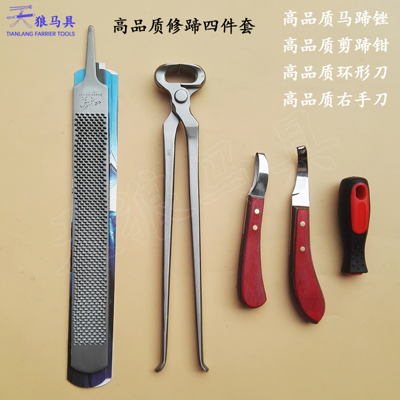
Ring knives have a hook-shaped blade, a handle with hole for fixing the rope and one or two rings. Inserting a knife into birth paths in a closed hand with a middle ring finger. Dissection of soft tissues the fetus is carried out with a movement of the hand on myself.
Hidden knives. Use knives of models Afanasiev and Malkmus. at first the blade is pushed forward by pressing the latch on the handle, the second - cutting part of the blade protrudes from the slot of the handle by pressing with the finger of the right hand on the blunt surface of the knife. Knives inject and removed from the birth canal in a closed with a thin rope attached.
Skin knife intended for cutting the skin limbs of the fetus during fetotomy in a closed way. Metal rod with a handle on the front edge bifurcated, a removable blade. Knife set against circular incision of the skin of the limb and moving forward cut it along the entire limb.
Obstetrics spatula used to separate fetal skin from tissues during fetotomy closed way. Obstetrics chisel serves to destroy the bone tissue of the head, spine and fetal pelvis.
Fetotom of Avrutis and Beskhlebnov consists of a metal frame-clamp, two thick-walled rubber tubes and wire saw. Used for separating limbs and head, as well as dissection fetal body. With the help of a sawmill a wire saw is circled around the part fetus to be separated. After of this, both ends of the saw are stretched with a mandrin through rubber tubes, attach handles and alternately pulling them set the saw in motion.
Fetotom Afanasiev has two metal tubes, interconnected on both ends and middle, as well as wire saw. Split tubes are designed to reduce the length of the fetotome and are convenient for packaging and sterilization.
Most often instruments used in childbirth in animals are collected in obstetric kits. More common in veterinary clinics use obstetric I. N. Afanasyev's set. It consists of: sterilizer, metal collapsible box, looper, 20 m cotton cord for production of obstetric ropes, two rope handles, obstetric stick, obstetric shovel (spatula), hook long folding for fixing the fetus behind pelvic bones through the anus, two eye hooks, stick handle, knife hidden with two blades, fetotom with mandrin, ten four-meter wire saws, sawmill for wire bypass saws around the fruit, two saw holders. The metal box allows disinfect all instruments assembled form.
Preparation obstetric instruments . Toolkit that can needed during obstetric care, sterilize 30 min in 2% sodium bicarbonate solution. For sterilizers are used for this purpose. large sizes. If you have a set Afanasyev's instruments are boiled in sterilizer included. The metal box of the set is assembled and fill with a solution of furacilin 1 : 5000, where load tools. As it gets dirty disinfectant is changed during operation.
Obstetric instruments, preparation and rules for their use in childbirth. Obstetrics, gynecology and artificial insemination of farm animals
Obstetrics, gynecology and artificial insemination of farm animals
test
During childbirth, one should strive to remove the fetus by hand without the use of tools.
Obstetric instruments should be used in cases where it is not possible to extract the fetus without them. Obstetric instruments, depending on their purpose, are divided into three groups: 1) auxiliary; 2) for repulsion and extraction of the fetus; 3) for fetotomy.
Auxiliary tools. Auxiliary tools include loop guides (Fig. 64) by Zwick, Afanasiev, Lindgorst.
Zwick's loop guide is 25 cm long. It is used to loop a rope around the neck, limb or torso of the fetus.
The Afanasiev loop guide is the most convenient compared to other loop guides, it can be easily moved between parts of the fruit that are in close contact with each other.
Lindhorst loop guide is an oval ring of round unpolished iron 14 cm long and 4 cm wide.
Fetal repulsion instruments. These include obstetric clubs (Fig. 65) Gunther, Kühn, Becker and Kaiser. The obstetric stick must be used carefully in a necessarily under the control of the hand, introduced simultaneously with it into the uterus, in order to protect it from injury in the event of slipping of the stick.
Gunther's stick is a metal rod about 1 m long and 1-1.5 si thick. The front end of the stick has the shape of a semicircular fork, and the rear end is equipped with a handle. The stick rests on the chest, shoulder or ischial notch of the fetus.
Kühn's stick at the ends of the semicircular fork has one each. hole through which the rope is passed. When the end of the rope is pulled, the hook is pressed tightly against the necessary part of the fetus, as a result of which it cannot slip off when pushed away. The stick is used both for repulsion and for retrieving the fetus.
Wecker's stick secures the fruit part with a rope. The anterior end of the stick has a spherical shape, thereby protecting the uterus from damage.
The Kaiser's stick, due to the presence of fork-shaped processes at the front end of the movable structure, very firmly fixes the body part (limb or chest) of the fetus to be repelled, which prevents the stick from slipping.
Fetal Retrieval Instruments. Various tools have been proposed for fetal extraction: obstetric ropes, hooks and forceps. The main tool used for these purposes are obstetric ropes.
Obstetric ropes should be smooth and strong, 0. 5-0.7 cm thick and 2-3 m long. They are only allowed to be used in obstetric care. Before use, the ropes must be sterilized by boiling, as a result of which they become soft and elastic. During operation, they are periodically immersed in a 2% solution of creolin or lysol.
Before applying the rope to one or another part of the fetus, a single or double obstetric loop is made from it. Obstetric loops (Fig. 66} are most often applied to the limbs, head or lower jaw. On the limbs, loops must be put on above the fetlock joints, which contributes to their stronger fixation. For ease of tension, the end of the obstetric loop is fixed on a special stick.
On the head of the fetus the rope is applied in the form of a loop or various halters.When applying a loop on the head of a live fetus, one must be careful not to slip it around the neck.To prevent this, the loop is put on in such a way that it goes behind the ears and passes through the mouth or around the head of the fetus with the capture of one ear.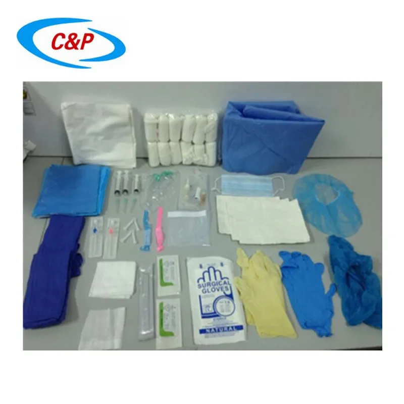
Obstetric hooks (Fig. 67) have also been widely used in practice. These include double hooks of Krey-Shogtper, Afanasyev, Dekkver, Garms eye hooks, anal.
Cray-Schottler, Afanasiev and Dekkwer hooks are very handy. They are inserted into the uterus in a closed form and fixed to the spine, neck, lower back, skin and other parts of the fetus. The advantages of these hooks are that they close when torn off and do not cause injury to the birth canal.
Garms ophthalmic hooks are used for malposition of the fetal head. For live fruits, hooks are inserted only into the inner corners of the eyes. Before inserting the hook, a rope is tied to the end of it.
When removing dead fetuses, hooks are inserted into any strong tissue (skin, tendons, bone holes). In order to avoid injury to the mucous membrane of the uterus, when inserting the hook, cover it with the hand, and in order to hold the hook in the tissues if they break when pulled on the rope, press the ring of the hook to the palm with the fingers of the hand in the uterus.
Anal hooks are used when removing a dead fetus from the birth canal in breech presentation. The hook is inserted into the anus of the fetus and strengthened by the front edge of the pubic fusion.
Obstetrical forceps (Fig. 68) are used to extract fetuses. In large animals, it is most convenient to use Talnd's forceps. They consist of two crosswise curved branches.
Their front ends have teeth that overlap one another, and the back ends have rings. Forceps are used in cases where it is not possible to extract the fetus with the help of loops and hooks.
In pigs, it is very convenient to remove fruits with Witt or Walch tongs, and in sheep with de Bruin tongs.
In large breed bitches, de Bruin's forceps can be used, and in small breed bitches, Veit's forceps, fenestrated forceps, forceps, and others.
Fetotomy instruments. A large number of different felttoms have been proposed for dissection of the fetus in the uterine cavity. The following is a description of only those tools that have received the most widespread use.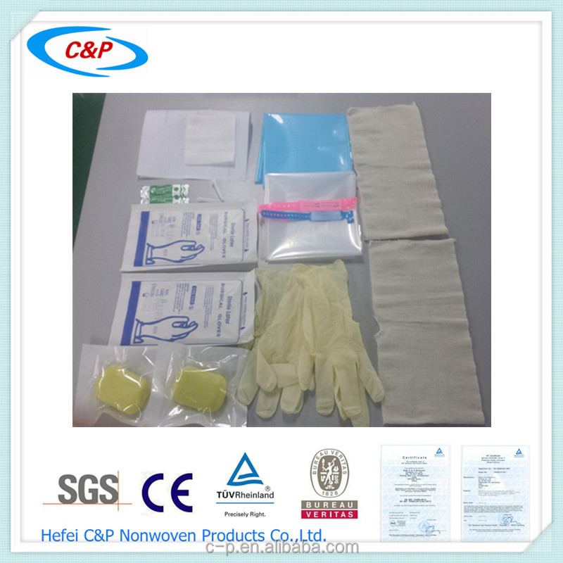
Ring knives (Fig. 69) models Gunter, Tapken, Holvekk are used to dissect the soft tissues of the fetus. The ring or rings of the knife are put on the middle finger, and the rest of the fingers cover the blade when it is inserted into the birth canal and brought out.
Hidden knives (Fig. 70) come in two models: the blade of the Afanasiev model extends forward from the handle, while the knife of the Malkmus model protrudes through the gap between the side plates of the handle. The closed knife is inserted into the uterine cavity with an obstetric rope threaded through the hole in the handle. Pressing the finger of the right hand on the blunt surface of the rein, and pulling the rope with the left hand, the necessary soft parts of the fetus are dissected.
Obstetrical spatulas (Fig. 71) are used to separate skin from underlying tissues in closed fetotomy techniques.
Obstetrical chisels (fig. 72) are used for crushing bones when separating the spine or head of the fetus.
Obstetrical saws and chain knives are used for cutting skin, muscle and bone tissue. Of these tools, the most convenient are the Van Staa wire saw (Fig. 73) and the Lindhorst and Masha chain knives (Fig. 74).
Complex fetotomes (Fig. 75) are used to separate the limbs, the head, or to dissect the fetal body. The most convenient are the fetotomes of Pflyants, Tigensen, Avrutis and Beskhlebnov, Afanasyev. These instruments are essential when performing a fetotomy.
Fetotoi Pflants. It consists of a chain knife, a chain, a rod and a frame with a geared shaft attached to it. To carry out a fetotomy, they take the end of the chain in their hands and introduce it into the uterine cavity with the help of a rope and a looper. A chain knife attached at one end to the ear of the frame is fastened to the stump. The knife is circled around the * part of the fetus to be removed. The free end of the chain is passed through the eye of the frame and the guide ring, and then put on the hook of the shaft. The obstetrician directs the chain knife of the fetotom, and the assistant, holding the frame, winds the chain onto the shaft. When the chain is twisted, the loop of the chain knife is shortened, and the frame, moving into the birth canal, rests with its eye on the fetal tissue. A stretched chain knife cuts through all the captured parts of the fetus and is pulled by the chain into the eye of the fetotom.
Tiegensen's fetotome consists of two interconnected metal tubes, through which a steel wire saw is pulled using a special mandrin, the free end of which was previously circled around the part of the fetus adjacent to the separation. With the help of alternating tension of the handles, sawing movements are performed. The fetotom is simple and safe for the woman in labor and the obstetrician, since the saw is immersed in the tubes of the device.
Fetotom Afanasyev consists of two metal tubes connected in parallel at the ends. The cutting part is a wire saw, which is inserted into the tubes using a saw guide. The method of its application is the same as the previous fetotom. Fetotom is portable and easy to use.
Fetotome of Avrutis and Beskhlebnov consists of two rubber tubes, a metal head, a wire saw and a metal conductor. Fetotom is used in the same way as the previous ones.
The disadvantage of this fetotome is that when using it, the obstetrician must always fix the metal head in the uterine cavity by hand.
BASIC RULES OF DELIVERY ASSISTANCE
The main task of obstetrics is to save the life of the mother and fetus. When solving this problem, the following rules must be observed.
1. Obstetric care should be provided in a timely manner.
2. When providing obstetric care, great attention should be paid to the observance of asepsis and antisepsis, which play an important role in the prevention of various postpartum complications in the puerperal.
3. When the birth canal is dry, to facilitate repulsion and subsequent extraction (extraction) of the fetus, it is necessary to introduce vaseline oil, a decoction of flaxseed, a 4-5% solution of carboxymenyl cellulose, egg white or any other mucous substance into the uterine cavity .