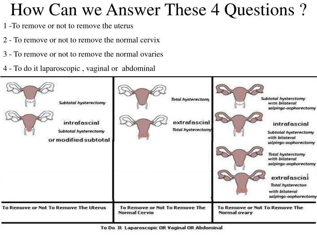
What does a normal uterus look like
Jaw-dropping image showing what a uterus looks like has people talking
Content note: Graphic image below.
A recent social media post is sparking debate – and misinformation – about the size of a ‘normal’ uterus.
Shape-shifting lady bits
The quite graphic – scroll on with this graphic content warning – update features a very small uterus and ovaries which have been removed from a post-menopausal woman via a radical hysterectomy. (This procedure involves the removal of the uterus and other reproductive organs and is performed to treat a number of medical conditions.)
In the accompanying image, a doctor’s hand holds the uterus and ovaries – and they look very, very tiny.
Commenters in their thousands marvelled at the photo, believing that their own uterus might look like this petite example. But chances are it won’t and this photo purporting to be the norm is actually far from it.
Also? It’s kind of weird that we are celebrating the size of uteruses now! Isn’t there enough body image pressure without marvelling at petite reproductive organs?!
What is ‘normal’?
The now viral post, shared by the Institute of Mums Facebook page, goes on to detail just how adaptable a uterus is – true! – and provide some statistics on its changing size.
For the record a ‘normal-sized’ uterus is pear-shaped and apparently around 7.6 cm long, approximately 4.5 cm wide and 3.0 cm thick.[1][2] It typically weighs around 60 grams.
The uterus obviously changes size during pregnancy, stretching to accommodate the baby or babies. During menopause, the uterus shrinks in size as a woman ages getting smaller the further she gets from menopause.
The doctor who posted the original image on Instagram confirms this is a particularly tiny uterus and not really the norm for your average woman.
Anyone else surprised by how small it is?!! ❤️This incredible visual from @docshawarma (via @vbmaternitywellnessllc) —…
Posted by Institute of Mums on Saturday, 6 July 2019
The size of a drumstick
“After you give birth, the uterus immediately starts contracting to shrink itself,” the repost continues.
That part, at least, is true. Anyone who has had a baby will be familiar with those postpartum contraction feels, especially if you are able to breastfeed your baby. The uterus cleverly keeps sizing down in the days and weeks after birth.
While lots of people went for the information shared in this post hook, line and sinker, a number of nurses piped up to flag the photo as misleading.
“As an OR nurse I have NEVER seen a uterus that small on any women,” one wrote under the image. “I am having a hard time buying that is a grown woman’s uterus – and trust me I have assisted on over 100 hysterectomies in 30 years. ”
”
“I actually thought it looked like teen or toddler size,” nurse Tracie continued.
“I agree,” another commenter posted. “I’ve seen many hysterectomy’s never seen them this small.”
“Absolutely agree, I‘m a surgical assistant,” someone else commented.
“As a student nurse in theatre on gynae ops, wombs I saw removed were more the size and shape of a chicken drumstick,” another Facebook user wrote.
“This is way too small and I think it’s photoshopped, I’ve seen lots of hysterectomies and the uterus is usually like a pear, not a walnut,” someone else confirmed.
So there you have it. This photo shows some amazing reproductive organs, but the average woman of childbearing age will sport much larger versions of those depicted.
View this post on Instagram
Cuteruses Before Duderuses, Ovaries Before Brovaries . . . .
. . . . . #uterus #cuterus #obgyn #residency #ovaries #instagay #gaydoc #lgbtinmedicine #surgery #hysterectomy #prochoice #endometriosis
A post shared by Ram Sharma, MD, MPH (@docshawarma) on
Posted on by Pip Lincolne
Get more babyology straight to your inbox
Join now
Anatomy of pregnancy and birth - uterus
Anatomy of pregnancy and birth - uterus | Pregnancy Birth and Baby beginning of content5-minute read
Listen
What does the uterus look like?
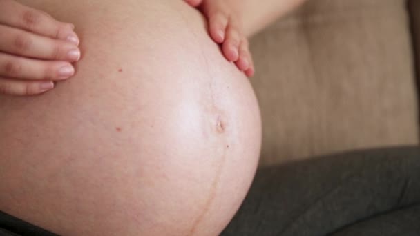
One of the most recognised changes in a pregnant woman’s body is the appearance of the ‘baby bump’, which forms to accommodate the baby growing in the uterus.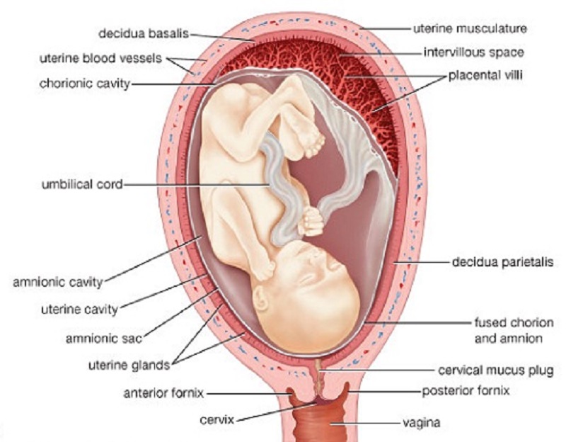 The primary function of the uterus during pregnancy is to house and nurture your growing baby, so it is important to understand its structure and function, and what changes you can expect the uterus to undergo during pregnancy.
The uterus (also known as the ‘womb’) has a thick muscular wall and is pear shaped. It is made up of the fundus (at the top of the uterus), the main body (called the corpus), and the cervix (the lower part of the uterus ). Ligaments – which are tough, flexible tissue – hold it in position in the middle of the pelvis, behind the bladder, and in front of the rectum.
The uterus wall is made up of 3 layers. The inside is a thin layer called the endometrium, which responds to hormones – the shedding of this layer causes menstrual bleeding. The middle layer is a muscular wall. The outside layer of the uterus is a thin layer of cells.
Illustration showing the female reproductive system.The size of a non-pregnant woman's uterus can vary. In a woman who has never been pregnant, the average length of the uterus is about 7 centimetres. This increases in size to approximately 9 centimetres in a woman who is not pregnant but has been pregnant before. The size and shape of the uterus can change with the number of pregnancies and with age.
How does the uterus change during pregnancy?
During pregnancy, as the baby grows, the size of a woman’s uterus will dramatically increase. One measure to estimate growth is the fundal height, the distance from the pubic bone to the top of the uterus. Your doctor (GP) or obstetrician or midwife will measure your fundal height at each antenatal visit from 24 weeks onwards. If there are concerns about your baby’s growth, your doctor or midwife may recommend using regular ultrasound to monitor the baby.
Fundal height can vary from person to person, and many factors can affect the size of a pregnant woman’s uterus. For instance, the fundal height may be different in women who are carrying more than one baby, who are overweight or obese, or who have certain medical conditions. A full bladder will also affect fundal height measurement, so it’s important to empty your bladder before each measurement. A smaller than expected fundal height could be a sign that the baby is growing slowly or that there is too little amniotic fluid. If so, this will be monitored carefully by your doctor. In contrast, a larger than expected fundal height could mean that the baby is larger than average and this may also need monitoring.
As the uterus grows, it can put pressure on the other organs of the pregnant woman's body. For instance, the uterus can press on the nearby bladder, increasing the need to urinate.
How does the uterus prepare for labour and birth?
Braxton Hicks contractions, also known as 'false labour' or 'practice contractions', prepare your uterus for the birth and may start as early as mid-way through your pregnancy, and continuing right through to the birth. Braxton Hicks contractions tend to be irregular and while they are not generally painful, they can be uncomfortable and get progressively stronger through the pregnancy.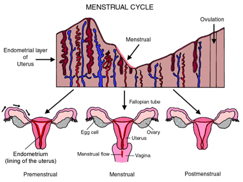
During true labour, the muscles of the uterus contract to help your baby move down into the birth canal. Labour contractions start like a wave and build in intensity, moving from the top of the uterus right down to the cervix. Your uterus will feel tight during the contraction, but between contractions, the pain will ease off and allow you to rest before the next one builds. Unlike Braxton Hicks, labour contractions become stronger, more regular and more frequent in the lead up to the birth.
How does the uterus change after birth?
After the baby is born, the uterus will contract again to allow the placenta, which feeds the baby during pregnancy, to leave the woman’s body. This is sometimes called the ‘after birth’. These contractions are milder than the contractions felt during labour. Once the placenta is delivered, the uterus remains contracted to help prevent heavy bleeding known as ‘postpartum haemorrhage‘.
The uterus will also continue to have contractions after the birth is completed, particularly during breastfeeding.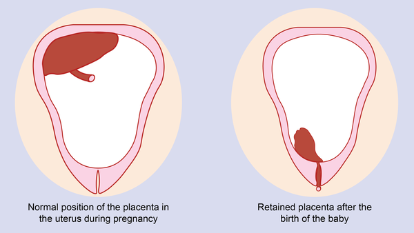 This contracting and tightening of the uterus will feel a little like period cramps and is also known as 'afterbirth pains'.
Read more here about the first few days after giving birth.
Sources:
The Royal Australian and New Zealand College of Obstetricians and Gynaecologists (Labour and birth), StatPearls Publishing (Anatomy, Abdomen and Pelvis), Department of Health (Clinical practice guidelines: Pregnancy care), Better Health Channel Victoria (Pregnancy stages and changes), Mater Mother's Hospital (Labour and birth information), Royal Australian and New Zealand College of Obstetricians and Gynaecologists (The First Few Weeks Following Birth), Queensland Health (Queensland Clinical Guidelines – maternity and neonatal), King Edward Memorial Hospital (Fundal height: Measuring with a tape measure), Royal Hospital for Women (Fetal growth assessment (clinical) in pregnancy), MSD Manual (Female internal genital organs)Learn more here about the development and quality assurance of healthdirect content.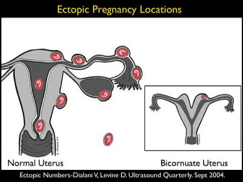
Last reviewed: October 2020
Back To Top
Related pages
- Anatomy of pregnancy and birth - perineum and pelvic floor
- Anatomy of pregnancy and birth - pelvis
- Anatomy of pregnancy and birth - cervix
- Anatomy of pregnancy and birth - abdominal muscles
- Anatomy of pregnancy and birth
Need more information?
Prolapsed uterus - Better Health Channel
The pelvic floor and associated supporting ligaments can be weakened or damaged in many ways, causing uterine prolapse.
Read more on Better Health Channel website
Uterus, cervix & ovaries - fact sheet | Jean Hailes
This fact sheet discusses some of the health conditions that may affect a woman's uterus, cervix and ovaries.
Read more on Jean Hailes for Women's Health website
Dilatation and curettage (D&C)
A D&C is an operation to lightly scrape the inside of the uterus (womb).
Read more on WA Health website
Ectopic pregnancy
An ectopic pregnancy occurs when a fertilised egg implants outside the uterus (womb)
Read more on WA Health website
Placental abruption - Better Health Channel
Placental abruption means the placenta has detached from the wall of the uterus, starving the baby of oxygen and nutrients.
Read more on Better Health Channel website
Pelvic Floor | Family Planning NSW
The pelvic floor is a group of muscles in the pelvic area that support the bladder, bowel and uterus (womb).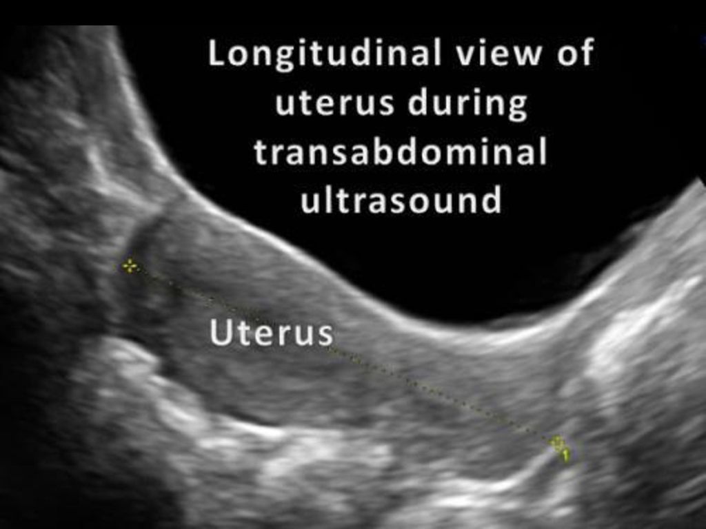
Read more on Family Planning NSW website
Mirena IUD | Hormonal IUD Mirena | IUD Mirena insertion | IUD Mirena cost | Mirena IUD Melbourne - Sexual Health Victoria
The hormonal intrauterine device (IUD) is a small contraceptive device that is put into the uterus (womb) to prevent pregnancy.
Read more on Sexual Health Victoria website
Endometriosis | Your Fertility
Endometriosis is a condition where the tissue that lines the uterus also grows in other areas of the body
Read more on Your Fertility website
Pelvic Floor Muscle Damage - Birth Trauma
The pelvic floor muscles are a supportive basin of muscle attached to the pelvic bones by connective tissue to support the vagina, uterus, bladder and bowel.
Read more on Australasian Birth Trauma Association website
What is Ectopic Pregnancy? | Ectopic Pregnancy Symptoms | How do I know if I have had an Ectopic Pregnancy? | Ectopic Pregnancy Treatment - Sexual Health Victoria
Ectopic pregnancy is a pregnancy that develops outside the uterus, usually in one of the fallopian tubes. In almost all cases, the embryo dies.
Read more on Sexual Health Victoria website
Disclaimer
Pregnancy, Birth and Baby is not responsible for the content and advertising on the external website you are now entering.
OKNeed further advice or guidance from our maternal child health nurses?
1800 882 436
Video call
- Contact us
- About us
- A-Z topics
- Symptom Checker
- Service Finder
- Linking to us
- Information partners
- Terms of use
- Privacy
Pregnancy, Birth and Baby is funded by the Australian Government and operated by Healthdirect Australia.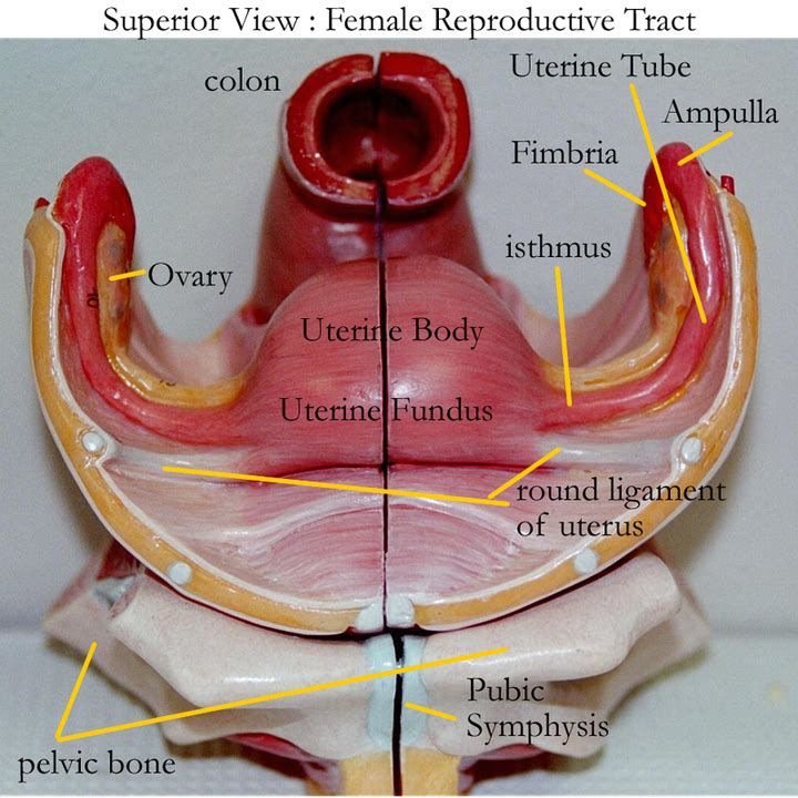
Pregnancy, Birth and Baby is provided on behalf of the Department of Health
Pregnancy, Birth and Baby’s information and advice are developed and managed within a rigorous clinical governance framework. This website is certified by the Health On The Net (HON) foundation, the standard for trustworthy health information.
This site is protected by reCAPTCHA and the Google Privacy Policy and Terms of Service apply.
This information is for your general information and use only and is not intended to be used as medical advice and should not be used to diagnose, treat, cure or prevent any medical condition, nor should it be used for therapeutic purposes.
The information is not a substitute for independent professional advice and should not be used as an alternative to professional health care. If you have a particular medical problem, please consult a healthcare professional.
Except as permitted under the Copyright Act 1968, this publication or any part of it may not be reproduced, altered, adapted, stored and/or distributed in any form or by any means without the prior written permission of Healthdirect Australia.
Support this browser is being discontinued for Pregnancy, Birth and Baby
Support for this browser is being discontinued for this site
- Internet Explorer 11 and lower
We currently support Microsoft Edge, Chrome, Firefox and Safari. For more information, please visit the links below:
- Chrome by Google
- Firefox by Mozilla
- Microsoft Edge
- Safari by Apple
You are welcome to continue browsing this site with this browser. Some features, tools or interaction may not work correctly.
90,000 photos of ultrasound of the uterus, ovaries, fallopian tubes. SERVICESWhat does a normal ultrasound picture look like in healthy women?
Nominal queen
Fig. 1. Normal uterus.
Second cycle phase. Myometrium is homogeneous.
M-ECHO thickness corresponds to the day of the cycle.
When assessing the condition of the uterus with ultrasound, you can determine:
- The position of the uterus.
Normally, the uterus is either deviated towards the bladder, that is, anteriorly (this position of the uterus is called anteflexio), or deviated towards the rectum, that is, backwards, - (retroflexio). - Dimensions of the uterus (longitudinal, anteroposterior and transverse). The average size of a normal uterus in length is from 4.0 to 6.0 cm, anterior-posterior from 2.7 to 4.9 mm. The size of the body of the uterus varies depending on the woman's age, constitution and obstetric and gynecological history.
- The condition of the endometrium (its thickness varies depending on the day of the menstrual cycle).
Immediately after the end of menstruation, the endometrium is visualized as a strip 1-2 mm thick. In the second phase of the cycle, the thickness of the endometrium (M-ECHO) can be from 10 to 14 mm on average. - State of the myometrium.
Normally, the myometrium should be homogeneous and not have pathological formations in its structure (myomas, adenomyosis, etc.)
Normal ovaries
Pic. 2. Normal ovary with follicular apparatus.
There is no dominant follicle, since the study was carried out on the 3rd day of the menstrual cycle.
When assessing the state of the ovaries by ultrasound, the following is determined:
- The position of the ovaries.
Normally located on the sides of the uterus, most often asymmetrically, at a small distance from the corners of the uterus. The shape of the ovaries is usually oval, while the right and left ovaries are not at all identical to each other. - Dimensions of the ovaries (longitudinal, anterior-posterior and transverse).
The average size of normal ovaries in length is from 2.4 to 4.0 cm, anterior-posterior from 1.5 to 2.5 mm. - Structure of the ovaries.
Normally, the ovaries consist of a capsule and follicles of varying degrees of maturity (in the first phase of the cycle). In the second phase of the cycle, as a rule, the corpus luteum is visualized - a sign of ovulation that has occurred. The number of follicles may not be the same on the left and right. The maturing follicle is detected already in the first phase of the cycle and reaches its maximum size by ovulation, an average of about 20 mm.
 The content of the dominant follicle is homogeneous because it contains follicular fluid and the capsule is thin. After ovulation, a corpus luteum is formed at the site of the dominant follicle, which, as a rule, has a reticulate echostructure (it contains adipose tissue) and also a thin capsule - 1-2 mm. Most often in shape, this formation is oval or irregularly shaped.
In postmenopausal women, the ovaries are normally either not visualized or located as fibrous bands.
Normal fallopian tubes
Normally, the fallopian tubes are not visible on ultrasound.
Early pregnancy
Fig. 3. Uterine pregnancy 7-8 weeks.
The size of the fetal egg and embryo correspond to the delay in menstruation.
During pregnancy, only the fetal egg is visualized in the uterine cavity in the early stages, later, the embryo appears. The size of the fetal egg and embryo should correspond to the gestational age for menstruation.
It is also obligatory to assess the fetal heartbeat, which, as a rule, appears after 10-14 days of delayed menstruation.
M-echo thickness, standard. 2D.
Ultrasound of normal uterus. 2D.
Ultrasound of a normal uterus. 2D.
During pregnancy, the yellow body of pregnancy should be visualized in one of the ovaries, which controls the development of this pregnancy and ensures the vital activity of the fetus in the early stages (before the formation of the placenta).
Next:
» Pathology of the pelvic organs
Infertility treatment services at the clinic "I'm healthy!"
- Consultation MD, professor obstetrician-gynecologist
- Consultation of c.m.s., obstetrician-gynecologist
- Expert ultrasound of the pelvic organs with Doppler
- Expert ultrasound of all organs
- Consultation by relevant specialists on the results of the examination
Step-by-step procedures for diagnosing infertility
Prices for services
Reviews
Alina Thank you very much to the doctor Natalya Viktorovna Dmitrieva for her sensitive heart and for her high professionalism! And most importantly, for her faith in my pregnancy!!! I came to her after trying unsuccessfully to get pregnant for two years.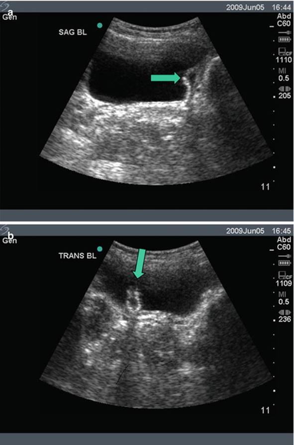 And now, I can rightfully say - NEVER GIVE UP EVER! I am now a mother and this is largely the merit of Natalia Viktorovna! More Victoria I liked it very much at the reception of Dr. Aleshkina. Very friendly, courteous and explained everything in detail. Sincerely wants to help. Gave appointments, contacts of colleagues. I will be watching her. More Kati On behalf of our entire family, I would like to wish the clinic prosperity and many years of work! Your doctors are doing the most important job! More Svetlana I express my admiration for the doctor Ekaterina Dmitrievna Dubinskaya! She is an incredibly knowledgeable specialist and also a wonderful, sympathetic person!!! I am happy that I found out about her and got to her appointment and treatment! More Tatiana I had 13 IVF attempts in 8 years resulting in a single embryo at your clinic in 2019year.
 You implanted an embryo on October 29, 2021. Now he is a cheerful boy Misha, almost 3 months old, was born on July 5, 2022 by COP at 39 weeks in the Filatov hospital. Natalya Viktorovna, Without the achievements of modern medicine and without you in my story, I would never have become a mother. I am immensely grateful to you and your assistants for working with me, your professionalism, for believing in a miracle (together with me :), when absolutely no one believed anymore, for my great happiness to become a mother!❤️ More Viggo When problems started in the male part, I found your clinic in the search. We made an appointment with the doctor Lychagin. After examination and tests, he suggested treatment by shock wave therapy. After completing a course of 7 procedures, my problems were gone. Gratitude !! More Faith The clinic is good, modern, professors work in "I'm healthy", they sort out difficult cases related to pregnancy and women's health in general.
 I had a colposcopy here and the procedure was painless. More Galina I express my gratitude to the Doctors for their professionalism and indifference. Bringing hope back to people is worth a lot. I am expecting my twins and I am happy-) Even the problem with my husband's card did not affect anything. More Regina Aksenova In the clinic, each doctor has his own individual approach. It has its own academic base. Professors and academicians work. I came from Kazakhstan and received a full range of infertility treatment. I was able to conceive a child and my happiness is immeasurable. Thanks to everyone who helped me with this! More Ana Mariam Gareginovna, good afternoon! I wanted to share the news with you - we became the parents of the most charming boy on August 23, I reached 38 weeks and 1 day! I still do not believe that thanks to Natalya Viktorovna, our dream is already close to us! Please pass on words of gratitude and love to Natalya Viktorovna! ❤️❤️❤️❤️❤️ I’m even scared to think what would have happened if we hadn’t met your entire team: Pyatykh, who told us about Lychagin, then Andrey Sergeevich sent us to Natalya Viktorovna ❤️❤️❤️❤️ thank you very much for handing me over to Victoria Viktorovna Gnipova, I gave birth with Anna Alexandrovna Dyakonova.
 These people are just the angels of my pregnancy!!! Such a chain of professionals! thank you for such happiness❤️❤️❤️❤️ More
Leave your details.
Our specialists will contact you to clarify the details.
Your application has been sent!
We will call you as soon as possible.
Thank you for contacting the Clinic "I'm healthy!"
Leave your details.
Our specialists will contact you to clarify the details.
Your application has been sent!
We will call you as soon as possible.
Thank you for contacting the Clinic "I'm healthy!"
Ultrasound of uterus
Dmitrieva Olga Nikolaevna
Chief physician of "Polyclinika.ru" on Frunzenskaya, neurologist, ENMG specialist
reviews
Clinic
m. Frunzenskaya
Stolbova Tatyana Sergeevna 1905, internist, gastroenterologist
reviews
Clinic
m. Street 1905 Goda
Yuliy Sergeevich Gromov
Head physician of "Polyclinic.ru" on Sukharevskaya, surgeon
reviews
Clinic
m. Sukharevskaya
Agarkova Elena Valentinovna
Head doctor "Polyclinika.ru" in Zelenograd, gastroenterologist
reviews
Clinic
Zelenograd
Aydinov Gennadiy Ivanovich
Chief doctor of Polyclinic.ru Academician Yangelya, therapist
reviews
Clinic
m. st. Academician Yangel
Godulyan Aleksey Viktorovich
chief physician of "Polyclinika. ru" at Krasnye Vorota, endocrinologist, KMN
reviews
Clinic
m. Red Gate
Kupriyanova Olga Sergeevna
Chief doctor "Polyclinika.ru" on Avtozavodskaya, general practitioner
reviews
Clinic
m. Avtozavodskaya
Sumina Evgenia Yuryevna
Chief physician of "Polyanka.ru" in Polyanka, neurologist
reviews
Clinic
m. Polyanka
Tolstikova Anna Pavlovna
Chief physician of "Polyclinic.ru" on Taganskaya, general practitioner
reviews
Clinic
m.