What can radiation do to a fetus
Radiation and Pregnancy: Information for Clinicians
This overview provides physicians with information about prenatal radiation exposure as an aid in counseling pregnant women.
How to use this document
This information is for clinicians. If you are a patient, we strongly advise that you consult with your physician to interpret the information provided, as it may not apply to you. Information on radiation exposure during pregnancy for members of the public can be found on the Health Information for Specific Groups webpage.
CDC recognizes that providing information and advice about radiation to expectant mothers falls into the broader context of preventive healthcare counseling during prenatal care. In this setting, the purpose of the communication is always to promote health and long-term quality of life for the mother and child.
This page is also available as a PDF pdf icon[365 KB]
Radiation exposure to a fetus
Most of the ways a pregnant woman may be exposed to radiation, such as from a diagnostic medical exam or an occupational exposure within regulatory limits, are not likely to cause health effects for a fetus. However, accidental or intentional exposure above regulatory limits may be cause for concern.
Although radiation doses to a fetus tend to be lower than the dose to the mother, due to protection from the uterus and surrounding tissues, the human embryo and fetus are sensitive to ionizing radiation at doses greater than 0.1 gray (Gy). Depending on the stage of fetal development, the health consequences of exposure at doses greater than 0.5 Gy can be severe, even if such a dose is too low to cause an immediate effect for the mother. The health consequences can include growth restriction, malformations, impaired brain function, and cancer.
Estimating the Radiation Dose to the Embryo or Fetus
Health effects to a fetus from radiation exposure depend largely on the radiation dose. Estimating the radiation dose to the fetus requires consideration of all sources external and internal to the mother’s body, including the following:
- Dose from an external source of radiation to the mother’s abdomen.

- Dose from inhaling or ingesting a radioactive substance that enters the bloodstream and that may through the placenta.
- Dose from radioactive substances that may concentrate in maternal tissues surrounding the uterus, such as the bladder, and that could irradiate the fetus.
Most radioactive substances that reach the mother’s blood can be detected in the fetus’ blood. The concentration of the substance depends on its specific properties and the stage of fetal development. A few substances needed for fetal growth and development (such as iodine) can concentrate more in the fetus than in corresponding maternal tissue.
Consideration of the dose to specific fetal organs is important for substances that can localize in specific organs and tissues in the fetus, such as iodine-131 or iodine-123 in the thyroid, iron-59 in the liver, gallium-67 in the spleen, and strontium-90 and yttrium-90 in the skeleton.
Radiation experts can assist in estimating the radiation dose to the embryo or fetus
Hospital medical physicists and health physicists are good resources for expertise in estimating the radiation dose to the fetus.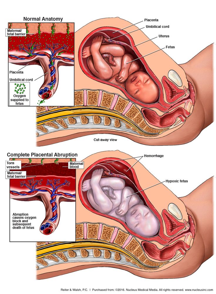 In addition to the hospital or clinic’s specialized staff, physicians may access resources from or contact the following organizations for assistance in estimating fetal radiation dose.
- The National Council on Radiation Protection and Measurementsexternal icon’ Report No. 174, “Preconception and Prenatal Radiation Exposure: Health Effects and Protective Guidance” [NCRP2013] provides detailed information for assessing fetal doses from internal uptakes.
- The International Commission on Radiological Protection’s “Publication 84: Pregnancy and Medical Radiation”external icon [ICRP2000] provides fetal dose estimations from medical exposures to pregnant women.
- The Conference of Radiation Control Program Directorsexternal icon maintains a list of state Radiation Control/Radiation Protection program contact information.
- The Health Physics Societyexternal icon maintains a list of active certified Health Physicists.
- The American Association of Physicists in Medicineexternal icon provides information resources.

Once the fetal radiation dose is estimated, potential health effects can be assessed.
Potential Health Effects of Prenatal Radiation Exposure (Other Than Cancer)
Table 1 summarizes the potential non-cancer health risks of concern. This table is intended to help physicians advise pregnant women who may have been exposed to radiation, not as a definitive recommendation. The indicated doses and times post-conception are approximations.
| Acute Radiation Dose* to the Embryo/Fetus | Time Post Conception (up to 2 weeks) | Time Post Conception (3rd to 5th weeks) | Time Post Conception (6th to 13th weeks) | Time Post Conception (14th to 23rd weeks) | Time Post Conception (24th week to term) |
|---|---|---|---|---|---|
<0.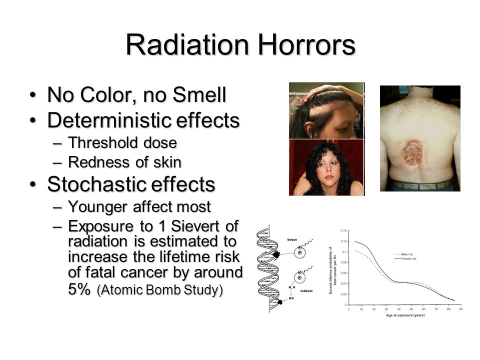 10 Gy 10 Gy (10 rads) | Noncancer health effects NOT detectable | ||||
| 0.10–0.50 Gy (10–50 rads) | Failure to implant may increase slightly, but surviving embryos will probably have no significant (non-cancer) health effects. | Growth restriction possible | Growth restriction possible | Noncancer health effects unlikely | |
| > 0.50 Gy (50 rads) The expectant mother may be experiencing acute radiation syndrome in this range, depending on her whole-body dose. | Failure to implant will likely be high, depending on dose, but surviving embryos will probably have no significant (non- cancer) health effects. | Probability of miscarriage may increase, depending on dose. Probability of major malformations, such as neurological and motor deficiencies, increases. Growth restriction is likely | Probability of miscarriage may increase, depending on dose. Growth restriction is likely. | Probability of miscarriage may increase, depending on dose. Growth restriction is possible, depending on dose. (Less likely than during the 6th to 13th weeks post conception) Probability of major malformations may increase | Miscarriage and neonatal death may occur, depending on dose. § |
| 8th to 25th Weeks Post Conception: The most vulnerable period for intellectual disability is 8th to 15th weeks post conception Severe intellectual disability is possible during this period at doses > 0.5 Gy Prevalence of intellectual disability (IQ<70) is 40% after an exposure of 1 Gy from 8th to 15th week Prevalence of intellectual disability (IQ<70) is 15% after an exposure of 1 Gy from 16th to 25th week Table adapted from Table 1.1. of the National Council on Radiation Protection and Measurements’ Report No. | |||||
Gestational age and radiation dose are important determinants of potential non-cancer health effects. The following points are of particular note.
- During the first 2 weeks post-conception, the health effect of concern from an exposure of ≥ 0.1 Gy is the possibility of death of the embryo. Because the embryo is made up of only a few cells, damage to one cell, the progenitor of many other cells, may cause the death of the embryo, and the blastocyst may fail to implant in the uterus. Embryos that survive, however, are unlikely to exhibit congenital abnormalities or other non-cancer health effects, no matter what dose of radiation they received.
- In all stages post-conception, radiation-induced non-cancer health effects are not detectable for fetal doses below about 0.10 Gy.
Carcinogenic Effects of Prenatal Radiation Exposure
Radiation exposure to an embryo/fetus may increase the risk of cancer in the offspring, especially at radiation doses > 0.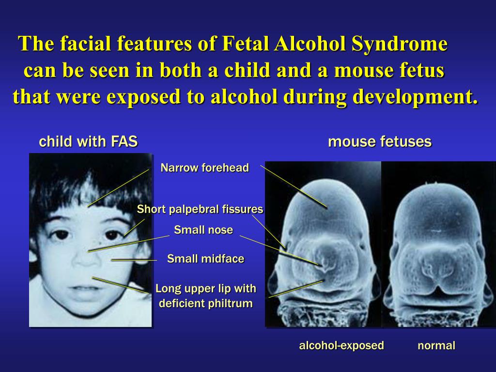 1 Gy, which are well above typical doses received in diagnostic radiology. However, attempting to quantify cancer risks from prenatal radiation exposure presents many challenges. These challenges include the following:
- The primary data for the risk of developing cancer from prenatal exposure to radiation come from the lifespan study of the Japanese atomic bomb (A-bomb) survivors. [Preston et al. 2008]. The analysis of that cohort includes cancer incidence data only up to the age of 50 years. This precludes making lifespan risk estimates as a result of prenatal radiation exposure.
- From the Japanese lifespan study [Preston et al. 2008], it can be concluded that for those exposed
in early childhood (birth to age 5 years), the theoretical risk of an adult-onset cancer by age 50 is approximately ten-fold greater than the risk for those who received prenatal exposure. Therefore, the risk following prenatal exposure may be considerably lower than for radiation exposure in early childhood
[NCRP2013].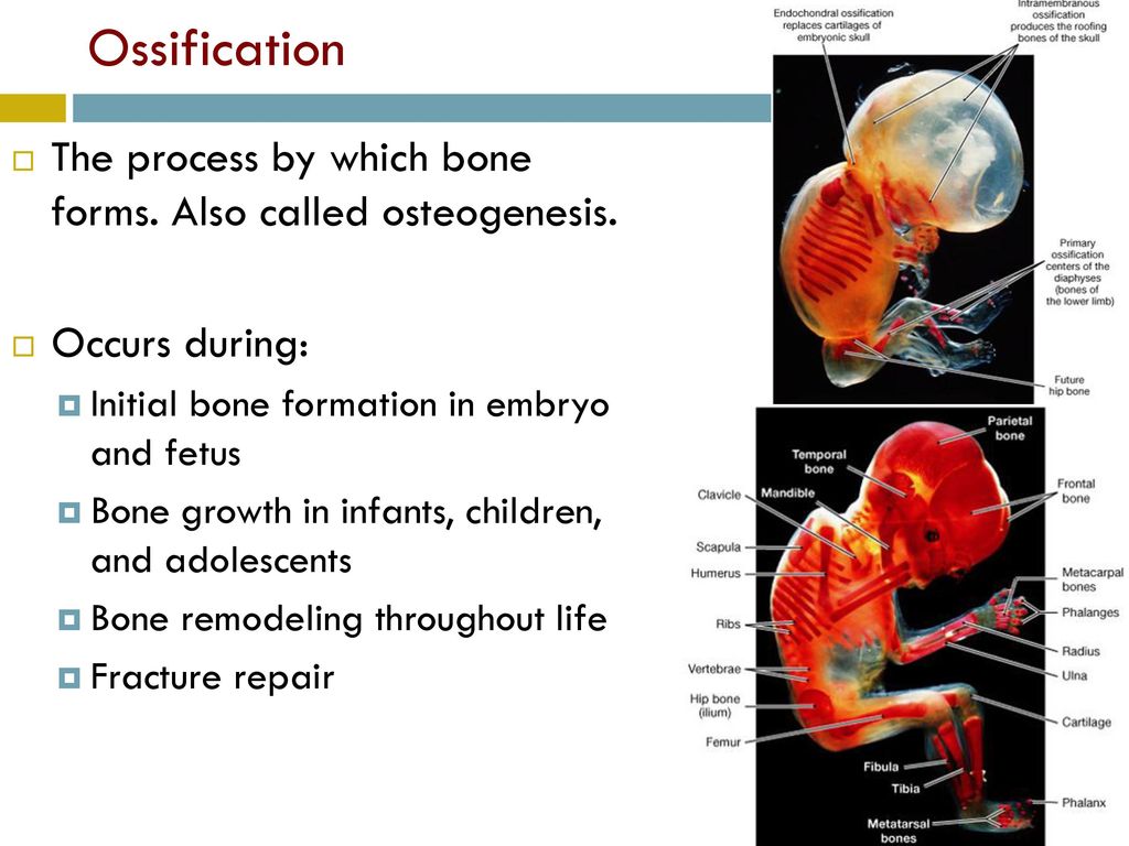
- No reliable epidemiological data are available from studies to determine which stage of pregnancy is the most sensitive for radiation-induced cancer in the offspring [NCRP2013].
The lifespan study of the Japanese A-bomb survivors is continuing as the cohort ages. Future analyses of the accumulating data should provide a better understanding of the lifetime risk of cancer from prenatal and early childhood radiation exposure.
References
ICRP2000] International Commission on Radiological Protection. 2000. Valentin, J. (2000). Pregnancy and medical radiation. Oxford: Published for the International Commission on Radiological Protection.
[NCRP2013] National Council on Radiation Protection and Measurements. 2013. Preconception and Prenatal Radiation Exposure Health Effects and Protective Guidance. (2013). Bethesda: National Council on Radiation Protection & Measurements.
Preston DL, Cullings H, Suyama A, Funamoto S, Nishi N, Soda M, Mabuchi K, Kodama K, Kasagi F, Shore RE.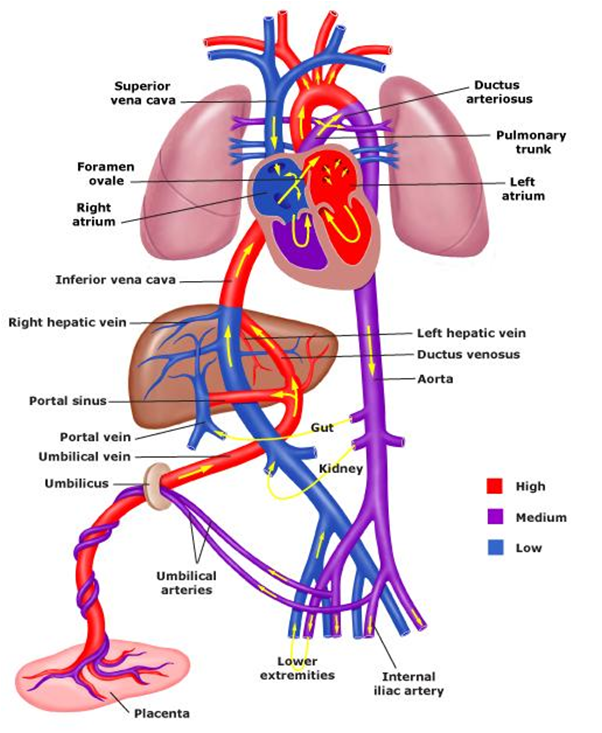 2008. Solid cancer incidence in atomic bomb survivors exposed in utero or as young children. J Natl Cancer Inst 100(6):428-436.
[UNSCEAR2013] United Nations Scientific Committee on the Effects of Atomic Radiation. 2013. Sources, effects and risks of ionizing radiation. Vol. II, Scientific Annex B: Effects of radiation exposure of children.
For more information on medical management and other topics on radiation emergencies:
Radiation Emergency Assistance Center/Training Siteexternal icon (REAC/TS) is a program uniquely qualified to teach medical personnel, health physicists, first responders and occupational health professionals about radiation emergency medical response.
Radiation Emergency Medical Management (REMM)external icon provides guidance to health care providers (primarily physicians) about clinical diagnosis and treatment of radiation injury during radiological and nuclear emergencies.
Conference of Radiation Control Program Directorsexternal icon
Health Physics Societyexternal icon
International Commission on Radiological Protectionexternal icon
National Council on Radiation Protection and Measurementsexternal icon
American Association of Physicists in Medicineexternal icon
Radiation exposure during pregnancy | Pregnancy Birth and Baby
beginning of content4-minute read
Listen
What is radiation?
If you’re pregnant or thinking about becoming pregnant, you might be worried about x-rays and other forms of radiation. It's important you discuss any concerns with your doctor and always tell a health professional that you are pregnant before you have any medical imaging.
Radiation is energy that travels through air, and some materials, as waves or tiny particles. We are exposed daily to radiation from different natural and artificial sources, such as the sun, microwaves and radio waves.
The type of radiation used in medical imaging is called ionising radiation. In Australia, people receive about 1,500 to 2,000 μSv of ionising radiation every year from natural sources. This level of radiation is not harmful.
A typical x-ray or CT scan of exposes you to 2,600 μSv of ionising radiation.
X-rays and some other forms of radiation can alter the molecules that make up the body. At high enough doses, radiation can kill cells and damage genes.
Medical procedures that give off radiation
If you are pregnant or think you might be pregnant, you should be cautious about medical procedures that use radiation, such as:
- x-rays
- CT scans
- mammography
- fluoroscopy
- nuclear medicine
- angiography
Tell your doctor and tell the radiology practice before you have any tests.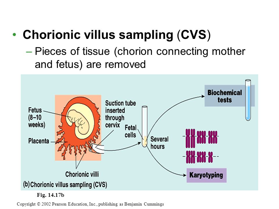 You can discuss whether they can be delayed or avoided, and whether there are alternative tests.
Tests that don’t use radiation, such as magnetic resonance imaging (MRI) or ultrasound, may be useful alternatives in some situations.
What are the effects of radiation on a mother and unborn baby?
Any harm to the developing baby will depend on:
- the radiation dose – smaller doses (amounts) are safer
- the age of the fetus – the further along you are in your pregnancy, the better
- where the radiation is administered – tests involving your abdomen or pelvis, or where the radiation is carried in your blood, pose a higher risk than other tests
Most radiation exposure during medical testing is unlikely to harm a developing baby. But sometimes, depending on the radiation dose and the developmental stage of the fetus, the effects can be serious and may result in:
- failure of the embryo to implant
- miscarriage
- abnormalities of the central nervous system
- congenital malformations
- slower than normal growth
- malformation
- cataracts
- childhood cancer
Accidental exposure to radiation
If you are accidently exposed to radiation from medical imaging while you are pregnant, you should talk to your doctor. The risk to your baby can be worked out using a formula and should be calculated by an expert. Most normal doses or a single exposure to radiation are not likely to be harmful to the baby.
If you are exposed to radiation in an emergency, you should follow instructions from emergency officials and seek medical attention as soon as possible.
Usually, the fetus receives less radiation than the mother. The mother’s abdomen partially protects the baby. However, if you swallow or breathe in radiation, it can cross over into the baby. The baby is most sensitive to radiation from 2 to 18 weeks of pregnancy.
If you are exposed to radiation while you are breastfeeding, you should feed your baby with pre-pumped and stored breast milk or formula, if possible. If there is no other source of food available to feed your baby, then continue to breastfeed, but wipe the breast and nipple thoroughly with soap and water before feeding.
Speak to your doctor or health professional about when to start breastfeeding again.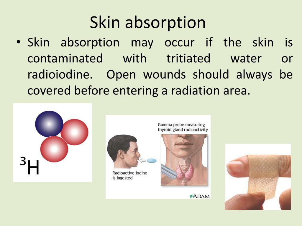
Radiotherapy for cancer
Radiotherapy, or radiation therapy, is a treatment for some sorts of cancers. Sometimes a pregnant woman is advised to have radiotherapy as a treatment for cancer during pregnancy. If you’re in this situation, you and your doctor can weigh up the benefits of the radiotherapy against any potential harm to your developing baby.
If you suspect you may be pregnant at any stage of radiotherapy, you should discuss with your doctor whether to continue the treatment.
Radioactive material can be passed to babies through breast milk. Breastfeeding mothers undergoing radiotherapy should ask a health professional for advice.
Radiation, pregnancy and the workplace
If you are pregnant or planning to become pregnant, and your work exposes you to radiation, it is important to discuss alternative roles with your employer.
Speak to a healthcare professional if you are worried about radiotherapy exposure during pregnancy.
Sources:
Department of Health (Prenatal Radiation Exposure), Inside Radiology (Radiation Risk of Medical Imaging During Pregnancy), Cancer Council NSW (Common questions about radiation), Centers for Disease Control and Prevention (Radiation emergencies and pregnancy), ANSTO (What is radiation?)Learn more here about the development and quality assurance of healthdirect content.
Last reviewed: June 2021
Back To Top
Related pages
- Medicines during pregnancy
This information is for your general information and use only and is not intended to be used as medical advice and should not be used to diagnose, treat, cure or prevent any medical condition, nor should it be used for therapeutic purposes.
The information is not a substitute for independent professional advice and should not be used as an alternative to professional health care. If you have a particular medical problem, please consult a healthcare professional.
Except as permitted under the Copyright Act 1968, this publication or any part of it may not be reproduced, altered, adapted, stored and/or distributed in any form or by any means without the prior written permission of Healthdirect Australia.
Support this browser is being discontinued for Pregnancy, Birth and Baby
Support for this browser is being discontinued for this site
- Internet Explorer 11 and lower
We currently support Microsoft Edge, Chrome, Firefox and Safari.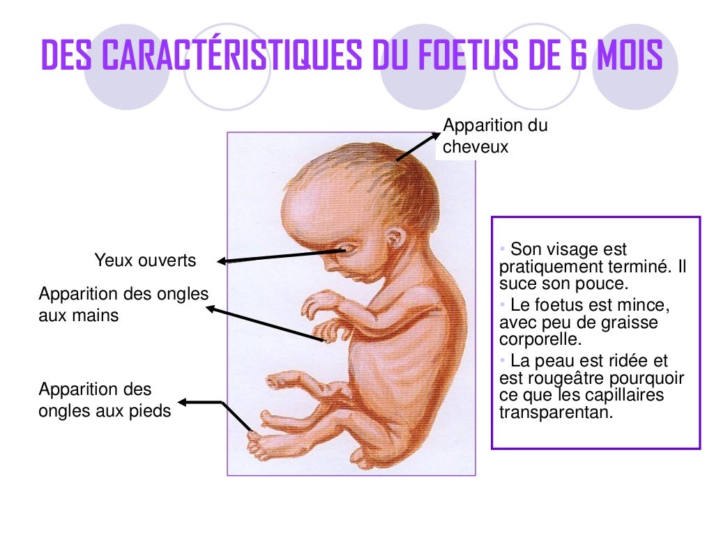 For more information, please visit the links below:
- Chrome by Google
- Firefox by Mozilla
- Microsoft Edge
- Safari by Apple
You are welcome to continue browsing this site with this browser. Some features, tools or interaction may not work correctly.
Ionizing radiation, health effects and protective measures
Ionizing radiation, health effects and protective measures- Healthcare issues »
- A
- B
- B
- G
- D
- E
- and
- 9000 O
- P
- R
- C
- T
- in
- Ф
- x
- C hours
- Sh
- K
- U
- I
- Popular Topics
- Air pollution
- Coronavirus disease (COVID-19)
- Hepatitis
- Data and statistics »
- Newsletter
- The facts are clear
- Publications nine0005
- Find the country »
- A
- B
- B
- G
- D
- E
- and
- L
- N
- O
- P
- With
- T
- U
- F
- x
- Sh
- e
- i
- i
004 b
- WHO in countries »
- Reporting
- Regions »
- Africa
- America
- Southeast Asia
- Europe
- Eastern Mediterranean
- Western Pacific
- Media Center
- Press releases
- Statements
- Media messages nine0005
- Comments
- Reporting
- Online Q&A
- Developments
- Photo reports
- Questions and answers
- Update
- Emergencies "
- News "
- Disease Outbreak News
- WHO data »
- Dashboards »
- COVID-19 Monitoring Dashboard
- Highlights "
- About WHO »
- General director
- About WHO
- WHO activities
- Where does WHO work?
- Governing Bodies »
- World Health Assembly
- Executive committee
- Main page/
- Media Center / nine0004 Newsletters/
- Read more/
- Ionizing radiation, health effects and protective measures
\n
\nAbove certain thresholds, exposure to radiation may impair tissue and/or organ function and may cause acute reactions such as reddening of the skin, hair loss, radiation burns, or acute radiation syndrome.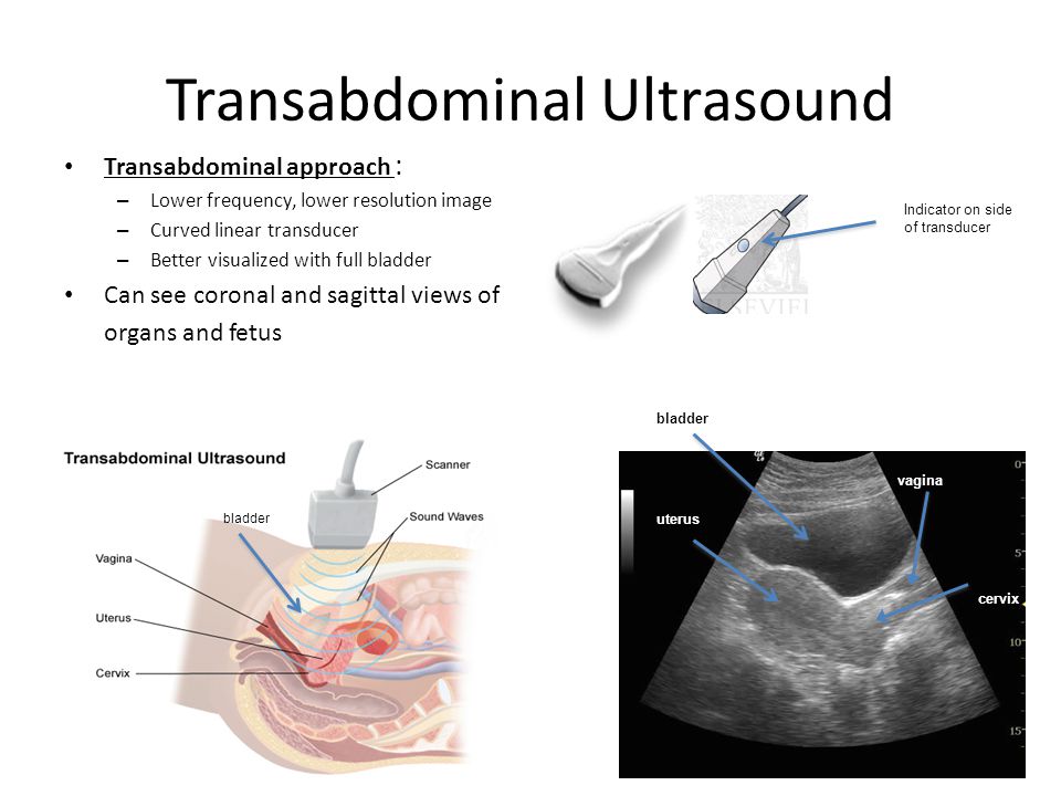 These reactions are stronger at higher doses and higher dose rates. For example, the threshold dose for acute radiation syndrome is approximately 1 Sv (1000 mSv). nine0285
\n
\nIf the dose is low and/or for a long period of time (low dose rate), the risk associated with this is significantly reduced, since in this case the probability of repair of damaged tissues increases. However, there is a risk of long-term consequences, such as cancer that may take years or even decades to appear. Effects of this type do not always appear, but their probability is proportional to the radiation dose. This risk is higher in the case of children and adolescents, as they are much more sensitive to the effects of radiation than adults. nine0285
\n
\nEpidemiological studies in exposed populations, such as atomic bomb survivors or radiotherapy patients, have shown a significant increase in the likelihood of cancer at doses above 100 mSv.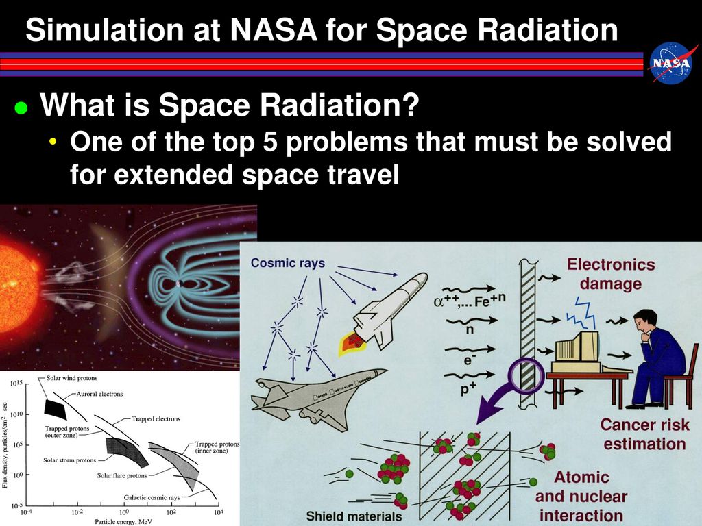 In some cases, more recent epidemiological studies in humans exposed as children for medical purposes (Childhood CT) suggest that the likelihood of cancer may be increased even at lower doses (in the range of 50-100 mSv) . nine0285
\n
\nPrenatal exposure to ionizing radiation can cause fetal brain damage at high doses in excess of 100 mSv between 8 and 15 weeks of gestation and 200 mSv between 16 and 25 weeks of gestation. Human studies have shown that there is no radiation-related risk to fetal brain development before 8 weeks or after 25 weeks of gestation. Epidemiological studies suggest that the risk of developing fetal cancer after exposure to radiation is similar to the risk after exposure to radiation in early childhood. nine0285
\n
WHO activities
\n
\nWHO has developed a radiation program to protect patients, workers and the public from the health risks of radiation exposure in planned, existing and emergency exposures.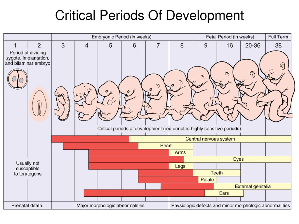 This program, which focuses on public health aspects, covers activities related to exposure risk assessment, management and communication.
\n
\nIn line with its core function of \"setting, promoting and monitoring norms and standards\", WHO collaborates with 7 other international organizations to revise and update international standards for basic safety related to radiation (SBB). WHO adopted new international PRSs in 2012 and is currently working to support the implementation of PRSs in its Member States. nine0285
\n
","datePublished":"2016-04-29T09:30:00.0000000+00:00","image":"https://cdn.who.int/media/images/default -source/imported/radiation/radiation-africa630x420-jpg.jpg?sfvrsn=e8581c1b_10","publisher":{"@type":"Organization","name":"World Health Organization: WHO","logo": {"@type":"ImageObject","url":"https://www.who.int/Images/SchemaOrg/schemaOrgLogo.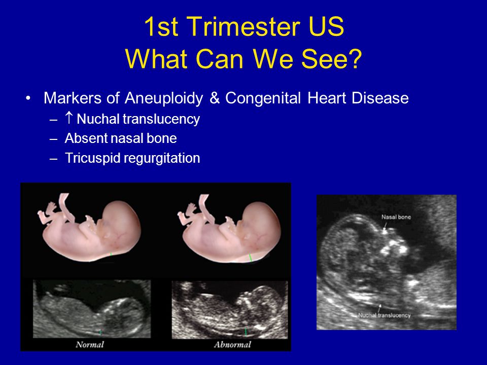 jpg","width":250,"height":60}},"dateModified ":"2016-04-29T09:30:00.0000000+00:00","mainEntityOfPage":"https://www.who.int/ru/news-room/fact-sheets/detail/ionizing-radiation-health -effects-and-protective-measures","@context":"http://schema.org","@type":"Article"}; nine0285
Key Facts
- Ionizing radiation is a form of energy released by atoms in the form of electromagnetic waves or particles.
- People are exposed to natural sources of ionizing radiation such as soil, water, plants, and man-made sources such as X-rays and medical devices.
- Ionizing radiation has numerous useful applications, including medicine, industry, agriculture, and scientific research. nine0005
- As the use of ionizing radiation increases, so does the potential for health hazards if it is used or restricted inappropriately.
- Acute health effects such as skin burn or acute radiation syndrome may occur when the radiation dose exceeds certain levels.
- Low doses of ionizing radiation may increase the risk of longer term effects such as cancer.

What is ionizing radiation? nine0303
Ionizing radiation is a form of energy released by atoms in the form of electromagnetic waves (gamma or x-rays) or particles (neutrons, beta or alpha). The spontaneous decay of atoms is called radioactivity, and the excess energy that results from this is a form of ionizing radiation. Unstable elements formed during decay and emitting ionizing radiation are called radionuclides.
All radionuclides are uniquely identified by the type of radiation they emit, the energy of the radiation, and their half-life. nine0285
Activity, used as a measure of the amount of radionuclide present, is expressed in units called becquerels (Bq): one becquerel is one decay per second. The half-life is the time required for the activity of a radionuclide to decay to half its original value. The half-life of a radioactive element is the time it takes for half of its atoms to decay. It can range from fractions of a second to millions of years (for example, the half-life of iodine-131 is 8 days, and the half-life of carbon-14 is 5730 years).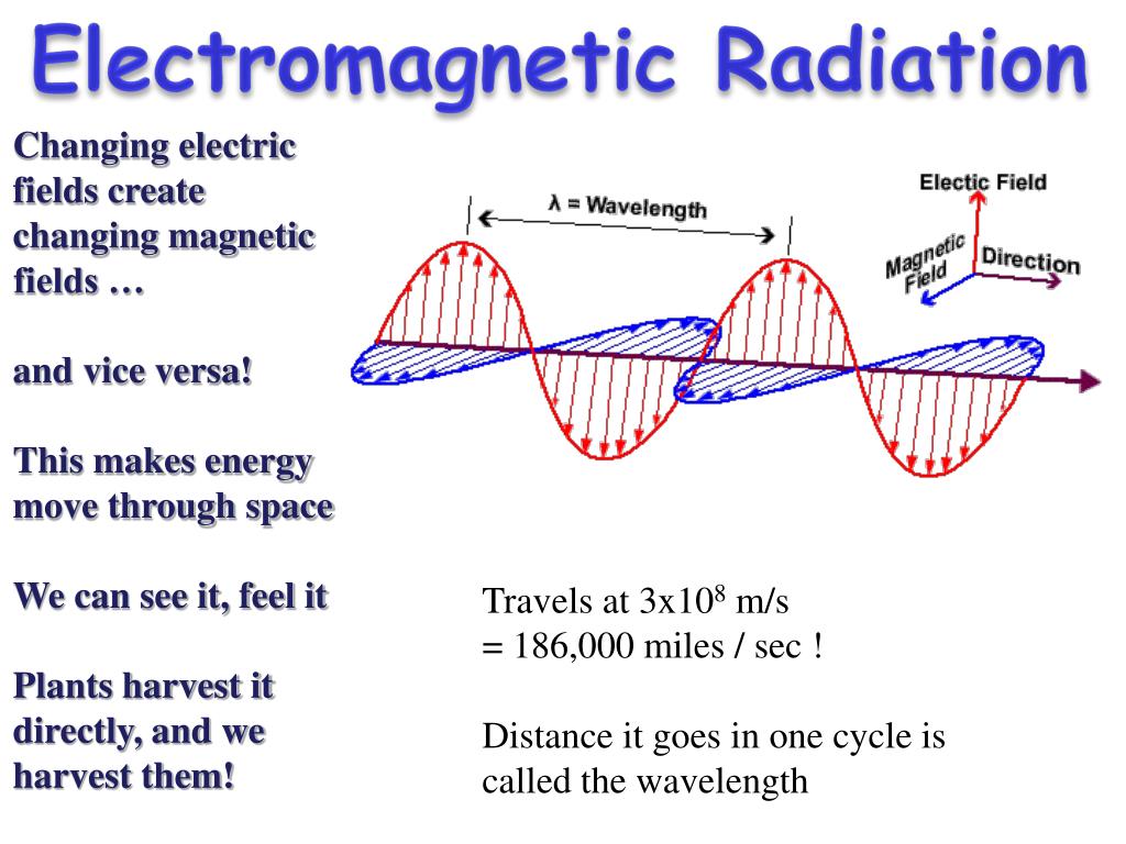 nine0285
Radiation sources
People are exposed to natural and artificial radiation every day. Natural radiation comes from numerous sources, including over 60 naturally occurring radioactive substances in soil, water and air. Radon, a naturally occurring gas, is formed from rocks and soil and is the main source of natural radiation. Every day people inhale and absorb radionuclides from air, food and water.
Humans are also exposed to natural radiation from cosmic rays, especially at high altitudes. On average, 80% of the annual dose that a person receives from background radiation is from naturally occurring terrestrial and space sources of radiation. The levels of such radiation vary in different rheographic zones, and in some areas the level can be 200 times higher than the global average.
Humans are also exposed to radiation from man-made sources, from nuclear power generation to the medical use of radiation diagnosis or treatment. Today, the most common artificial sources of ionizing radiation are medical devices, such as x-ray machines, and other medical devices. nine0285
Exposure to ionizing radiation
Exposure to radiation can be internal or external and can occur in a variety of ways.
Internal exposure to ionizing radiation occurs when radionuclides are inhaled, ingested, or otherwise enter the circulation (eg, injection, injury). Internal exposure stops when the radionuclide is excreted from the body, either spontaneously (with feces) or as a result of treatment. nine0285
External contamination can occur when radioactive material in the air (dust, liquid, aerosols) is deposited on skin or clothing. Such radioactive material can often be removed from the body by simple washing.
Exposure to ionizing radiation may also occur as a result of external radiation from a suitable external source (eg, such as exposure to radiation emitted by medical x-ray equipment). External exposure stops when the radiation source is closed, or when a person goes outside the radiation field. nine0285
People can be exposed to ionizing radiation in a variety of settings: at home or in public places (public exposure), in their workplace (occupational exposure), or in health care settings (patients, caregivers and volunteers).
Exposure to ionizing radiation can be classified into three types of exposure.
The first case is planned exposure, which is due to the intentional use and operation of radiation sources for specific purposes, for example, in the case of the medical use of radiation for the diagnosis or treatment of patients, or the use of radiation in industry or for scientific research purposes. nine0285
The second case is existing sources of exposure, where radiation exposure already exists and for which appropriate control measures need to be taken, such as exposure to radon in homes or workplaces, or exposure to natural background radiation in environmental conditions.
The last case is exposure to emergencies caused by unexpected events requiring prompt action, such as nuclear incidents or malicious acts. nine0285
Medical uses of radiation account for 98% of total radiation dose from all artificial sources; it accounts for 20% of the total impact on the population. There are 3,600 million diagnostic radiological examinations, 37 million nuclear procedures and 7.5 million therapeutic radiotherapy procedures worldwide each year.
Health effects of ionizing radiation
Radiation damage to tissues and/or organs depends on the received radiation dose or absorbed dose, which is expressed in grays (Gy). nine0285
Effective dose is used to measure ionizing radiation in terms of its potential to cause harm. Sievert (Sv) is a unit of effective dose, which takes into account the type of radiation and the sensitivity of tissues and organs. It makes it possible to measure ionizing radiation in terms of the potential for harm. Sv takes into account the type of radiation and the sensitivity of organs and tissues.
Sv is a very large unit, so it is more practical to use smaller units such as millisievert (mSv) or microsievert (µSv).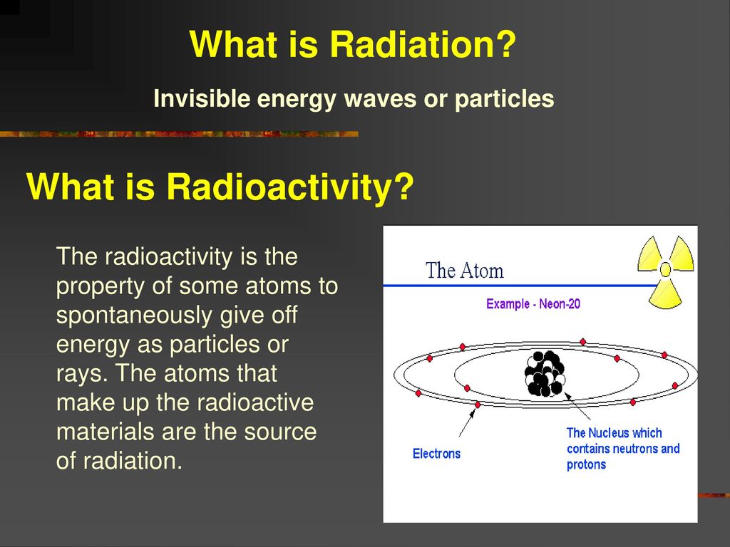 One mSv contains 1000 µSv, and 1000 mSv equals 1 Sv. In addition to the amount of radiation (dose), it is often useful to show the release rate of that dose, such as µSv/hour or mSv/year. nine0285
Above certain thresholds, exposure may impair tissue and/or organ function and may cause acute reactions such as reddening of the skin, hair loss, radiation burns, or acute radiation syndrome. These reactions are stronger at higher doses and higher dose rates. For example, the threshold dose for acute radiation syndrome is approximately 1 Sv (1000 mSv).
If the dose is low and/or a long period of time is applied (low dose rate), the resulting risk is significantly reduced, since in this case the likelihood of repair of damaged tissues increases. However, there is a risk of long-term consequences, such as cancer that may take years or even decades to appear. Effects of this type do not always appear, but their probability is proportional to the radiation dose. This risk is higher in the case of children and adolescents, as they are much more sensitive to the effects of radiation than adults.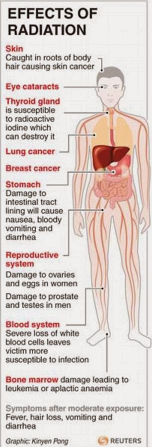 nine0285
Epidemiological studies in exposed populations, such as atomic bomb survivors or radiotherapy patients, have shown a significant increase in the likelihood of cancer at doses above 100 mSv. In some cases, more recent epidemiological studies in humans exposed as children for medical purposes (Childhood CT) suggest that the likelihood of cancer may be increased even at lower doses (in the range of 50-100 mSv) . nine0285
Prenatal exposure to ionizing radiation can cause fetal brain damage at high doses in excess of 100 mSv between 8 and 15 weeks of gestation and 200 mSv between 16 and 25 weeks of gestation. Human studies have shown that there is no radiation-related risk to fetal brain development before 8 weeks or after 25 weeks of gestation. Epidemiological studies suggest that the risk of developing fetal cancer after exposure to radiation is similar to the risk after exposure to radiation in early childhood. nine0285
WHO activities
WHO has developed a radiation program to protect patients, workers, and the public from the health hazards of radiation in planned, existing, and emergency exposures.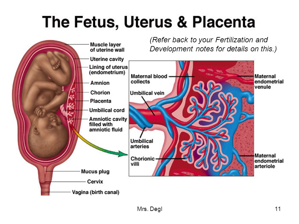 This program, which focuses on public health aspects, covers activities related to exposure risk assessment, management and communication.
Under its core function of "setting, promoting and monitoring norms and standards", WHO is collaborating with 7 other international organizations to revise and update international standards for basic radiation safety (BRS). WHO adopted new international PRSs in 2012 and is currently working to support the implementation of PRSs in its Member States. nine0285
- Consequences of the Chernobyl accident for health
What is radiation and how it affects health
Olga Badrina
9 173
High doses of radiation can destroy cells, tissues and organs and lead to serious consequences: burns, radiation sickness, oncological diseases. In the article, we figure out whether there is a safe dose of radiation, what the health consequences of exposure to radiation can be, and whether it is possible to protect yourself from it. nine0285
Content
What is radiation and how it affects health
Conditions of irradiation
Conditions of irradiation for women
Conditions of irradiation for men
Conditions of irradiation
radiation in medicine
explosionHow to protect yourself from radiation
What is radiation and how it affects health
Radiation is ionizing radiation that is produced when radioactive particles decay. nine0285
A person is in daily contact with radiation. Depending on the origin, its sources are divided into natural, artificial and man-made.
The natural radiation background surrounds a person everywhere: soil, water, air and even space. Every day, people breathe in air or consume a certain amount of radioactive molecules with water and food.
Artificial radiation background is mainly represented by medical radiation sources: x-ray machines, tomographs, fluorography machines, radiopharmaceuticals used for diagnostics and radiation therapy. nine0285
Approximately 80% of the annual dose of radiation a person receives from the environment, the remaining 20% falls on medical procedures: x-rays, computed tomography, and others.
There are also so-called man-made sources of radiation . These include the work of large industries, such as thermal power plants (CHP). In addition, sometimes major accidents at nuclear power plants (NPPs) act as man-made sources.
Depending on how, when and to what extent radiation affects a person, it can be neutral, beneficial or destructive. nine0285
Small doses of radiation to which a person is exposed daily do not affect health in any way, high doses can help cure cancer (radiation therapy), perform surgery on deep-lying tissues (stereotactic surgery) or, on the contrary, destroy healthy tissues.
Factors affecting the scale of potential harm from radiation
How ionizing radiation affects the body depends on many factors: the type of radiation and radioactive isotopes, tissue susceptibility, duration of exposure and some individual characteristics. nine0285
Type of radiation
-
Alpha particles are nuclei that do not penetrate deeper than 0.1 mm (about the thickness of a sheet of paper). The most dangerous when directly ingested with food or water, but cannot penetrate from the outside through the skin.
-
Beta particles are high-energy electrons that can penetrate up to 2 cm deep. Less dangerous than alpha particles, but due to their greater penetrating power, they can destroy the top layer of the skin and subcutaneous tissue, leading to serious burns. nine0285
-
Gamma rays are high-energy particles that can penetrate deep into tissues. A layer of lead is capable of temporarily delaying them. They lead to massive destruction of cells and tissues. It is this type of radiation that is most dangerous in a nuclear explosion.
Cell susceptibility to irradiation
The cells of the bone marrow and germ cells are the most sensitive to the damaging effects of radiation, the least - muscles and bones.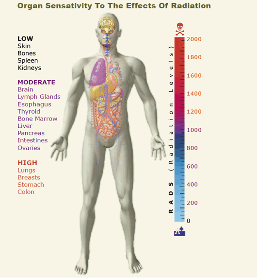
Dose and duration of exposure
A high, rapid single dose is more harmful than the same received in a week or month.
Individual characteristics
The severity of the effects of exposure also depends on age and some concomitant diseases. Thus, children are more susceptible to the effects of radiation than adults. In addition, diabetes and connective tissue diseases (rheumatoid arthritis, systemic lupus erythematosus, and others) can increase the sensitivity of cells to radiation damage. nine0285
Safe dose of radiation
Human exposure to radiation is called exposure.
Different units are used to measure received dose. In medicine, this is usually sievert (Sv) or millisievert (mSv) - the effective equivalent dose received by the whole body in a certain period of time (usually an hour).
In Russia, according to SanPiN, a safe radiation dose is 1 mSv per year, and the maximum dose is 5 mSv per year.

Compare:
- After the explosion at the Chernobyl nuclear power plant, the radiation level reached 2–3 mSv per hour.
- The radiation level 20 km from the Japanese Fukushima-1 nuclear power plant at the time of the accident was 0.161 mSv per hour.
- During a 2-3 hour flight, a person receives an average exposure of 0.02 mSv. The same dose can be obtained by taking 10-15 X-rays per day.
High doses of radiation (for example, above 50 mSv per day) can cause immediate destruction of cells, tissues and organs. Such exposure can be earned if you are close to the site of a nuclear bomb explosion, or at the time of an accident at a nuclear power plant. nine0285
Effects of radiation
Radiation can be neutral, beneficial or harmful. It all depends on the dose and area of irradiation.
So, small doses - up to 5 mSv per year - do not affect health in any way.
A flight from Khabarovsk to Moscow will "cost" a person about 0.
04 mSv of exposure. That's less than one chest x-ray.
Higher doses may help cure cancer if applied locally and for a short time. They are used in radiation therapy for cancer. The health benefits in this case outweigh the potential harm from exposure. nine0285
High doses of radiation can destroy cells, tissues and organs and lead to serious consequences.
Thus, an exposure dose of 1,000 mSv can lead to radiation sickness, 2,000 mSv increases the risk of developing cancer, and 3,000 mSv threatens the life of the irradiated person.
Local radiation injury
Typically, local injury occurs through direct contact with a radiation source, including as a result of radiation therapy in the treatment of cancer. Symptoms depend on the dose received. nine0285
So, with local irradiation, a person can lose hair at the site of exposure, the skin flakes off, and ulcers form on it.
Usually the symptoms of local radiation injury disappear without a trace as soon as the person finishes treatment.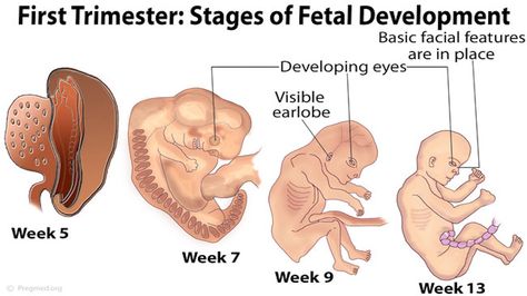
Radiation burns
Burns as a result of exposure to radiation can be mild - I or II degree: at the site of radiation, the skin may turn red, blisters filled with transparent contents appear on it. Such burns are usually accompanied by severe burning pain. nine0285
Very high doses of radiation can cause skin death at the site of radiation, up to damage to muscles and bones.
Radiation sickness
Radiation sickness develops with a single irradiation of 1,000 mSv. Such a dose can be obtained if one is near the site of a nuclear reactor explosion or if one takes 25,000 x-rays or 1,000 roentgens per day.
As a rule, radiation sickness is a consequence of nuclear disasters, it is impossible to get it in ordinary life, even if X-rays or fluorography are done regularly. nine0285
Radiation sickness was diagnosed in most people who caught the nuclear bombing of Hiroshima and Nagasaki and the accident at the Chernobyl nuclear power plant.
Depending on the absorbed dose of radiation, there are three types, or syndromes, of acute radiation sickness: bone marrow, intestinal and cerebral.
Hematopoietic (bone marrow) syndrome develops when exposed to a radiation dose of 700 mSv. As a result, the bone marrow is destroyed, the production of blood cells is disrupted, which makes it harder for the immune system to cope with even harmless infections, and the blood cannot clot properly. nine0285
Gastrointestinal (intestinal) syndrome occurs when exposed to about 10,000 mSv. In addition to the bone marrow, the digestive tract is also affected. As a result, dehydration occurs, electrolyte balance is disturbed, and severe infectious diseases develop. Death usually occurs within 2 weeks of exposure.
Cerebrovascular (cerebral) syndrome begins with exposure to 20,000 mSv. The production of blood cells is disrupted, intracranial pressure increases, damage to the brain and spinal cord develops.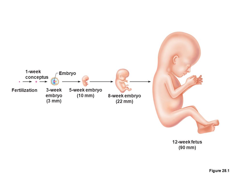 Death occurs within 3 days. nine0285
Regardless of the type of radiation sickness, it goes through three successive stages.
Stages of radiation sickness:
- Initial - primary symptoms (nausea, loss of appetite, vomiting, fatigue, diarrhea), which can occur both a few minutes and a few days after exposure.
- Asymptomatic - latent period. At this stage, a person gets better dramatically, he can look healthy for several hours or even weeks. nine0005
- Stage of peak (bright clinical manifestations) - specific symptoms develop that are characteristic of a particular radiation sickness syndrome. So, with bone marrow syndrome, massive poorly controlled bleeding and fever are observed, and with intestinal syndrome - dizziness, loss of consciousness and even coma.
After a period of vivid clinical manifestations, a person can either recover or die. It all depends on the dose of radiation and the state of health of the victim.
Stochastic effects
Stochastic, or so-called probable effects, are the consequences of exposure that do not have an exact dose threshold and may appear years after exposure to radiation.
Common stochastic effects:
- cancer,
- genetic mutations.
Under the influence of radiation, potentially carcinogenic particles are formed in the body - free radicals, which can damage the genetic material of cells. As a result, cells can begin to divide and grow uncontrollably, forming tumors. nine0285
It is known that 10 years after the nuclear bombing of Hiroshima and Nagasaki, cases of thyroid, breast and intestinal cancer have increased.
Considering that oncological diseases are related to the stochastic effects of radiation, it is difficult to identify a direct relationship between radiation dose and the occurrence of cancer (however, there are studies confirming it). In addition, when analyzing the causes of oncology, it is impossible to separate the influence of exposure itself and lifestyle, heredity, viruses and other environmental factors. nine0285
Consequences of exposure for women
Women who have been exposed to radiation are more likely to have chronic inflammatory diseases of the pelvic organs, as well as obstetric complications (ectopic pregnancy, placental insufficiency, preeclampsia, premature birth, miscarriage, stillbirth).
In addition, exposure to radiation during the 8th to 25th week of pregnancy can lead to impaired mental development of the fetus and fetal malformations.
At doses below 0.1 mSv, which are usually used during routine preventive examinations during childbearing, the risk of such complications does not increase. nine0285
Consequences of irradiation for men
In men who have been exposed to radiation, inflammatory and functional diseases of the reproductive system are more often recorded:
- varicocele - varicose veins of the testicle and spermatic cord;
- orchitis - inflammation of the testicle;
- prostatitis - inflammation of the prostate gland;
- erectile dysfunction.
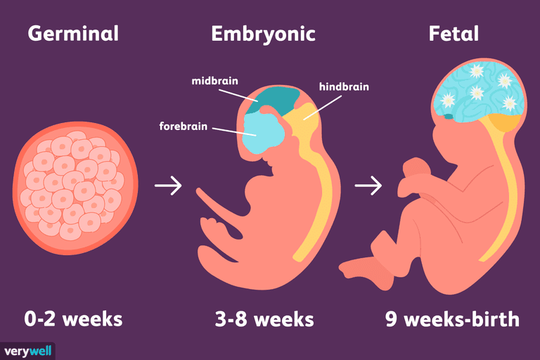
Exposure effects for children
The brain, lens of the eye and thyroid gland in children are more sensitive to radiation than in adults. The reasons for this are not fully understood, but doctors believe that the increased sensitivity of certain tissues in children is due to the high rate of cell growth and division. nine0285
Theoretically, genetic effects are also possible, but even among the 78,000 Japanese children who survived the atomic bombing of Hiroshima and Nagasaki, no increase in the number of cases of hereditary diseases was found.
Radiation in medicine
According to the requirements set out in SanPiN 2.6.1.1192-03, when performing preventive medical imaging procedures, which include x-rays, computed tomography, fluorography, and others, the radiation dose should not exceed 1 mSv per year. nine0285
This dose of radiation can be obtained if done in a year:
- 500 x-rays of the arm or leg,
- 80 x-rays of the jaw,
- 20 CT scans.
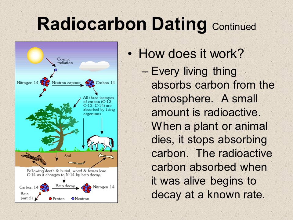
Even if one x-ray of the hand is taken daily for the whole year, plus 2 CT scans and 2 x-rays of the jaw are added to this, the exposure will still not exceed the permitted safe doses.
Medical Imaging
Medical imaging techniques include: radiography, computed tomography, fluorography, mammography, positron emission tomography, scintigraphy.
Radiography is a method of examining internal organs and bones using X-rays. As a result of radiography, an accurate image of the object being photographed is projected onto a special film or paper.
A simple x-ray of an arm or leg exposes a person to a radiation dose of 0.01 mSv on average
The dose of radiation to which a person is exposed during an X-ray is equivalent to several days or several years of exposure to natural radiation from the environment. The exact dose depends on the method of examination and the site of the body.
Fluorography is a type of radiography.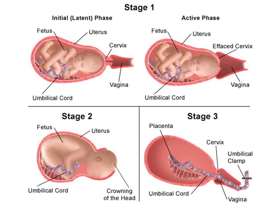 During the examination, the specialist takes an x-ray of the chest and lungs.
The dose of radiation that a person is exposed to during the procedure is higher than during x-rays, but does not harm health. nine0285
For one fluorography procedure, a person receives a radiation dose of not more than 0.1 mSv
Mammography is an X-ray method for examining breast tumors.
The dose of radiation that a person is exposed to during mammography is higher than during radiography and fluorography, but it is still safe for health.
Typically, a person receives a radiation dose of 0.4 mSv during one mammography procedure with scanning of two mammary glands
Scintigraphy, computed tomography and positron emission tomography are the so-called advanced X-ray examinations, because as a result, the doctor receives not a flat image, but a three-dimensional model.
Scintigraphy is an examination method that provides two-dimensional images.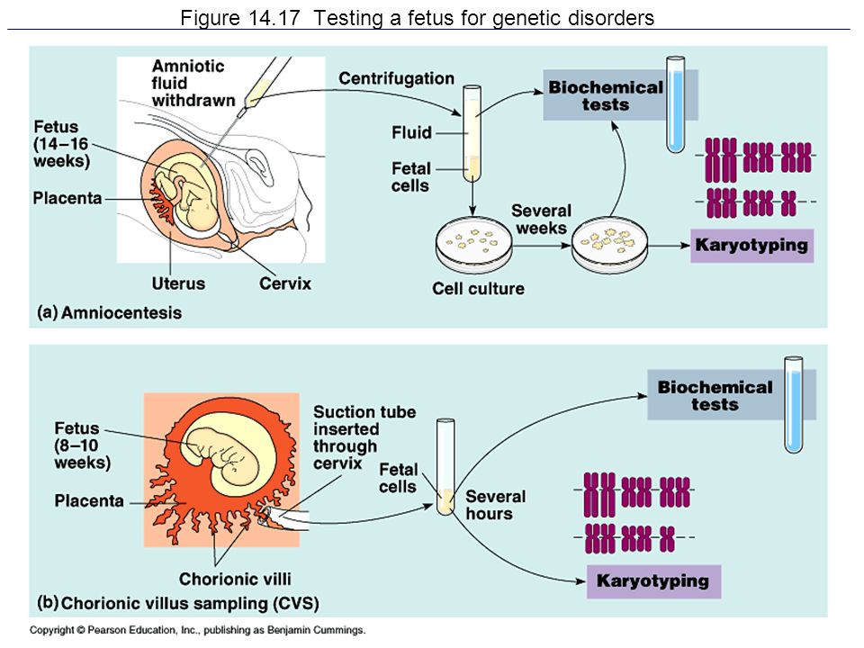 Computed and positron emission tomography are combined studies in which a computer processes several x-ray images at once and obtains a three-dimensional picture. nine0285
The radiation from a CT scan is between 0.4 and 0.7 mSv for a chest exam, and slightly higher for an abdominal scan . An X-ray tube is laid along the body of the tomograph, the rays from which pass through soft tissues, and the resulting images are transmitted to a computer. The computer processes the images, resulting in three-dimensional layered images. nine0285
Radiotherapy
Radiation therapy or radiotherapy is a treatment used to treat malignant tumors. In the course of radiation therapy with the help of radiation, tumor cells are targeted to destroy.
Radiation damages the DNA of cells, after which they lose the ability to divide and die. The course of treatment consists of several sessions that last from 5 to 15 minutes.
Radiation dose for radiation therapy depends on the location of the tumor and the stage of the cancer
Radiation therapy affects not only malignant, but also healthy cells that are located near the tumor, as well as healthy tissues through which the beam passes. As a rule, after treatment, the affected tissues are restored on their own.
Who should not be exposed to radiation
High doses of radiation are dangerous for all people, but some people are prohibited from even acceptable doses of radiation for medical procedures.
Radiography is strictly prohibited in the first half of pregnancy. nine0448Thus, radiography is strictly prohibited in the first half of pregnancy, except in cases where the risk to the mother outweighs the benefit to the child. For example, when the issue of abortion is being decided or a pregnant woman needs urgent help.
What threatens an accident at a nuclear power plant or a nuclear explosion
The consequences of such accidents will greatly depend on many factors, for example, on whether the nuclear power plant reactor is turned on or off, on the power of the station or a nuclear bomb, weather conditions.
The main danger is the release of radioactive elements: iodine, cesium, strontium, plutonium and their decay products.
nine0285
Iodine is the most volatile element, with a half-life of up to 8 days. All this time, it threatens people's health. The fact is that iodine can accumulate in the thyroid gland and lead to the formation of malignant tumors.
Thyroid cancer can develop without symptoms for years, so doctors recommend checking it regularly. Laboratory studies of the level of thyroid hormones and hypothalamus are suitable for this.
T3 common
How to protect yourself from radiation
According to WHO recommendations, there are three main ways to protect yourself from radiation:
- time,
- distance,
- shielding.
The less time a person is in a zone of strong radiation and the farther he is from him, the weaker the potential harm to health. Shielding involves the use of special protective screens, such as lead bunkers, through which radioactive particles do not penetrate. nine0285
If an accident occurs, it will be reported centrally: by radio, television and other communication channels.
In some cases, people may be asked to take potassium iodide supplements urgently.
To avoid thyroid damage during an emergency, WHO recommends drinking potassium iodide tablets, Lugol's solution, or 5% tincture of iodine.
Iodine fills the thyroid gland and does not allow radioactive iodine to be deposited. As a result, the dangerous element is excreted from the body with urine. nine0285
Iodine protects only the thyroid gland and does not protect against the destructive effects of radioactive elements on the skin and internal organs.
Drinking iodine just for prevention is not worth it, it is dangerous to health.Why can't I take iodine preparations for prophylaxis?
You should take iodine preparations to prevent radiation damage to the thyroid gland only once and after the announcement of an emergency, and not in advance. The fact is that such "prevention" can lead to inflammation of the thyroid gland - thyroiditis, the development of goiter and hyperthyroidism, in which the thyroid gland begins to secrete a large amount of hormones.
nine0285
A single dose of potassium iodide protects the thyroid gland for about a day. Do not take the drug again only after the recommendation of local health authorities.
Information checked
medical expertHandbook Diseases
Rate this article:
1
2
3
4
5
6
7
8 286 10
Useful article? Share on social networks:
IMPORTANT
The information in this section cannot serve as a sufficient basis for making a diagnosis or prescribing treatment. The decision on this should be made by the doctor on the basis of all the data available to him.
You may be interested
Peptic ulcer of the stomach and duodenum
Syphilis
Trichomoniasis
Ureaplasmosis
Gonorrhea (clap)
Gilbert's syndrome
Hereditary benign liver disease.

Learn more

 174, “Preconception and Prenatal Radiation Exposure: Health Effects and Protective Guidance” [NCRP2013].