Scan 15 weeks
15 Weeks Pregnant Ultrasound: Procedure, Abnormalities and More
Pregnant women find that they tend to get to grips with the situation by the 15th week of pregnancy, as most symptoms, which cause irritation, tend to subside by then. Gone are the morning sickness, constant nausea, fatigue, and general irritability. Still, the most important thing that expectant mothers fuss about is the health of their growing baby. The child will be growing within her, and scans are the only way for them to check up on how the foetus is developing. This is why the 15-week ultrasound scan is so important for the mothers. Read on to know the developmental signs and changes you will observe in the 15th week of ultrasound.
15-Week Ultrasound – Why You Need It
There are many reasons why a mother needs to know about the growth of her baby during the second trimester.
- Doctors do this scan to see if the baby is growing appropriately.
- You can see how much the baby has grown by this time.
- You might get to see a few expressions on the baby’s face. Nothing makes a mother happier than seeing her child smile (or frown) for the first time.
How to Prepare for the Scan
Preparing for this ultrasound scan does not include anything considerably different, compared to the earlier tests. Like in all ultrasound scans, you will be encouraged to drink a lot of water beforehand. This ensures that you have a full bladder, which will help the technician to get clear images of the child. So you can start preparing by drinking at least a litre (or more) of water 1-2 hours before the process is to begin so that you have the urge to urinate at the time of the scan. Also, you should not urinate again until after the test is complete.
15-Week Ultrasound – How Long It Takes to Complete the Scan
The scanning process is usually done within ten to fifteen minutes, as all ultrasound scans do not take much time. If there are any technical malfunctions, the process may take longer. However, in most cases, the test gets done within half an hour or so.
15-Week Ultrasound – How It Is Performed
Usually, the ultrasound examinations for pregnant women do not include any invasive procedures. You will be made to lie on your back, with your belly showing. The technician will apply jelly all over the stomach, for reducing friction while moving the probe. The probe is then pressed on top of your belly and moved around to get real-time images on a computer screen. The technician takes snapshots when he/she gets some images. The entire process does not last long unless the images are not clear enough. In such cases, a plastic probe emitting ultrasound waves will be inserted into your vagina, so that the technician can get clear images of the child. In all cases, the test does not take more than half an hour to complete.
15-Week Ultrasound – What Can Be Seen
The images of a baby at 15 weeks ultrasound scan can tell you and the doctor a lot about how the baby has been growing. You will be able to watch your child make a lot of funny expressions during the scan – the baby will grimace, frown, smile, and even squint. This is just the child trying out his new, developing muscles. You might even be able to see the child swallowing or sucking his thumb.
Arms and legs could be moving, too. The images will show the skin of the child, which will have a paper-like consistency. The blood vessels running underneath will be visible. The scalp of the child, along with the eyebrows is also visible at this stage. Your child may already have hair on his head. This temporary, fine hair is called the lanugo.
The skeletal system of the child is still developing, as the cartilage in place slowly transforms into the harder bone. However, worry not; the bones still stay flexible enough so that the child can pass through the birth canal without difficulty. They do not harden enough for the child to stand until he is a toddler. He might also be able to turn his head and make fists at this time.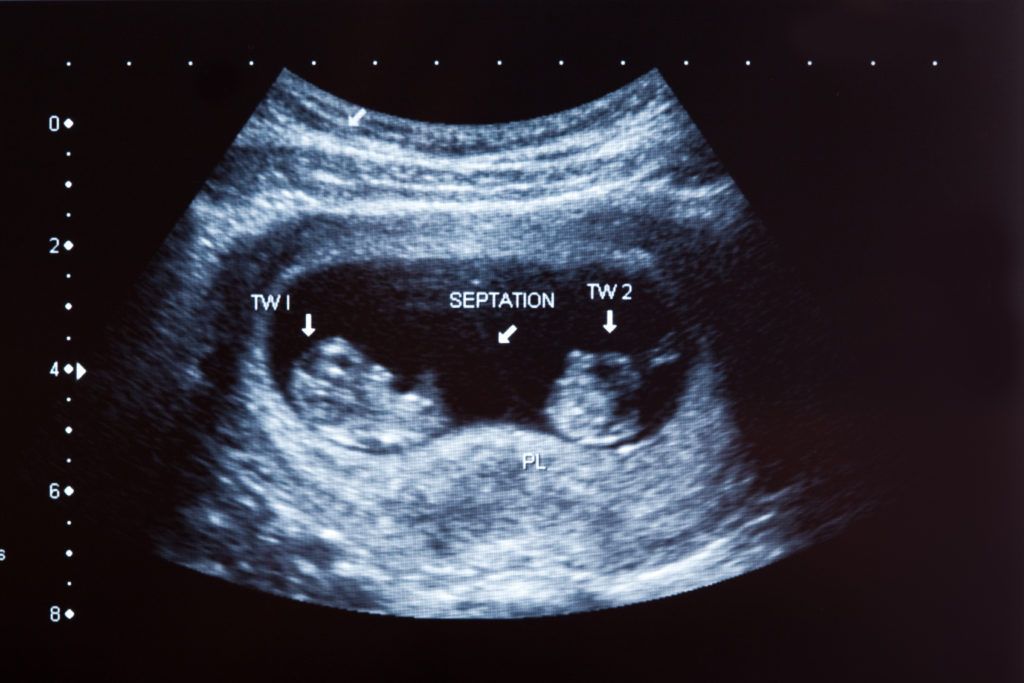 The development of external genitalia will also be present, so the doctor will be able to guess the gender of the baby with almost 80% accuracy.
15-Week Ultrasound – Identifying Abnormalities
The 15-week foetus ultrasound scan definitely gives the doctor a clear idea about how the baby has been growing, so he might advise you to have further tests if he detects anything out of place. Although the 15 week 3D ultrasound scan is quite comprehensive, it is still not enough to be sure.
There are no routine tests scheduled for mothers at this time of pregnancy, but women above the age of 35, who are pregnant may benefit from doing an amniocentesis and multiple marker screening tests. The former uses a sample of amniotic fluid from your womb, in order to check for genetic abnormalities in the child.
An ultrasound scan at the 15th week of pregnancy can really benefit both the mother and the child. The doctor can see how well the child has been growing, while the mother gets to see the child make expressions for the first time.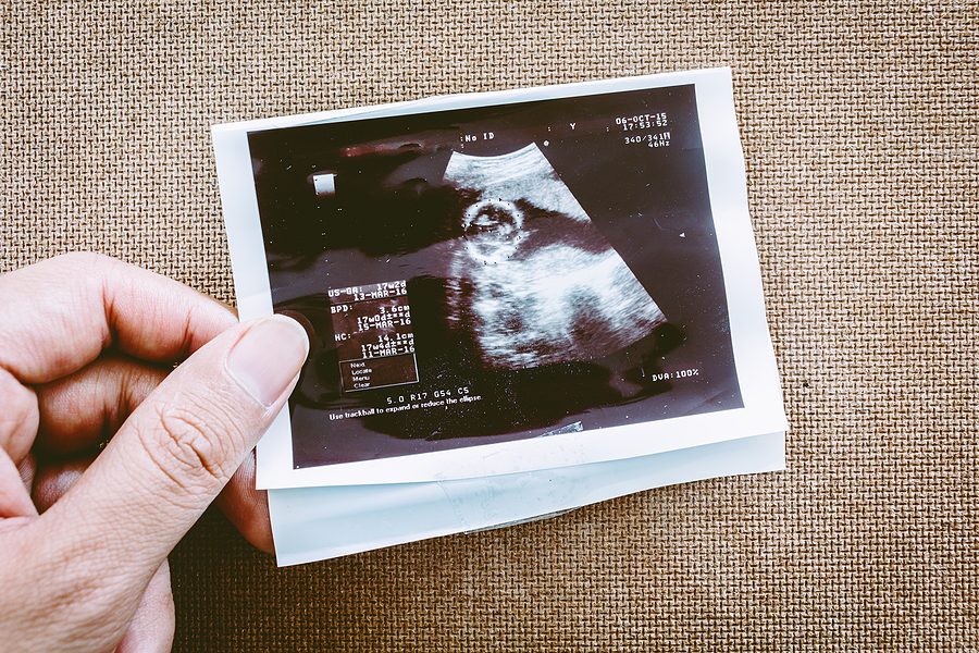 It can also help mothers detect any sort of abnormality in the development of the child at an early stage, so the scan at this time is a highly recommended one.
Previous Week: 14 Weeks Pregnant Ultrasound
Next Week: 16 Weeks Pregnant Ultrasound
12-14 Week Scan - Women's Imaging
This scan should be ideally performed between 12 weeks 5 days and 13 weeks 6 days of your pregnancy. Women’s Imaging conducts a detailed risk assessment for your baby in accordance with the Fetal Medicine Foundation. As well as checking that your baby is growing well and confirming your due date the main aim of the scan includes:
- To see if there are any structural abnormalities. Some can already be identified or suspected at this stage so a normal appearing scan is very reassuring for you.
- To check your uterus and ovaries to ensure they won’t be an issue during the pregnancy.
- To confirm the growth of the pregnancy and the due date.
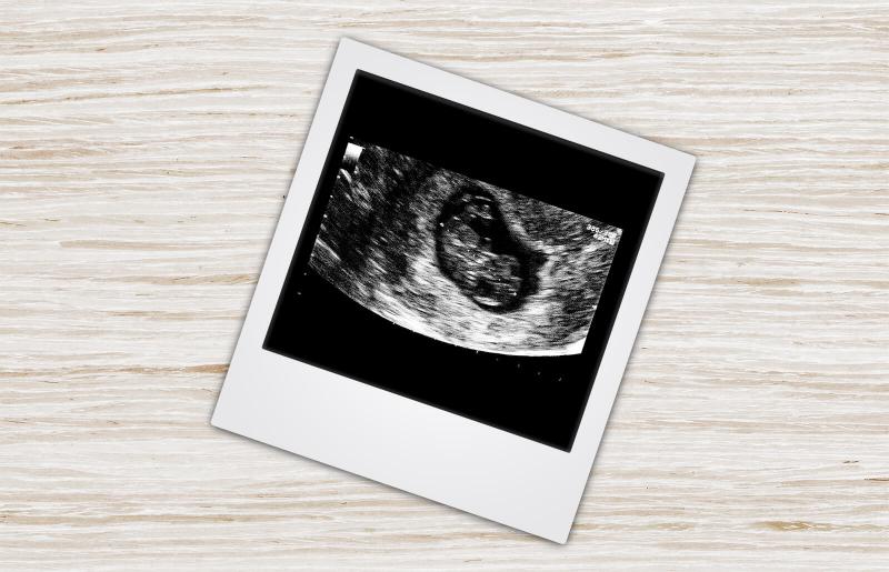
- To check where the placenta is lying, where the umbilical cord is in relation to the placenta and if there is sufficient fluid around the baby.
Risks
Ultrasound is safe to use throughout your pregnancy.
Sometimes we need to do a vaginal scan. If you are allergic to latex prior to the vaginal scan or you don’t know then a latex-free cover will be used on the probe.
Occasionally there is some discomfort from probe pressure on a full bladder or from the vaginal probe manipulation. If this is extremely painful please let us know.
Benefits
You will be able to see all of your developing baby
We are able to take some important measurements which allows us to give you an accurate risk assessment for your baby.
You have the option of getting the results of the scan on the day by our consultant. Please let reception know at the time of booking.
What is an ultrasound?
An ultrasound scan uses high-frequency sound waves to create images of the inside of your body and baby. Sound waves are used instead of radiation which makes them safe.
What do I need to do to have a risk assessment for my baby?
You will need to have a blood test done before coming to Women’s Imaging for your ultrasound scan. Your doctor will organise this for you. Ideally you should have your 1st Trimester bloods/ Maternal Serum done at 11 weeks gestation. Your doctor will organise a form for you.
We like to scan you between 12.5 and 13.6 weeks of pregnancy.
Why is a 12-14 weeks scan different at Women’s Imaging?
We offer the most sensitive and comprehensive risk assessment for your baby between 12.5-13.6 weeks which includes pre-eclampsia and fetal growth restriction. We are currently the only practice in Tasmania that does this. Traditional chromosomal abnormalities such as Down’s Syndrome continue to be assessed. Our sonographers are trained to the highest standards to perform these specialist measurements of you and your baby. They hold Certificates in Competence from the Fetal Medicine Foundation which is recognised by the Royal Australian and New Zealand College of Obstetricians and Gynaecologists.
We measure:
- Your baby’s nasal bone
- The thin layer of fluid at the back of the baby’s neck (the nuchal translucency)
- Your blood pressure (we take 2 measurements from both arms)
- The blood flow between you and your baby
We also examine:
- Your blood results
- Your age
- Your weight
- Whether you have had any previous problems in pregnancies (if relevant).
When you choose to have your First Trimester scan with us, you can be confident that the risk assessment results you receive are the best available in obstetric scanning today.
Women’s Imaging provides a comforting and safe environment with the latest state of the art equipment to ensure that you have the best possible care and experience.
What happens on the day?
When you arrive for your scan you will be asked to fill out a form about your pregnancy to date.
You will then have your blood pressure taken on both arms and your height and weight will be measured if you are not sure.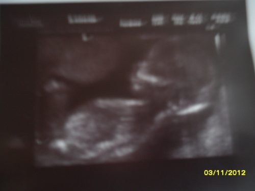 Sometimes your measurements are taken before you have your scan and sometimes after.
When the sonographer takes you through to the scanning room you will be asked to lie on the table and expose your tummy. A towel will be tucked into your pants to limit spread of the gel onto your clothes.
Clear gel is applied to your tummy and the sonographer moves the probe over your tummy recording images. The clear gel is water-soluble so it won’t stain your clothes. It’s just a little sticky!
Please come with a full bladder which will make it easier to obtain images of your baby. A full bladder pushes the uterus away from your lower pelvis thereby making it easier for us to see and scan your baby.
We will take measurements of your baby and check your placenta and ovaries.
Occasionally a vaginal scan is also performed to give us a better view of your baby. You will be able to watch the entire scan on our wall-mounted plasma screen monitors!
Once the scan is completed the sonographer will leave the room.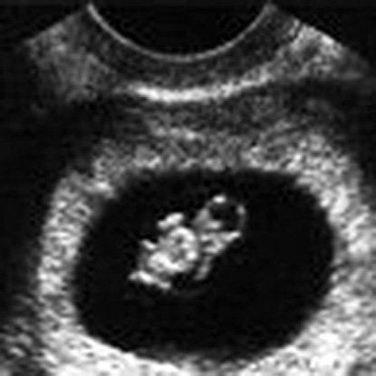 All of your measurements including your blood pressure and blood results will be reviewed by our radiologist or on-site obstetrician. Results will be sent to your referring doctor and they will give them to you at your next appointment.
You will be given your expected due date on the day and you will be given a online access card to view your images through our website.
How long will it take?
The 12-14 week scan takes approximately 45 minutes.
The 12-14 week scan and a consultation with our doctor on the day takes approximately 60 minutes.
(Please allow some extra time in your schedule when you come to see us for a 12-14 week scan. Occasionally we have to recalculate your measurements and this can take a few extra minutes)
fetal ultrasound at 15 weeks
- Fetal development by week
- Our apparatus
- other types...
- Pregnancy management
- Fetometry data at various times
The cost of ultrasound in the second trimester in the period from 14 to 26 weeks is 550 hryvnia. The price includes prenatal screening, biometric protocols, 3D/4D visualization. The cost of comprehensive prenatal screening according to PRISCA (ultrasound + free estriol + alpha-fetoprotein + beta-hCG with the calculation of the individual risk of chromosomal pathologies (for example, Down syndrome or Edwards syndrome) and developmental defects (for example, neural tube defect) - 1060 hryvnia.
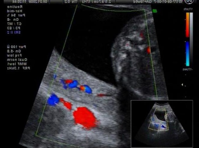
With fetal ultrasound at 15 weeks gestation , you can see how the baby actively moves its legs and kicks with its elbows, while you still do not feel it, the fetus is still small so that you can feel its tremors. The dimensions of the ultrasound of the fetus at 15 weeks of gestation approximately correspond to the average orange. Your baby can already hear your heartbeat and your voice. He is already ready to hear how much he is loved by you. This important achievement is possible due to the fact that the ears of the fetus are shifted to the sides, where they should be. With a normal fetal ultrasound at 15 weeks of gestation, it is not so easy to see the ears, but it is possible when performing 3d 4 d ultrasound during pregnancy. Fetal eyes at Fetal ultrasound at 15 weeks Pregnancy shifts forward to its usual place. In the fetus, eyebrows and eyelashes are stained, the hair on the head becomes thicker. A fetal ultrasound at 15 weeks gestation shows the baby moving its fingers and toes, making spontaneous breathing movements, preparing for life outside the womb. Of course, the fetus is still too immature for independent life, it still desperately needs its cozy home in your stomach. The fruit is already completely covered with lanugo - fluffy hairs that warm it and make it very cute. Lanugo lasts up to approximately 35 weeks of gestation.
The heart during ultrasound of the fetus at 15 weeks of pregnancy beats at a speed of 140-160 beats per minute and pumps 28 liters of blood per day!
The hair on the head takes on color, it is already possible to understand whether the father's forelock or mother's forelock has grown in the child.
In addition to breathing movements, with ultrasound of the fetus at 15 weeks of pregnancy, you can see how the child is intensively training for the main debut in his life - going out into the world. The fetus sucks fingers, actively swallows amniotic fluid - important skills for survival in the external environment.
What else needs to be checked during fetal ultrasound at 15 weeks of gestation?
Woman's cervix on fetal ultrasound at 15 weeks gestation
It is important to assess the condition of the cervix on fetal ultrasound at 15 weeks gestation. It is at this time that the first signs of isthmic-cervical insufficiency most often debut. Isthmic-cervical insufficiency is a problem of the cervix associated with the failure of the obturator function of the cervix. Roughly speaking, the cervix cannot hold the fetus inside the uterus, it opens ahead of time, and a miscarriage occurs.
The main causes of isthmic-cervical insufficiency:
- Traumatic gynecological operations (curettage, abortion, biopsy)
- Sexually transmitted infections - chlamydia, ureaplasmosis, mycoplasmosis, etc.
 - and associated chronic endocervicitis (inflammation inside the cervix).
 - Cicatricial deformity of the cervix after previous difficult labor with cervical ruptures.
Woman's ovaries on fetal ultrasound at 15 weeks gestation
A woman's ovaries are also evaluated during fetal ultrasound at 15 weeks' gestation. It is not always possible to evaluate unaltered ovaries (if everything is in order with the ovaries). Their visualization is difficult due to the enlarged pregnant uterus.
Uterus of a woman with fetal ultrasound at 15 weeks of pregnancy
In the structure of the uterus, the homogeneity of the muscle layer (myometrium) is necessarily assessed, where myomatous nodes can be visualized. Normally, with ultrasound of the uterus at 15 weeks of gestation, the myometrium is represented by a thin, about 2.5 cm, homogeneous structure.
Placenta on fetal ultrasound at 15 weeks gestation
Placenta on fetal ultrasound at 15 weeks gestation may have different locations.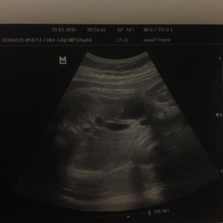 Most often, it is located on the anterior or posterior wall of the uterus. Sometimes the placenta reaches the cervix or overlaps the cervix. This location of the placenta on ultrasound of the fetus at 15 weeks of gestation is called placenta previa. This situation requires constant medical supervision. With placenta previa, there is a need to frequently undergo inpatient preventive treatment. Complete placenta previa will generally require almost the entire pregnancy after the diagnosis of bed rest, more often in a hospital setting. This tactic is due to the fact that the placenta during presentation is located in the most mobile and extensible part of the uterus - in the lower segment. This localization is dangerous by the threat of premature placental abruption, intrauterine fetal death and fatal uterine bleeding. Migration of the placenta (moving the placenta above the danger zone) takes place until the 28th week, but if an ultrasound at 15 weeks of gestation diagnoses complete placenta previa, the likelihood that the placenta will remain in this place is very high.
Fetal ultrasound at 15 weeks
Fetal ultrasound at 15 weeks gestation The heart has four chambers: two atria and two ventricles, just like in an adult. A feature of the fetal heart is the presence of an open oval window through which blood is drained. The foramen ovale will close after the first breath, but in some people it does not close completely. There is a complete septum between the atria in the normal ultrasound of the fetus at 15 weeks of gestation.
CNS on fetal ultrasound at 15 weeks gestation
The brain is fully assessed at fetal ultrasound at 15 weeks gestation . The ventricles of the brain, the cerebellum, the division of the brain into two hemispheres are evaluated.
Other organs during fetal ultrasound at 15 weeks of gestation
Presence and correct location of the stomach, bladder, kidneys, and intestines must be assessed during fetal ultrasound at 15 weeks of gestation.
Changes in the body of the expectant mother
Your heart is pumping 20% more blood at this stage of pregnancy than before pregnancy. Most often, a healthy woman feels completely normal. The compensatory capacity of the body is great.
The fundus of the uterus is located in the middle between the pubis and the navel. You can check this yourself if you lie on your back and gently touch your stomach with your palm. So even without an ultrasound of the fetus at 15 weeks of gestation, you can be calm that the pregnancy is developing.
Hormones make your gums vulnerable. You need to take better care of your oral hygiene to prevent bacterial gum disease. It's time to visit the dentist if you didn't have time to do it before pregnancy.
read more: 16th week of pregnancy
- Hydrotubation (echohydrotubation): examination of the patency of the fallopian tubes (ultrasonic hysterosalpingoscopy)
- Transvaginal
- Ovaries
- Uterus
- Mammary glands
Female ultrasound
- Duplex Scan
- Cerebral vessels
- Neck vessels (duplex angioscanning of the main arteries of the head)
- Veins of the lower extremities
Vascular ultrasound
- Transrectal (trusion): prostate
- Scrotum (testicles)
- Vessels of the penis
Male ultrasound
- Appendicitis
- Abdomen
- Gallbladder
- Stomach
- Intestines
- Bladder
- Soft tissue
- Pancreas
- Liver
- Kidney
- Joints
- Thyroid
- Echocardiography (ultrasound of the heart)
Organ ultrasound
- Varicose veins: ultrasound diagnosis of varicose veins
- Hypertension: Ultrasound diagnosis of hypertension
- Thrombosis: Ultrasound diagnosis of venous thrombosis
- Ultrasound diagnosis of chronic pancreatitis
- for kidney stones
- for cholecystitis
Ultrasound diagnostics of diseases
- Hip joints in newborns (if hip dysplasia is suspected)
Pediatric ultrasound
Ultrasound during pregnancy.
 Features, description of the method
 HomeArticlesUltrasound during pregnancy. Features, description of method
Prenatal ultrasound is a non-invasive diagnostic test that uses sound waves to create a visual image of the fetus, placenta, uterus, and other pelvic organs of a pregnant woman. Allows the doctor to obtain valuable information about the course of pregnancy and the health of the baby. During the procedure, an ultrasound machine or scanner transmits high-frequency sound waves. They pass through the uterus, are reflected from the baby. The computer captures the information received, translates it into a video image format that shows the shape, position and movements of the child. Rigid tissues, such as bones, appear white in the image, as they reflect a large number of sound waves. Soft tissues appear grey. The fluids surrounding the fetus appear black on the screen because the sound waves simply pass through them and are not reflected.
Timing of the procedure
The pregnancy doctor determines when a fetal ultrasound is required.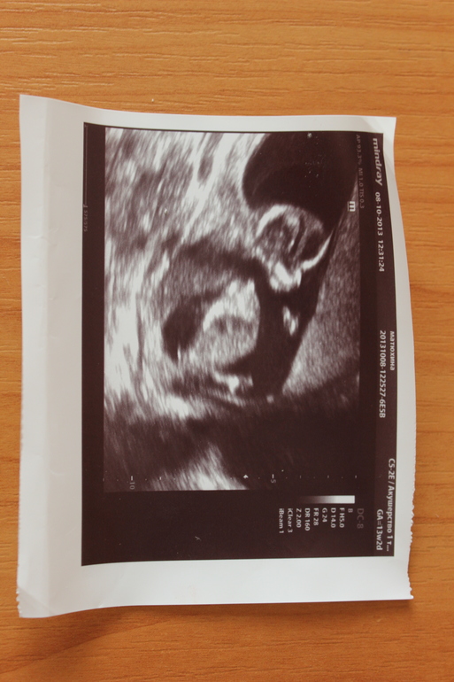
- As a rule, the first ultrasound is prescribed at 10-14 weeks of pregnancy. The purpose of the first scan is to check how many children a woman is carrying, whether they are developing normally. The procedure during the first trimester provides information about the basic anatomy of the head, abdominal wall, limbs, and checks whether the baby's heart is beating. The first ultrasound examination gives parents and the doctor a first impression of the baby. The resulting image can be printed and saved as a keepsake.
- 19-23 weeks. The size, gender of the fetus is revealed. There is an assessment of the growth rate of the child, the development of internal organs. The presence or absence of chromosomal abnormalities can already be said at this stage.
- 32-36 weeks. It is carried out for the purpose of diagnosing late anomalies. The date of birth is specified.
Purpose
Depending on the stage of pregnancy, an ultrasound examination is used to obtain the following information:
- Check that the baby's heart is beating
- Determine if a woman is pregnant with one child, twins or more
- Detect ectopic pregnancy (when the embryo begins to develop outside the uterus)
- Find cause of bleeding if present
- Accurately date pregnancy by measuring baby
- Assess the risk of having a baby with Down syndrome by measuring the fluid in the back of the neck of an embryo at 11-14 weeks of gestation
- Determine cause of abnormal blood test
- Assess the position of the baby, placenta, amount of amniotic fluid
- Examine the child, make sure that the organs are normal
- Diagnose violations
- Measure the child's growth rate based on previous scans.
- Determination of the sex of the child. In some cases, the child assumes an awkward position, and it is difficult to say a boy or a girl.
Procedure
No special training required. Your doctor may ask you to drink several glasses of water to keep your bladder full. This will help the specialist see the child better. The patient lies on his back on a chair. A special gel is applied to the abdomen to improve the conduction of sound. The doctor slowly moves a small transducer across the abdomen. The video image is displayed on the monitor.
Basic scan takes 15 to 20 minutes. A more detailed study is carried out using more sophisticated equipment within 30-90 minutes. When carrying out the procedure in early pregnancy, a vaginal scan is prescribed. Prenatal ultrasound is a painless procedure that does not cause discomfort.
3-D and 4-D scanning
New technologies make it possible to obtain a three-dimensional image of the fetus. In terms of clarity and clarity, the result is similar to a photograph. The procedure may be helpful in detecting birth defects in the fetus.
Norm of indication
- 3-4 weeks: size 2-4 mm;
- 5 weeks: size 7-8 mm; there is a pulsation, heart contractions
- 6 weeks: size 12-13 mm; heart rate 120-130 beats / min; bumps appear in place of future arms and legs
- 7 weeks: 18-19 mm; width 6-8 mm; heart rate up to 200 beats / min; the spine is formed, the bones of the skull
- 8 weeks: internal organs begin to differentiate
- 10-11 weeks: embryo movements become visible, nasal bone is formed;
- 12 weeks: nuchal space thickness 2-3 mm;
- 15th week: active formation of bones and central nervous system; length 10 cm; weight 70 g;
- 17 weeks: length 12 cm; weight 100g; rapid development;
- 18-19 weeks: hears, distinguishes noises; reacts to light organs and tissues are formed; size 20 cm; weight 200g.

