Red mark on baby skin
Birthmarks - red Information | Mount Sinai
Strawberry mark; Vascular skin changes; Angioma cavernosum; Capillary hemangioma; Hemangioma simplex
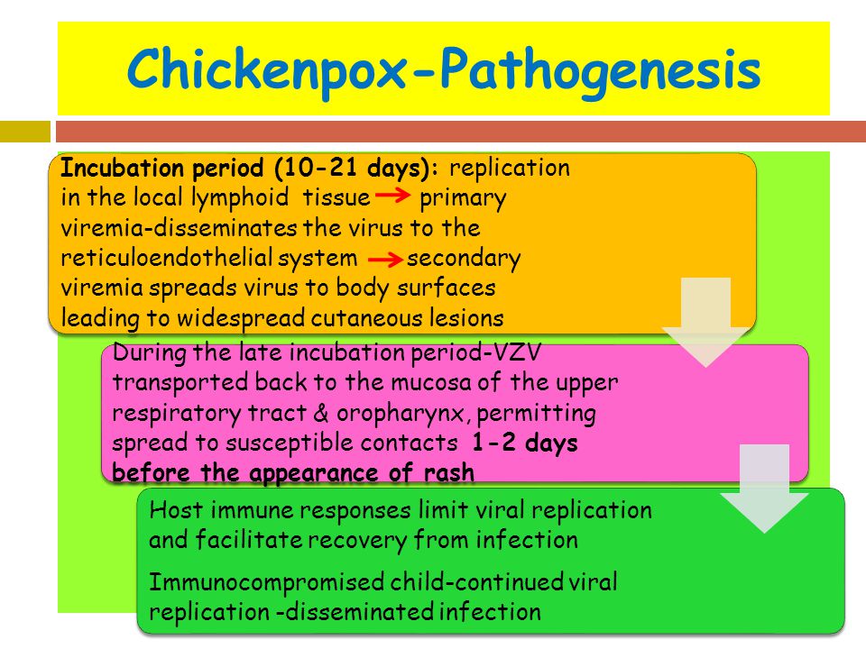
Red birthmarks are skin markings created by blood vessels close to the skin surface. They develop before or shortly after birth.
A stork bite is a vascular lesion quite common in newborns consisting of one or more pale red patches of skin. Most often stork bites appear on the forehead, eyelids, tip of the nose, upper lip or back of the neck. They are usually gone within 18 months of birth.
Hemangiomas are tumors made up of dilated blood vessels that usually appear shortly after birth, although they may be present at birth. Hemangiomas on the face can be disfiguring and may interfere with visual development or cause obstruction of the airway.
This child has a juvenile hemangioma (strawberry hemangioma) on the chin. These may begin as flat, red spots and later become larger and elevated. Juvenile hemangiomas often go away (involute) spontaneously.
Causes
There are two main categories of birthmarks:
- Red birthmarks are made up of blood vessels close to the skin surface. These are called vascular birthmarks.
- Pigmented birthmarks are areas in which the color of the birthmark is different from the color of the rest of the skin.

Hemangiomas are a common type of vascular birthmark. Their cause is unknown. Their color is caused by the growth of blood vessels at the site. Different kinds of hemangiomas include:
- Strawberry hemangiomas (strawberry mark, nevus vascularis, capillary hemangioma, hemangioma simplex) may develop several weeks after birth. They may appear anywhere on the body, but are most often found on the neck and face. These areas consist of small blood vessels that are very close together.
- Cavernous hemangiomas (angioma cavernosum, cavernoma) are similar to strawberry hemangiomas, but they are deeper and may appear as a red-blue spongy area of tissue filled with blood.
- Salmon patches (stork bites) are very common. Up to half of all newborns have them. They are small, pink, flat spots made up of small blood vessels that can be seen through the skin. They are most common on the forehead, eyelids, upper lip, between the eyebrows, and on the back of the neck. Salmon patches may be more noticeable when an infant cries or during temperature changes.

- Port-wine stains are flat hemangiomas made of expanded tiny blood vessels (capillaries). Port-wine stains on the face may be associated with Sturge-Weber syndrome. They are most often located on the face. Their size varies from very small to over half of the body's surface.
Symptoms
The main symptoms of birthmarks include:
- Marks on the skin that look like blood vessels
- Skin rash or lesion that is red
Exams and Tests
A health care provider should examine all birthmarks.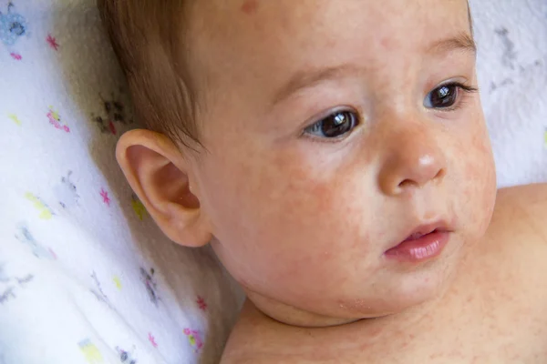 Diagnosis is based on how the birthmark looks.
Tests to confirm deeper birthmarks include:
- Skin biopsy
- CT scan
- MRI of the area
Treatment
Many strawberry hemangiomas, cavernous hemangiomas, and salmon patches are temporary and do not need treatment.
Port-wine stains may not need treatment unless they:
- Affect your appearance
- Cause emotional distress
- Are painful
- Change in size, shape, or color
Most permanent birthmarks are not treated before a child reaches school age or the birthmark is causing symptoms. Port-wine stains on the face are an exception. They should be treated at a young age to prevent emotional and social problems. Laser surgery can be used to treat them.
Concealing cosmetics may hide permanent birthmarks.
Oral or injected cortisone may reduce the size of a hemangioma that is growing quickly and affecting vision or vital organs.
Other treatments for red birthmarks include:
- Beta-blocker medicines
- Freezing (cryotherapy)
- Laser surgery
- Surgical removal
Outlook (Prognosis)
Birthmarks rarely cause problems, other than changes in appearance. Many birthmarks go away on their own by the time a child reaches school age, but some are permanent. The following development patterns are typical for the different types of birthmarks:
- Strawberry hemangiomas usually grow quickly and stay the same size.
 Then they go away. Most strawberry hemangiomas are gone by the time a child is 9 years old. However, there may be a slight change in color or puckering of the skin where the birthmark was.
 - Some cavernous hemangiomas go away on their own, usually as a child is about school age.
- Salmon patches often fade as the infant grows. Patches on the back of the neck may not disappear. They usually are not visible as hair grows.
- Port-wine stains are often permanent.
Possible Complications
The following complications can occur from birthmarks:
- Emotional distress because of appearance
- Discomfort or bleeding from vascular birthmarks (occasional)
- Interference with vision or bodily functions
- Scarring or complications after surgery to remove them
When to Contact a Medical Professional
Have your provider look at all birthmarks.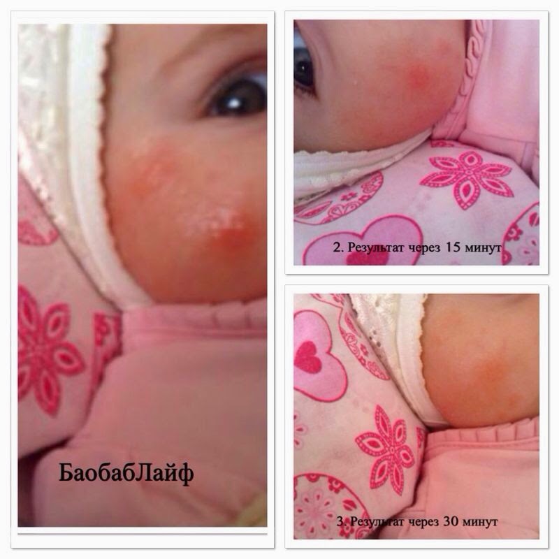
Prevention
There is no known way to prevent birthmarks.
Dinulos JGH. Vascular tumors and malformations. In: Dinulos JGH, ed. Habif's Clinical Dermatology. 7th ed. Philadelphia, PA: Elsevier; 2021:chap 23.
Folpe AL, Kozakewich HP. Benign vascular tumors and malformations. In: Goldblum JR, Folpe AL, Weiss SW, eds. Enzinger and Weiss's Soft Tissue Tumors. 7th ed. Philadelphia, PA: Elsevier; 2020:chap 20.
Patterson JW. Vascular tumors. In: Patterson JW, ed. Weedon's Skin Pathology. 5th ed. Philadelphia, PA: Elsevier Limited; 2021:chap 39.
Last reviewed on: 11/10/2020
Reviewed by: Ramin Fathi, MD, FAAD, Director, Phoenix Surgical Dermatology Group, Phoenix, AZ. Also reviewed by David Zieve, MD, MHA, Medical Director, Brenda Conaway, Editorial Director, and the A.D.A.M. Editorial team.
Infantile Hemangiomas: About Strawberry Baby Birthmarks
Hemangiomas are clusters of extra blood vessels on a baby's skin. They may be there when a baby is born, or form within a few weeks or months of birth. Some may look like rubbery, bumpy red "strawberry" patches while others resemble deep bruises. Seeing a hemangioma develop can be worrisome for new parents.
The American Academy of Pediatrics (AAP) guidelines say it's important to identify and begin monitoring infantile hemangiomas right after they appear―when they tend to change most quickly.
While hemangiomas can vary a lot in size, appearance, and placement, they are universally benign (non-cancerous). Most will go away on their own without causing any problems. Some hemangiomas―particularly those on the face or those that are very large―need treatment early to prevent them from interfering with body functions or causing permanent scars. Thankfully, there are excellent treatments available today to can prevent these problems if treated early on.
About Infantile Hemangiomas:
Infantile hemangiomas appear after a baby is born, typically within a month. Roughly 4% to 5% of all infants get them, although they are more common in Caucasians, girls, twins, and preterm or low-birth-weight babies. Infantile hemangiomas typically go through a period of rapid growth, followed by more gradual fading and flattening.
There are different types of infantile hemangiomas:
Superficial hemangiomas have been called "strawberry marks," because they can resemble the surface of berries. They may begin as small white, pink, or red areas on the skin that quickly change into brighter red, raised lesions.
 Superficial hemangiomas may be focused in one spot or spread out over a larger area.
Deep hemangiomas have a smooth surface and form under the skin. They may have a bluish tint and resemble bruises. Some cause the skin to look swollen.
Mixed hemangiomas are a combination of superficial and deep growths.
Are there other birthmarks like it?
Some of the other marks that can show up on a baby's skin include port wine stains and "stork bites." These also are caused when more blood than usual floods the capillaries under the skin. Port wine stains turn a reddish-purple and are often permanent; like hemangiomas, stork bites usually disappear, but can remain if they're on the back of the neck.
What to do if you think your baby has an infantile hemangioma:
Infantile hemangiomas usually become noticeable by 4 weeks of age. They may start out looking like a tiny bump or scratch. But many then grow especially fast between 5 and 7 weeks old. If you think your baby might have a hemangioma, it's best to contact your baby's pediatrician right away. He or she probably will want to see your baby within a short timeframe. According to the AAP, the best "window of opportunity" to be evaluated and start treatment if needed is about 1 month of age.
Your pediatrician will want to know:
Size: Is the hemangioma small (3/4" or less across) or larger?
Location: Is it located on the face or in the diaper area, or in a different area?
Number: Is there one or more than one? If more than one, how many?
If your doctor can't see you in the office right away, they may ask you to send photos or recommend a telemedicine visit to have a look without delay.
What to expect next:
It's important to continue monitoring the hemangioma until it stops growing. Superficial hemangiomas typically reach their full size by 5 months of age, although
deep hemangiomas sometimes keep growing a while longer. In some cases, your pediatrician may give a referral to an infantile hemangioma specialist with expertise in
pediatric dermatology, hematology-oncology, otolaryngology, or
plastic surgery.
Superficial hemangiomas typically reach their full size by 5 months of age, although
deep hemangiomas sometimes keep growing a while longer. In some cases, your pediatrician may give a referral to an infantile hemangioma specialist with expertise in
pediatric dermatology, hematology-oncology, otolaryngology, or
plastic surgery.
By the time a baby is 6-18 months old, most hemangiomas begin to slowly improve. In a process called "involution," the hemangioma will become less red and more grey or whitish and gradually flatten and shrink from the center outward.
Each case is different. Most hemangiomas have finished or almost finished flattening and shrinking by 4 to 5 years of age. |
When does a hemangioma need to be treated?
Whether a hemangioma needs treatment depends on the age of the baby, where the hemangioma is located and how fast it is growing, whether it becomes sore or scabby, and the risk of it causing medical complications with a child's health and well-being.
There are 3 main reasons for treatment:
Medical problems. In rare cases and depending where they are located and how fast they are growing, hemangioma may begin to interfere with vital functions. Hemangiomas near the child's eyes, nose or mouth, for example, can affect the child's ability to see, eat, breath or hear well. In rare cases, hemangiomas grow inside the body, which may need to be monitored with imaging tests.
Skin breakdown. Sometimes, skin on the hemangioma's surface breaks down and becomes an open sore (called an ulcer) that could lead to bleeding, infection, or scarring.
Permanent skin changes. Changes in the skin's texture or color can remain even after the hemangioma has gone away. This can be a concern, especially for hemangiomas on the child's face. Large hemangiomas on facial features such as the nose or lip can also distort growth.
What kinds of treatments are available for hemangiomas?
If a baby's hemangioma risks causing problems, medications can be applied directly to the skin or taken by mouth. The goal is to keep them from getting any bigger during their period of rapid growth, or to make them shrink more quickly. Laser procedures or surgery may be an option in some cases, although it generally is avoided during early infancy to avoid increased anesthesia risks.
Systemic treatments
Propranolol, a beta blocker medication used for many years to treat high blood pressure, is now commonly given by mouth as an effective treatment for problem hemangiomas. To avoid a growth rebound, the pediatrician may recommend therapy continue until your child's first birthday. The drug must be used with close observation by your healthcare provider to watch for possible side effects and complications.
Oral steroids have been largely replaced by safer and more effective options, but are still used in select cases, determined by the healthcare provider.
Localized treatments
Topical medications applied directly on the skin may be used for small, superficial hemangiomas.
 Prescription creams or ointments containing beta-blockers are the most effective topical treatment option to help stop growth and sometimes shrink and fade hemangiomas. In some cases, steroid creams may be prescribed for smaller, thinner hemangiomas.
Steroid injections can be given directly into the hemangioma to help slow its growth. This works best for smaller, localized hemangiomas.
Other treatments
Surgery is usually only considered for smaller hemangiomas in areas where they may cause problems, or for small hemangiomas with broken skin. Because surgery will always leave a scar itself--and because most hemangiomas get better with time--early surgery is only recommended for a small minority of cases. Surgery can also repair extra skin or scars left by a hemangioma, but usually is delayed until a child is between 3 and 5 years old.
Laser treatment may be helpful in some cases to stop bleeding or to help heal hemangiomas with open sores (ulcers).
 They can also help to remove some of the redness or texture changes that may be left behind after the hemangioma improves.
Remember:
Contact your pediatrician if you notice anything developing on your baby's skin. Your baby's first few well-child visits are also a great time to bring it up. Few hemangiomas cause any trouble, and most go away on their own. But prompt evaluation, monitoring and treatment, when needed, can help ensure problem hemangiomas have as little impact as possible on your child.
Additional Information:
Baby Birthmarks & Rashes
Management of Infantile Hemangiomas (AAP Clinical Practice Guideline)
HemangiomaEducation.org
The information contained on this Web site should not be used as a substitute for the medical care and advice of your pediatrician. There may be variations in treatment that your pediatrician may recommend based on individual facts and circumstances.
Plaques on the skin - causes, diseases, diagnosis and treatment
- INVITRO
- Library
- Symptoms
- Plaques on the skin
Fungus
Allergy
Psoriasis
Keratoma
Mycosis
Nevus
Melanoma
6603 November 16
IMPORTANT!
The information in this section should not be used for self-diagnosis or self-treatment.
 In case of pain or other exacerbation of the disease, only the attending physician should prescribe diagnostic tests. For diagnosis and proper treatment, you should contact your doctor.
For a correct assessment of the results of your analyzes in dynamics, it is preferable to do studies in the same laboratory, since different laboratories may use different research methods and units of measurement to perform the same analyzes.Plaques on the skin: the causes of occurrence, in what diseases they occur, diagnosis and methods of treatment.
Definition
A plaque is a pathological element with clear edges that rises above the skin surface or merges with it, more than 5 mm in size.In dermatology, many types of plaques are distinguished - about 70 diseases occur with the formation of these elements, which makes the plaque one of the most common rashes.
Plaque varieties
The shape of the plaques are round, oval and irregular in shape. Over time, the shape, surface and appearance of this element may change.
 Due to the occurrence of plaques, they can be both a manifestation of skin diseases and a symptom of diseases of internal organs and systems (autoimmune reactions, liver diseases, oncological processes, allergic reactions).
Plaques are dry, smooth, red, brown, gray-white, etc.
Possible causes of plaques
Dry plaques on the skin in adults may be a manifestation of the following diseases: plaques with severe itching.
- Allergic reactions are characterized by the appearance on the skin of smooth dry plaques, pink spots, blisters, which are very itchy and cause severe discomfort. They can develop both when the skin comes into contact with the allergen, and when it gets on the mucous membranes (for example, with urticaria, hay fever, food and contact allergies).
- Psoriasis is a chronic non-infectious skin disease in which scaly dry plaques form on the elbows, knees, scalp, prone to fusion and accompanied by mild itching.

- Dry plaques form on the skin if it is exposed to stress for a long time with the loss of its protective functions.
- Diseases of the digestive tract, accompanied by malabsorption syndrome (impaired absorption of vitamins and trace elements in the small intestine), chronic diseases of the liver and other organs, in which substances that are not normally present in the dermis accumulate, also lead to the appearance of dry plaques.
- Solar keratoma is a precancerous condition, which is characterized by the presence of many light grayish plaques on the skin.
- Drug toxidermia is an allergic reaction accompanied by the appearance of elements in the form of plaques on the skin. In severe cases, Lyell's syndrome or Stevens-Johnson syndrome, toxic epidermal necrolysis, may develop.

- Dühring's dermatitis (herpetiform) is a chronic skin disease with no established etiology, which is characterized by recurrent appearance of a rash of various morphologies on the skin, accompanied by severe skin itching and burning.
- Mycosis fungoides is a primary T-cell lymphoma of the skin, a malignant lymphoid lesion, primarily of the skin. Itchy red plaques appear on the skin, resembling eczema. In the initial stages, they respond well to treatment with hormonal ointments, but the disease itself requires more complex therapy.
- In children, the appearance of red spots and plaques on the skin is most often associated with an allergic reaction to food.
- Becker's nevus is an anomaly in the development of the dermis, when dark plaques with an uneven surface appear on the skin, on which hair can begin to grow over time.

- Pigmentary nevus - "birthmark", may rise above the skin, has a brown or dark color.
- Melanoma is the most malignant skin tumor characterized by rapid metastasis. It develops mainly from nevi and moles. If the nature of the surface, the boundaries of the mole change, its size increases, bleeding occurs, you should immediately contact a dermatologist or oncologist to exclude the development of melanoma.
- Basal cell skin cancer is more often localized on the head, face, neck, does not metastasize, is characterized by slow growth.
- Senile keratoma occurs in elderly people, most likely due to a lack of vitamins, an abundance of animal fats consumed, skin sensitivity to ultraviolet radiation due to a violation of its protective functions. Typical localization - face, neck, open areas of the body.
- Seborrheic keratoma is a yellowish plaque on the skin that eventually transforms into a dark brown growth that tends to flake off, itch severely, crack, bleed, and can serve as an entryway for infection.
- change in the shape of the plaque - the edges have become uneven;
- change in the surface of the plaque - cracks, ulcerations appeared;
- change in the size of the plaque - it began to grow rapidly above the surface of the skin or actively spread through it;
- discoloration of the plaque - in cases of malignancy, an uneven color of the formation is usually observed with areas of darker and lighter shades;
- the appearance of bleeding - both contact and spontaneous;
- enlargement of regional (nearby) lymph nodes.
- Clinical guidelines.
 Dermatitis herpetiformis // Russian Society of Dermatovenerologists and Cosmetologists. 2016.
 - Clinical guidelines. Urticaria in children // Union of Pediatricians of Russia; Russian Association of Allergists and Clinical Immunologists. 2018.
- Clinical guidelines. Toxidermia // Russian Society of Dermatovenerologists and Cosmetologists. 2016.
- Clinical guidelines. Familial hypercholesterolemia // National Society for the Study of Atherosclerosis. 2018.
-
Gastrointestinal bleeding
502 29 September
-
Intestinal colic
4436 28 September
-
Exanthema
4494 12-th of September
- Local infantile hemangiomas
- Segmental infantile hemangiomas
- Special shapes
- Active growth phase: the first stage lasts from 6 to 9 months
- Growth arrest phase: tumor size no longer changes
- Regression phase (gradual reverse development of hemangioma, that is, the tumor begins to “resolve”, the recovery process is underway): as a rule, recovery ends by the 9th year of the child's life.
- superficial skin infantile hemangiomas. They grow on the surface of the skin (flat) or may protrude above the skin (convex, i.e. do not grow deep)
- deep subcutaneous hemangiomas. They grow deep under the skin
- combined hemangiomas. That is, a mixed type, when an infantile hemangioma simultaneously grows as a superficial skin and deep subcutaneous
The appearance of red plaques on the skin indicates their good blood supply. Possible causes of this condition may be the following nosologies:
Brown plaques occur when melanin is deposited in the affected area of the dermis, which causes a brown (dark) color. Possible causes may be the following diseases:
Which doctors to contact
With the formation of plaques on the skin, it is necessary to contact a dermatologist to determine the causes of the appearance of this element of the rash.
Plaque diagnostics and examinations
For the diagnosis of fungal skin lesions, scraping from the affected area is used for subsequent microscopic examination.
The development of an allergic reaction requires seeking medical help to identify the allergen, prescribing antihistamines, and sometimes hormonal drugs. In clinical cases of allergy, along with skin tests, analyzes are performed using various sets of common allergens and triggers: a panel for respiratory allergens, for food allergens, and for a combination of both.
Respiratory Panel
Synonyms: Comprehensive Respiratory Allergen Test Panel; Respiratory allergens panel, Allergen respiratory profile, Allergy testing. Brief description of the study "Panel respira...
Up to 5 business days
Available with house call
5 515 RUB
Add to cart
Food Panel, IgE
Food Allergen Panel: hazelnut, peanut, Walnut, almond, cow's milk, egg white, egg ...
Up to 5 working days
Available with house call
5 515 RUB
Add to cart
Food and Respiratory Panel
Panel different allergens: A mixture of grass allergens: fragrant spikelet; perennial rye; timothy; rye cultivated; Woolly buckthorn (GP3) IgE . ..
..
Up to 5 business days
Available with house call
5 515 RUB
Add to cart
In psoriasis, seeing a dermatologist and a rheumatologist can help reduce the symptoms of the disease if appropriate therapy is prescribed. For the diagnosis, it is usually sufficient to examine, determine, the skin manifestations of psoriasis are so characteristic, but if necessary, a differential diagnosis is carried out, including a clinical blood test, feces for the presence of worm eggs and protozoa, and a histological examination of the skin.
Clinical blood test: general analysis, leukoformula, ESR (with microscopy of a blood smear in the presence of pathological changes)
Synonyms: Complete blood count, UAC. Full blood count, FBC, Complete blood count (CBC) with differential white blood cell count (CBC with diff), Hemogram. Brief description of the study CBC: general...
Up to 1 business day
Available with house call
RUB 810
Add to cart
Fecal test for helminth eggs
Synonyms: Feces on worm eggs; Analysis of feces for eggs of worms. Ova and Parasite Exam; O&P; Stool O&P test. Brief description of the study "Analysis of feces for eggs of helminths&ra...
Up to 1 business day
Available with house call
RUB 570
Add to cart
Fecal analysis for protozoa (PRO stool)
Synonyms: Analysis of faeces for protozoa. Parasite Exam, feces; Parasitic Examination, fecal. Brief description of the study "Analysis of feces for protozoa" Analysis of feces for cis...
Up to 1 business day
Available with house call
RUB 570
Add to cart
Histological examination of biopsy material and material obtained during surgical interventions (endoscopic material; tissues of the female reproductive system; skin, soft tissues; hematopoietic and lymphoid tissue; bone and cartilage tissue)
Taking biomaterial is paid separately. According to the requirements of clause 17 of the Rules for conducting pathological and anatomical studies, approved. Order of the Ministry of Health of Russia. ..
..
Up to 5 business days
Available with house call
2 880 RUB
Add to cart
Diseases of the stomach and intestines can also lead to plaque formation on the skin. To identify the pathology of the gastrointestinal tract, it is enough to refer to therapist or gastroenterologist, conduct a number of endoscopic examinations (gastroscopy, and, if necessary, colonoscopy), ultrasound of the abdominal organs, perform some screening blood tests for diseases of the liver, intestines, stomach.
Gastroscopy
Examination of the mucosa of the upper gastrointestinal tract with the possibility of biopsy or endoscopic removal of small pathological ...
RUB 4,440 Sign up
Colonoscopy
Endoscopic examination of the colon to look for abnormalities, perform biopsies, and remove small polyps and tumors.
RUB 5,740 Sign up
Comprehensive ultrasound examination of the abdominal organs (liver, gallbladder, pancreas, spleen)
Scanning of the internal organs of the abdominal cavity to assess its functional state and the presence of pathology.
RUB 2,890 Sign up
Liver function tests: screening
Up to 1 business day
Available with house call
RUB 1,935
Add to cart
Diagnosis of celiac disease: intolerance to cereal proteins (gluten)
Up to 8 working days
Available with house call
7 520 RUB
Add to cart
Gastropanel
Up to 9 working days
Available with house call
RUB 4,760
Add to cart
To clarify the diagnosis of keratoma, a skin biopsy is performed and epithelium scraping is performed, followed by microscopic and histochemical examination.
Histological examination of biopsy material and material obtained during surgical interventions (endoscopic material; tissues of the female reproductive system; skin, soft tissues; hematopoietic and lymphoid tissue; bone and cartilage tissue)
Taking biomaterial is paid separately. According to the requirements of clause 17 of the Rules for conducting pathological and anatomical studies, approved. Order of the Ministry of Health of Russia...
Up to 5 working days
Available with house call
2 880 RUB
Add to cart
Examination of scrapings and impressions of tumors and tumor-like formations
Material for research. Imprints and scrapings are obtained from pathological lesions of the skin and mucous membranes (except for the cervix and cervical canal). Relative to test...
Up to 2 working days
Available with house call
RUB 1,030
Add to cart
If atypical cells are detected in scrapings or biopsies, immediately contact oncologist.
If xanthoma appears on the skin, it is recommended to consult a cardiologist, take blood tests for lipid profile and blood glucose levels, and screen for diabetes.
Lipid profile screening
Up to 1 business day
Available with house call
1 355 RUB
Add to cart
Glucose (in the blood) (Glucose)
Research material Serum or blood plasma. If it is not possible to centrifuge the sample 30 minutes after collection for serum/plasma separation...
Up to 1 business day
Available with house call
335 RUB
Add to cart
Diabetes management: advanced
Up to 1 business day
Available with house call
RUB 5 820
Add to cart
What should I do if plaque appears on the skin?
Any newly appeared neoplasms should be shown to a dermatologist. Their cosmetic removal without prior consultation with a specialist is fraught with serious consequences.
In addition, there are symptoms that require immediate medical attention:
Plaque treatment
When plaques of an allergic nature appear on the skin, antihistamines are prescribed, in cases of a severe course of the disease, glucocorticosteroids. In addition, it is important to follow a hypoallergenic diet.
Mycotic plaques require antifungal drugs, both local (ointments, creams) and systemic (tablets). Taking these drugs is associated with a high risk of side effects, and therefore it is possible only after consulting a doctor, accurate verification of the diagnosis and confirmation of the etiology of the disease.
Treatment of psoriasis is multi-stage and complex, it involves constant monitoring by a rheumatologist, taking cytostatics and other drugs, using ointments and shampoos to improve skin condition, using antihistamines to reduce itching, including physiotherapy and a hypoallergenic diet in the treatment regimen.
When confirming the presence of diseases of the gastrointestinal tract, properly selected therapy can stop the appearance of new plaques on the skin, as well as prevent the development of complications of the underlying disease.
Sources:
IMPORTANT!
The information in this section should not be used for self-diagnosis or self-treatment. In case of pain or other exacerbation of the disease, only the attending physician should prescribe diagnostic tests. For diagnosis and proper treatment, you should contact your doctor.
For a correct assessment of the results of your analyzes in dynamics, it is preferable to do studies in the same laboratory, since different laboratories may use different research methods and units of measurement to perform the same analyzes.
Recommendations
Show more
Thyrotoxicosis
Bronchitis
Pulmonary edema
Bronchial asthma
Influenza
Diffuse goiter
Allergy
Tumor
Wheezing
The appearance of wheezing occurs when there is a violation of the passage of air through the bronchial tree. Whistling when breathing can be a sign of serious illness.
More
Fungus
Diarrhea
Allergy
Diabetes mellitus
Hemorrhoids
Intertrigo
Intertrigo: causes of occurrence, in which diseases it occurs, diagnosis and methods of treatment.
More
Fungus
Cystitis
Thrush
Colpitis
Urine flakes
Urine flakes: causes, diseases, diagnosis and treatment.
More
Diarrhea
Pancreatitis
Allergy
Colitis
Rotavirus
Salmonellosis
Loose stools
Loose stools: causes, diseases in which it develops, methods of diagnosis and treatment.
More
HIV
Fungus
COPD
Bronchial asthma
Cystic fibrosis
Emphysema
Sarcoidosis
Rheumatoid arthritis
Scleroderma
Pneumothorax
Pneumothorax: causes of occurrence, in what diseases it occurs, diagnosis and methods of treatment.
More
Nothing found
Try editing your query or select a doctor or service from the list.
Doctor not found
Try changing your query or select doctor from the list
Medical office not found
Try changing your query or select medical office from the list
Therapist Traumatologist-orthopedist Endocrinologist Urologist Gynecologist Ultrasound doctor Cardiologist Pediatrician
Nothing found
Try changing the query
Thank you!
You have successfully made an appointment
Detailed information has been sent to your e-mail
What are the types of infantile (infantile) hemangiomas?
Erstellt am 2017/10/19, last modified: 2017/10/19 https://kinderkrebsinfo.de/doi/e195094
Contents
Typically, infantile hemangiomas (that is, hemangiomas in newborns and infants, and many doctors use the term infantile hemangiomas) appear in the first days or weeks after the birth of a child. First, the first symptoms appear (specialists talk about the precursor forms of hemangiomas) - dilated subcutaneous vessels in a limited area of \u200b\u200bthe skin (telangiectasia in the language of specialists), or, for example, very pale spots, or spots of bright red or cyanotic color, or changes in the skin, similar to red birthmarks (doctors talk about vascular nevus). The classic infantile hemangioma at the birth of a child does not yet look like a tumor, it becomes one after some time.
Good to know: infantile hemangioma goes through three stages:
Local infantile hemangiomas
90% of all infantile hemangiomas are local. This means that they have clear boundaries and grow from one central point.
Local infantile hemangiomas are divided into:
Usually, at birth, an infantile hemangioma is not yet visible in a child. But then, during repeated examinations in the first weeks after birth, a small spot of red color becomes noticeable. Some infantile hemangiomas do not change for weeks or months. Others begin to grow rapidly and grow to enormous size. Most infantile hemangiomas (60%) appear in the head and neck.
Segmental infantile hemangiomas
Segmental infantile hemangiomas (that is, a large hemangioma will grow in a certain area of the body) are less common than local ones. They can appear both in the head and neck, and in the region of the lumbar spine and in the coccyx. Usually the size of segmental infantile hemangiomas is larger than that of local forms. In addition, they often appear when abnormal development of blood vessels or internal organs begins in the body (in this case, doctors talk about malformations or developmental anomalies). Characteristic of segmental hemangiomas is that they are very large and cover a certain department (segment) of the body. They are almost invisible at birth. But they can grow very quickly and then the baby often has various health problems.
For example, segmental infantile hemangiomas in the face or shoulder region are associated with the so-called PHACES syndrome (P.H.A.C.E.S. syndrome is a set of several congenital malformations, each letter of the abbreviation denotes a specific malformation). First, anomalies in the development of the chest, aorta, as well as heart defects and cysts in the brain (in the language of specialists, the so-called Dandy-Walker variant) are found in the baby, and then a segmental hemangioma appears. Another complication is a tendency to form ulcers and a tendency to frequent infections.
Hemangiomas that grow in the perineum are part of the PELVIS (in the pelvis) and SAKRAL (in the sacrum) syndromes. They are accompanied by skin growths, as well as anomalies in the development of the bladder, spinal cord and spinal cord membranes, and abnormal development of the anus.
In rare cases, infantile hemangiomas can also grow in the area of internal organs, such as the liver or kidneys.
Special shapes
"Rapid Involuting Congenital Hemangioma / abbreviated as RICH":
These forms may also be referred to as congenital rapidly regressing hemangiomas. Already at the birth of a child, they are fully developed (doctors say "fully formed") and quickly (in English " rapid " ) completely disappear, usually by the third year of a baby's life (doctors often use the term "involution “, from the English term "involuting" , which means reverse development of ).