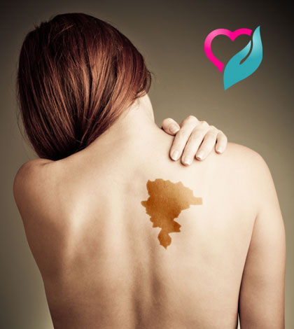
Red birthmark on back of head
Birthmarks in Infants | Johns Hopkins Medicine
A baby's skin coloring can vary greatly, depending on the baby's age, race or ethnic group, temperature, and whether or not the baby is crying. Skin color in babies often changes with both the environment and health. Some of these differences are just temporary. Others, such as certain birthmarks, may be permanent.
Birthmarks are areas of discolored and/or raised skin that are present at birth or within a few weeks of birth. Birthmarks are made up of abnormal pigment cells or blood vessels.
Although the cause of birthmarks is not known, most of them are harmless and do not require treatment. Babies with birthmarks should be examined by your child's health care provider, especially if they are:
-
Located in the middle of the back, along the spine (may be related to spinal cord problems)
-
Large birthmarks on the face, head or neck
-
Interfering with movement of activity, for example a birthmark on the eyelid that may interfere with vision
Some common birthmarks include:
| Birthmark | What it looks like |
|---|---|
| Stork bites, angel kisses, or salmon patches | These are small pink or red patches often found on a baby's eyelids, between the eyes, upper lip, and back of the neck. |
| Congenital dermal melanocytosis (also known as Mongolian spots) | Congenital dermal melanocytosis refers to areas of blue or purple-colored, typically on the baby's lower back and buttocks. These can occur in darker-skinned babies of all races. The spots are caused by a concentration of pigmented cells. They usually disappear in the first 4 years of life. |
| Strawberry hemangioma | This is a bright or dark red, raised or swollen, bumpy area that looks like a strawberry. Hemangiomas are formed by a concentration of tiny, immature blood vessels. Most of these occur on the head. They may not appear at birth, but often develop in the first 2 months. Strawberry hemangiomas are more common in premature babies and in girls. These birthmarks often grow in size for several months, and then gradually begin to fade. They may bleed or get infected in rare cases. Nearly all strawberry hemangiomas completely disappear by 9 years of age. Most of these occur on the head. They may not appear at birth, but often develop in the first 2 months. Strawberry hemangiomas are more common in premature babies and in girls. These birthmarks often grow in size for several months, and then gradually begin to fade. They may bleed or get infected in rare cases. Nearly all strawberry hemangiomas completely disappear by 9 years of age. |
| Port-wine stain | A port-wine stain is a flat, pink, red, or purple colored birthmark. These are caused by a concentration of dilated tiny blood vessels called capillaries. They usually occur on the head or neck. They may be small, or they may cover large areas of the body. Port-wine stains do not change color when gently pressed and do not disappear over time. They may become darker and thicker when the child is older or as an adult. Port-wine stains on the face may be associated with more serious problems. Skin-colored cosmetics may be used to cover small port-wine stains. The most effective way of treating port-wine stains is with a special type of laser. This is done when the baby is older by a plastic surgery specialist. The most effective way of treating port-wine stains is with a special type of laser. This is done when the baby is older by a plastic surgery specialist. |
| Congenital moles | These common moles (less than 3 inches in diameter) occur in about 1 out of every 100 newborns. They increase in size as the child grows, but usually don't cause any problems. Your child's health care provider will watch them closely as rarely they can develop into a cancerous mole. |
Birthmarks - NHS
Birthmarks are coloured marks on the skin that are present at birth or soon afterwards. Most are harmless and disappear without treatment, but some may need to be treated.
Types of birthmark
There are many different types of birthmark.
Flat, red or pink areas of skin (salmon patches or stork marks)
Credit:
DR P. MARAZZI/SCIENCE PHOTO LIBRARY https://www.sciencephoto.com/media/263023/view
Salmon patches:
- are red or pink patches, often on a baby's eyelids, head or neck
- are very common
- look red or pink on light and dark skin
- are easier to see when a baby cries
- usually fade by the age of 2 when on the forehead or eyelids
- can take longer to fade when on the back of the head or neck
Raised red lumps (strawberry marks or haemangiomas)
Credit:
Mediscan / Alamy Stock Photo https://www. alamy.com/capillary-haemangioma-image1683612.html?pv=1&stamp=2&imageid=B90D6561-3E1C-43EF-8BBC-6E247DC47070&p=17774&n=0&orientation=0&pn=1&searchtype=0&IsFromSearch=1&srch=foo%3dbar%26st%3d0%26pn%3d1%26ps%3d100%26sortby%3d2%26resultview%3dsortbyPopular%26npgs%3d0%26qt%3dATB09D%26qt_raw%3dATB09D%26lic%3d3%26mr%3d0%26pr%3d0%26ot%3d0%26creative%3d%26ag%3d0%26hc%3d0%26pc%3d%26blackwhite%3d%26cutout%3d%26tbar%3d1%26et%3d0x000000000000000000000%26vp%3d0%26loc%3d0%26imgt%3d0%26dtfr%3d%26dtto%3d%26size%3d0xFF%26archive%3d1%26groupid%3d%26pseudoid%3d788068%26a%3d%26cdid%3d%26cdsrt%3d%26name%3d%26qn%3d%26apalib%3d%26apalic%3d%26lightbox%3d%26gname%3d%26gtype%3d%26xstx%3d0%26simid%3d%26saveQry%3d%26editorial%3d1%26nu%3d%26t%3d%26edoptin%3d%26customgeoip%3d%26cap%3d1%26cbstore%3d1%26vd%3d0%26lb%3d%26fi%3d2%26edrf%3d0%26ispremium%3d1%26flip%3d0%26pl%3d
Strawberry marks:
- are blood vessels that form a raised red lump on the skin
- appear soon after birth
- usually look red on light and dark skin
- are more common in girls, premature babies (born before 37 weeks), low birth weight babies, and multiple births, such as twins
- get bigger for the first 6 to 12 months, and then shrink and disappear by the age of 7
- sometimes appear under the skin, making it look blue or purple
- may need treatment if they affect vision, breathing, or feeding
Red, purple or dark marks (port wine stains)
Credit:
MID ESSEX HOSPITAL SERVICES NHS TRUST / SCIENCE PHOTO LIBRARY https://www. sciencephoto.com/media/1026532/view
Port wine stains:
- are red, purple or dark marks and usually on the face and neck
- are present from birth
- look like very dark patches on dark skin
- usually affect one side of the body, but can affect both
- can sometimes be made lighter using laser treatment (it's most effective on young children)
- can become darker and lumpier if not treated
- can be a sign of Sturge-Weber syndrome and Klippel-Trenaunay syndrome, or macrocephaly-capillary malformation, but this is rare
Flat, light or dark brown patches (cafe-au-lait spots)
Credit:
DR P. MARAZZI / SCIENCE PHOTO LIBRARY https://www. sciencephoto.com/media/146602/view
Cafe-au-lait spots:
- are light or dark brown patches that can be anywhere on the body
- are common, with many children often having 1 or 2
- look darker on dark skin
- can be different sizes and shapes
- may be a sign of neurofibromatosis type 1 if a child has 6 or more spots
Blue-grey spots
Credit:
SCIENCE PHOTO LIBRARY https://www.sciencephoto.com/media/520677/view
These birthmarks:
- can look blue-grey on the skin like a bruise
- are often on the lower back, bottom, arms or legs
- are there from birth
- are most common on babies with darker skin
- do not need treating and will usually go away by the age of 4
- are not a sign of a health condition
If your baby is born with a blue-grey spot it should be recorded on their medical record.
Brown or black moles (congenital moles or congenital melanocytic naevi)
Credit:
BSIP SA / Alamy Stock Photo https://www.alamy.com/stock-photo-congenital-naevus-49287995.html?pv=1&stamp=2&imageid=1DB6D728-88CC-4DAF-9C9B-CE3F27AAAA65&p=165781&n=0&orientation=0&pn=1&searchtype=0&IsFromSearch=1&srch=foo%3dbar%26st%3d0%26pn%3d1%26ps%3d100%26sortby%3d2%26resultview%3dsortbyPopular%26npgs%3d0%26qt%3dCT579F%26qt_raw%3dCT579F%26lic%3d3%26mr%3d0%26pr%3d0%26ot%3d0%26creative%3d%26ag%3d0%26hc%3d0%26pc%3d%26blackwhite%3d%26cutout%3d%26tbar%3d1%26et%3d0x000000000000000000000%26vp%3d0%26loc%3d0%26imgt%3d0%26dtfr%3d%26dtto%3d%26size%3d0xFF%26archive%3d1%26groupid%3d%26pseudoid%3d788068%26a%3d%26cdid%3d%26cdsrt%3d%26name%3d%26qn%3d%26apalib%3d%26apalic%3d%26lightbox%3d%26gname%3d%26gtype%3d%26xstx%3d0%26simid%3d%26saveQry%3d%26editorial%3d1%26nu%3d%26t%3d%26edoptin%3d%26customgeoip%3d%26cap%3d1%26cbstore%3d1%26vd%3d0%26lb%3d%26fi%3d2%26edrf%3d0%26ispremium%3d1%26flip%3d0%26pl%3d
Congenital moles:
- are brown or black moles caused by an overgrowth of pigment cells in the skin
- look darker on dark skin
- can become darker, raised and hairy, particularly during puberty
- may develop into skin cancer if they're large (the risk increases the larger they are)
- do not need to be treated unless there's a risk of skin cancer
Information:
Find out about other types of birthmark:
The Birthmark Support Group has information about other types of birthmark and getting help and support.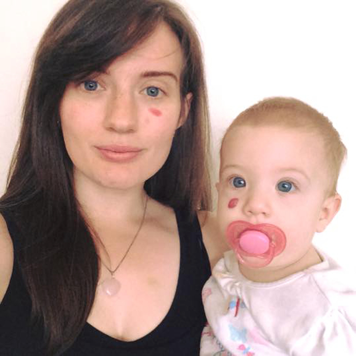
Non-urgent advice: See a GP if:
- you're worried about a birthmark
- a birthmark is close to the eye, nose, or mouth
- a birthmark has got bigger, darker or lumpier
- a birthmark is sore or painful
- your child has 6 or more cafe-au-lait spots
- you or your child has a large congenital mole
The GP may ask you to check the birthmark for changes, or they may refer you to a skin specialist (dermatologist).
Treatment for birthmarks
Most birthmarks do not need treatment, but some do. This is why it's important to get a birthmark checked if you're worried about it.
A birthmark can be removed on the NHS if it's affecting a person's health. If you want a birthmark removed for cosmetic reasons, you'll have to pay to have it done privately.
Possible treatments for birthmarks include:
- medicines – to reduce blood flow to the birthmark, which can slow down its growth and make it lighter in colour
- laser therapy – where heat and light are used to make the birthmark smaller and lighter (it works best if started between 6 months and 1 year of age)
- surgery – to remove the birthmark (but it can leave scarring)
Page last reviewed: 04 February 2020
Next review due: 04 February 2023
Spot on the back of the head in a child
07/12/2021 Reading time: 2 min 72559
When a child is born with a red spot on the back of the head or back of the neck, it is said to have been carelessly carried by a stork, and the spot itself is called a stork bite.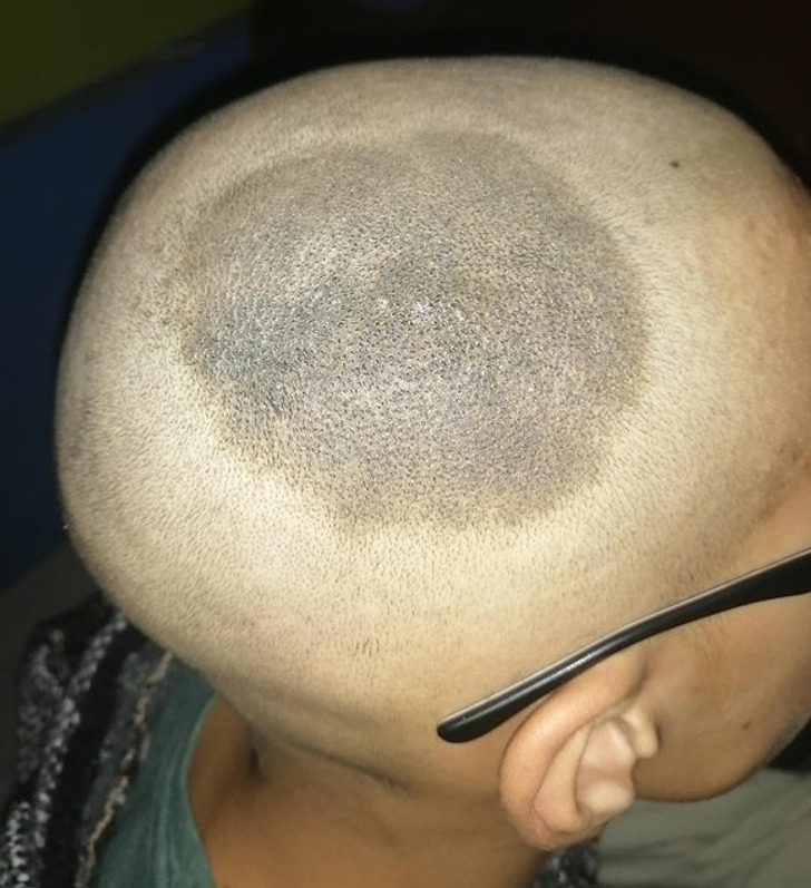 It is clear that this is a joke explanation. But where, then, are the marks, if the stork has nothing to do with it? The answer must be sought not in folk legends, but in medical sources.
Spot of a stork. What is it and why is it formed?
In the medical literature, the stork's spot has a completely non-fabulous scientific name: Unna's nevus or a simple nevus. It can be located not only on the back of the head or on the back of the neck, but also on the forehead, eyelids of the baby. In this case, it is called the "kiss of an angel."
There is an opinion that these spots are the result of traumatic childbirth, in which the baby's head was squeezed for a long time, but scientists refute it. They say that a simple nevus is still being laid in utero. It is formed due to a failure in the laying of blood vessels: in this area they are slightly expanded and blood flow is increased in them, so the skin here is more pink. Another version - a stork spot is formed in the place where the child's head is pressed against the mother's sacrum in the later stages. Due to pressure, the capillaries dilate and a stain results: have you ever woken up with red marks from the pillow on your face? It's about the same here.
Be that as it may, almost half of babies are born with a simple nevus on the face. You shouldn't be afraid of him. It's not a disease. There is no fault of the mother that the child is born with a mark on the back of the head, and she cannot somehow prevent her appearance.
Will the spot on the back of the head go away on its own?
A simple nevus is the most harmless deviation in the formation of skin capillaries. During the first year of a baby's life, the spot begins to lighten until it matches the color of the usual skin tone. For some time it can still manifest itself after intense screaming or strong exertion, and usually disappears by the age of two.
But if we talk specifically about the stork spot - a simple nevus located on the back of the head or the back of the neck - then it may not completely disappear, but only turn pale. Fortunately, since it is not located in the most noticeable area, the damage to the appearance is minimal.
Should I be treated?
As we remember, a simple nevus is not a disease, which means that it does not require treatment. Be patient, and the stork's spot will disappear on its own, or at least turn very pale. There are no creams, ointments or lotions that would speed up this process, so the doctor will not recommend anything to you, except for observation. And without a prescription, it is strictly forbidden to use any medicines, as well as traditional medicine recipes, when it comes to young children.
When should I see a doctor?
If the spot on the back of the child's head is a simple nevus, he will not require a visit to the doctor. Most likely, they will show it to you in the hospital and explain that there is no need to worry.
Another thing is if another pathology of vascular development was mistaken for a stork spot: a flaming nevus or hemangioma.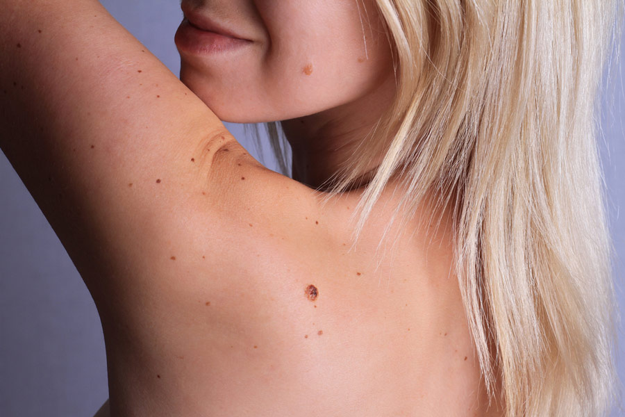 These conditions require a different approach to monitoring and sometimes need to be treated.
If the condition of the spot worries you for some reason: for example, it does not turn pale for a long time or, on the contrary, it began to darken, increase in size, peel off, become inflamed - be sure to show the child to the doctor to clarify the diagnosis and, if necessary, prescribe treatment.
Summing up:
- Stork spot, or, scientifically, Unna's nevus (simple nevus) is a bright pink or red patch of skin on the back of the head of a newborn. The capillaries on it are expanded, and the blood flow is increased, hence the intense color. This spot is formed in utero and does not depend on the course of childbirth.
- The stork spot may disappear by the age of two, or it may turn pale and remain on the skin forever.
- It is not necessary to treat a simple nevus, observation is enough.
- You should consult a doctor if the condition of the spot worsens: it becomes brighter, larger, inflamed, peeling or rash appears.
 The doctor will clarify the diagnosis and prescribe treatment.
(14 ratings; article rating 3.5)
What are the types of infantile (infantile) hemangiomas?
Erstellt am 2017/10/19, last modified: 2017/10/19 https://kinderkrebsinfo.de/doi/e195094
Contents
- Local infantile hemangiomas
- Segmental infantile hemangiomas
- Special forms
Typically, infantile hemangiomas (that is, hemangiomas in newborns and infants, and many doctors use the term infantile hemangiomas) appear in the first days or weeks after the birth of a child. First, the first symptoms appear (specialists talk about the precursor forms of hemangiomas) - dilated subcutaneous vessels in a limited area of \u200b\u200bthe skin (telangiectasia in the language of specialists), or, for example, very pale spots, or spots of bright red or cyanotic color, or changes in the skin, similar to red birthmarks (doctors talk about vascular nevus).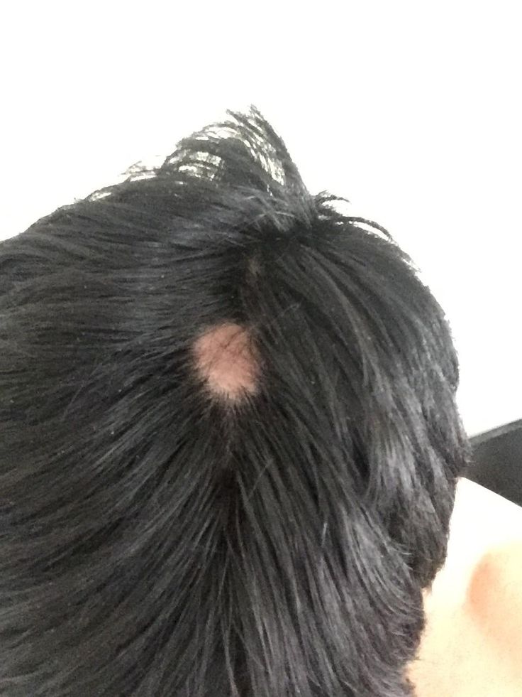 The classic infantile hemangioma at the birth of a child does not yet look like a tumor, it becomes one after some time.
Good to know: infantile hemangioma goes through three stages:
- Active growth phase: the first stage lasts from 6 to 9 months
- Growth stop phase: tumor size no longer changes
- Regression phase (gradual reverse development of hemangioma, that is, the tumor begins to “resolve”, the recovery process is underway): as a rule, recovery ends by the 9th year of the child's life.
Local infantile hemangiomas
90% of all infantile hemangiomas are local. This means that they have clear boundaries and grow from one central point.
Local infantile hemangiomas are divided into:
- superficial skin infantile hemangiomas. They grow on the surface of the skin (flat) or may protrude above the skin (convex, i.e. do not grow deep)
- deep subcutaneous hemangiomas.
 They grow deep under the skin
 - combined hemangiomas. That is, a mixed type, when an infantile hemangioma simultaneously grows as a superficial skin and deep subcutaneous
Usually, at birth, an infantile hemangioma is not yet visible in a child. But then, during repeated examinations in the first weeks after birth, a small spot of red color becomes noticeable. Some infantile hemangiomas do not change for weeks or months. Others begin to grow rapidly and grow to enormous size. Most infantile hemangiomas (60%) appear in the head and neck.
Segmental infantile hemangiomas
Segmental infantile hemangiomas (that is, a large hemangioma will grow in a certain area of the body) are less common than local ones. They can appear both in the head and neck, and in the region of the lumbar spine and in the coccyx. Usually the size of segmental infantile hemangiomas is larger than that of local forms. In addition, they often appear when abnormal development of blood vessels or internal organs begins in the body (in this case, doctors talk about malformations or developmental anomalies). Characteristic of segmental hemangiomas is that they are very large and cover a certain department (segment) of the body. They are almost invisible at birth. But they can grow very quickly and then the baby often has various health problems.
For example, segmental infantile hemangiomas in the face or shoulder region are associated with the so-called PHACES syndrome (P.H.A.C.E.S. syndrome is a set of several congenital malformations, each letter of the abbreviation denotes a specific malformation). First, anomalies in the development of the chest, aorta, as well as heart defects and cysts in the brain (in the language of specialists, the so-called Dandy-Walker variant) are found in the baby, and then a segmental hemangioma appears. Another complication is a tendency to form ulcers and a tendency to frequent infections.
Hemangiomas that grow in the perineum are part of the PELVIS (in the pelvis) and SAKRAL (in the sacrum) syndromes. They are accompanied by skin growths, as well as anomalies in the development of the bladder, spinal cord and spinal cord membranes, and abnormal development of the anus.
In rare cases, infantile hemangiomas can also grow in the area of internal organs, such as the liver or kidneys.
Special shapes
"Rapid Involuting Congenital Hemangioma / abbreviated as RICH":
These forms may also be referred to as congenital rapidly regressing hemangiomas. Already at the birth of a child, they are fully developed (doctors say “fully formed”) and quickly (in English " rapid " ) completely disappear, usually by the third year of a baby's life (doctors often use the term "involution “ , from the English term "involuting" , which means reverse development of ).
"Congenital non-involuting hemangioma (Non involuting congenital hemangioma / abbreviated NICH)"
These infantile hemangiomas may also be referred to as congenital nonregressive hemangiomas. They don't disappear on their own, but they don't grow either.
Benign neonatal hemangiomatosis
A child has many small hemangiomas on the skin that look like small millet.