Downs baby ultrasound
3 questions to ask about Down syndrome testing during pregnancy | Your Pregnancy Matters
×
What can we help you find?Refine your search: Find a Doctor Search Conditions & Treatments Find a Location
Appointment New Patient Appointment
or Call214-645-8300
MedBlog
Your Pregnancy Matters
September 10, 2019
Your Pregnancy Matters
Robyn Horsager-Boehrer, M. D. Obstetrics and Gynecology
In July, Olympic gold medal-winning gymnast Shawn Johnson East and her husband, NFL player Andrew East, shared a personal video about their pregnancy – and their doctor's concerns that their developing baby might have Down syndrome.
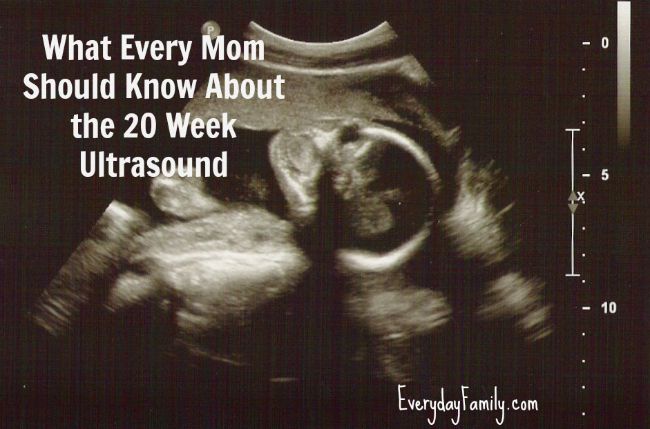
Around the eight-minute mark of the video, Shawn and Andrew discuss the moment their doctor discovered two concerning findings at their 20-week ultrasound: the baby's kidneys were dilated (a little more urine visible than normal), and the umbilical cord had just two blood vessels instead of three, which is also called having a single umbilical artery.
Neither condition is rare – 3% of normal fetuses have mild increases in fluid in their kidneys and about 1% of all fetuses have a single umbilical artery.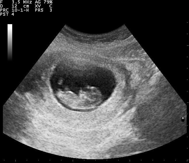 However, having the two findings might be indicative of Down syndrome.
I was struck by her use of the word “helpless” to describe how they felt in that moment, adding to the stress they already were feeling having previously gone through a miscarriage. Shawn said that as "control freak" athletes and new parents, it was difficult to accept that there was nothing they could do to improve their baby's health. However, they could pursue genetic testing if they wanted to know for sure. They opted for testing, and the couple recently shared that their testing came back negative for genetic anomalies – their baby does not have Down syndrome.
As a maternal-fetal medicine specialist (MFM), I often see patients who have to make decisions like Shawn and Andrew did. Their story inspired me to share three basic questions to ask if your provider is concerned your baby might have Down syndrome.
1. Why do you think my baby is at risk for Down syndrome?
There are multiple reasons your doctor might be concerned about Down syndrome, the most common of which is the age of the mother. Research has shown that as women age, their eggs tend to develop more chromosomal anomalies.
Your doctor might also be concerned if you had an abnormal screening, such as:
- Cell free fetal DNA screen. Also known as non-invasive prenatal testing or screening (NIPT/NIPS), this screening examines DNA fragments in a pregnant woman's blood for potential indicators of a genetic condition. Many patients are choosing to have this test as part of routine prenatal care in the first trimester, even if they are at low risk for Down syndrome.
- First trimester nuchal translucency screen. This ultrasound exam measures the amount of fluid at the back of the baby's neck. Excessive fluid can indicate a risk for Down syndrome. Adding in results from blood work gives you a numerical risk for having a baby with Down syndrome.
- Second trimester multiple marker screen. This test measures risk factors associated with levels of specific substances in a pregnant woman's blood that come from the placenta, the baby, or both.

- Ultrasound. As in Shawn's case, certain physical findings seen on an ultrasound might be associated with Down syndrome.
2. Do you want to know for sure before delivery?
It comes down to your level of concern about the results. I've had plenty of patients who opt for Down syndrome screening and, when the results show elevated risk, decide not to pursue diagnostic testing. That decision is completely OK.
However, you might want to have more certainty whether your child has a genetic condition and potentially which one. Some patients choose to undergo genetic testing to get more information and feel more prepared, while others prefer not to know for their own personal reasons. It's entirely the parents' choice.
If you do want to know more, we can perform further testing. Talk to your provider, who can recommend the most appropriate test. Certain diagnostic tests are available only during certain trimesters and some carry a risk of pregnancy loss.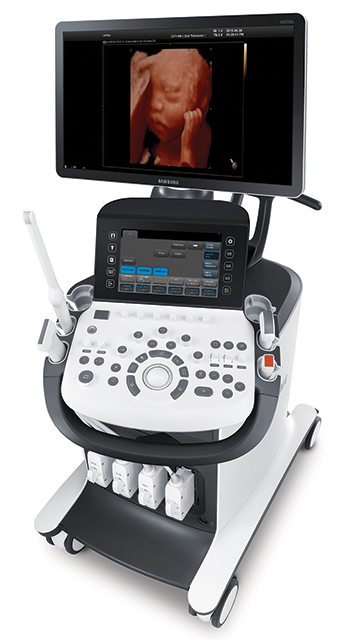 Also, keep in mind that some tests require a longer waiting period to return results.
3. What options are available for my stage of pregnancy?
Below are the diagnostic testing options available during the first and second trimester if screening suggests an increased risk for Down syndrome. We do not screen for Down syndrome after the second trimester, but invasive testing can be performed in the third trimester if new ultrasound findings that are worrisome are found later in pregnancy.
In the first trimester, you can consider a diagnostic test such as chorionic villus sampling, which is a biopsy of placental tissue. We usually don’t recommend having another screening test after you've already had a positive one. However, I’m seeing more patients select cell free fetal DNA screening to avoid the risks of invasive testing.
If your pregnancy was considered low risk for Down syndrome but you had an abnormal NIPT, I’d recommend using this positive predictive value calculator from the National Society of Genetic Counselors to estimate the risk. Enter the condition for which your test came back positive (such as Down syndrome) and the age you’ll be at delivery. The calculator will give you the probability that the NIPT result was right and that your baby has Down syndrome. Young women are often surprised to learn that even though their test came back abnormal, the true risk of their baby having Down syndrome is less than 50%.
In the second trimester, you can consider amniocentesis (analyzing the fluid around the baby) or fetal blood sampling. These tests provide the most complete information but do carry a small risk (less than 1 in 300 for amniocentesis) of pregnancy loss. As such, many patients who have an abnormal second trimester screen opt for less-invasive NIPT over diagnostic testing.
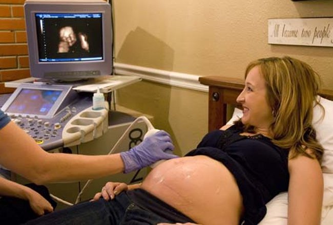
Dr. Robyn Horsager-Boehrer explains step-by-step what obstetricians are looking for when they conduct 18- to 20-week ultrasounds on pregnant women. You'll see as they check for birth defects such as Down syndrome and spina bifida.
A word about abnormal ultrasound screenings
Counseling patients about abnormal ultrasound findings is more complex than discussing next steps after abnormal screening results. Recommendations in this scenario must be tailored to the specific abnormality the provider sees.
Some abnormalities, such as mild fluid collections in the kidneys and a small or absent nose bone are considered soft markers for Down syndrome, which means the trait is associated with but not necessarily indicative of the condition. Some of these markers are more indicative of a higher risk of Down syndrome, such as increased fluid buildup in the back of the neck.
If you have your full ultrasound report and want to learn more, consider entering the findings in this Down syndrome risk calculator from Perinatology. Here, you can use the cumulative findings to estimate the risk for the condition and help you decide whether to pursue genetic testing.
We typically don’t suggest another round of non-invasive testing when we see major anomalies unless we have a good idea of which genetic condition might be affecting the baby. Diagnostic testing for total number of chromosomes, as well as changes like small additions or deletions, will gives you more information in the setting of fetal abnormalities.
However, rapid advances in non-invasive testing are changing recommendations. If you're concerned, talk with an MFM about next steps that might include advanced diagnostic testing.
Like Shawn Johnson East, you might be nervous or worried about your pregnancy due to positive results from a Down syndrome screening. Whether you want to know more or you'd prefer not to pursue further testing, you can reach out to us for information, answers, and support at any point during your pregnancy.
To visit with an maternal-fetal medicine specialist, call 214-645-8300 or request an appointment online
Your Pregnancy Matters
- Robyn Horsager-Boehrer, M.
 D.
D.
November 15, 2022
Your Pregnancy Matters
- Robyn Horsager-Boehrer, M.D.
November 7, 2022
Mental Health; Your Pregnancy Matters
- Robyn Horsager-Boehrer, M.
 D.
D.
October 11, 2022
Prevention; Your Pregnancy Matters
- Robyn Horsager-Boehrer, M.D.
October 4, 2022
Mental Health; Your Pregnancy Matters
- Meitra Doty, M.
 D.
D.
September 27, 2022
Your Pregnancy Matters
- Robyn Horsager-Boehrer, M.D.
September 20, 2022
Men's Health; Women's Health; Your Pregnancy Matters
- Yair Lotan, M.
 D.
D.
September 6, 2022
Your Pregnancy Matters
August 29, 2022
Your Pregnancy Matters
- Patricia Santiago-Munoz, M.D.
August 23, 2022
More Articles
Ultrasound scan spots Down's syndrome
Ultrasound scan spots Down's syndrome
Download PDF
- Published:
- Kendall Powell
Nature (2002)Cite this article
-
55k Accesses
-
1 Altmetric
-
Metrics details
Nasal bone holds key to safer test for birth defects.
You have full access to this article via your institution.
Download PDF
Download PDF
Women over 35 have a 1-in-300 chance of having a Down's syndrome baby. Credit: © ISUOG
An ultrasound scan could save many mothers the decision over whether to have an amniocentesis and risk losing a baby.
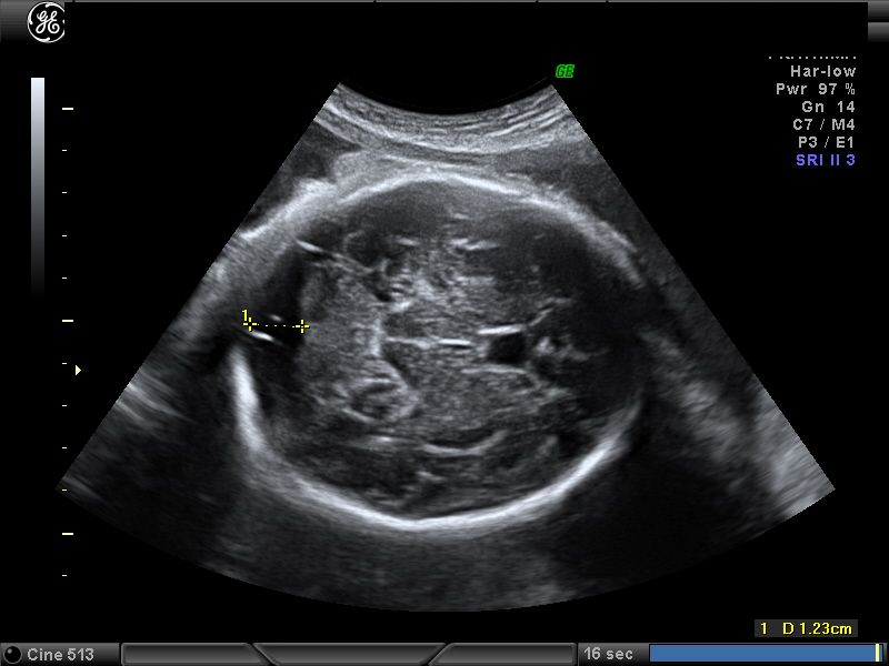
Nearly two-thirds of 15-22-week-old fetuses with Down's syndrome lack a nasal bone, fetal-medicine specialist Kypros Nicolaides, of King's College, London, and his colleagues found1.
For normal fetuses, the figure is 1%. This makes it unlikely that the test would wrongly diagnose Down's syndrome.
The nasal scan is more accurate than previous ultrasound markers, such as the length of leg bones. Some UK and US doctors are already using the nose-bone scan in combination with other tests.
The detection of Down's syndrome requires a sample of fetal cells from the fluid surrounding the fetus. This test, called an amniocentesis, causes miscarriage in about 1% of women who have it.
The nose-bone scan will reduce these tests, says Nicolaides. Women over 35 have a 1-in-300 chance of having a Down's syndrome baby. Each year, in the United States, 375,000 such women are encouraged to have an amniocentesis.
The scan could save many mothers the agonizing decision over whether to have an amniocentesis and risk losing a normal baby. "If the nasal bone is present, then the risk of having a baby with Down's syndrome is automatically reduced to one-third of what it was before the scan," says Nicolaides.
The nasal bone can be a good marker for Down's syndrome, agrees Rosemary Reiss, who practises maternal fetal medicine at Brigham and Women's Hospital in Boston, Massachusetts.
But the proportion of false alarms jumps to 8% for patients of African descent, says Reiss. Differences among ethnic groups will have to be understood for screening to succeed, she says.
Testing time
The nasal-bone test gives similar results in fetal scans as early as 11-14 weeks2. The 15-22-week point coincides with the traditional check-up for high-risk mothers. "This is a simple scan that women have anyway," says Nicolaides.
The new test will help give patients a better idea of the risk they face, says Richard O'Shaughnessy, director of the fetal treatment programme at Ohio State University. He suggests combining it with blood tests and another ultrasound marker that looks for fluid collecting behind the neck.
References
Cicero, S. et al. Nasal bone hypoplasia in trisomy 21 at 15-22 weeks' gestation. Ultrasound in Obstetrics and Gynecology, Published online, doi:10.1002/uog.19, (2002).
Cicero, S.
 , Curcio, P., Papageorghiou, K. H., Sonek, J.. & Nicolaides, A.B. Absence of nasal bone in fetuses with trisomy 21 at 11-14 weeks of gestation: an observational study. Lancet , 358, 1665 - 1667, (2001).
Article Google Scholar
Download references
Authors
- Kendall Powell
View author publications
You can also search for this author in PubMed Google Scholar
Rights and permissions
Reprints and Permissions
About this article
Children's ultrasound in Nizhny Novgorod
Specify the availability of this service in a particular branch by tel. (831) 4-30-01-30
Ultrasound is the safest, least invasive and most informative method of modern diagnostics of general diseases. With the help of ultrasound, doctors have the opportunity to look beyond the boundaries of ordinary perception, assess the state of all organs and systems from the inside in the smallest detail and make a diagnosis with high accuracy even for a small patient.
Ultrasound diagnostics is based on the physical properties of an ultrasonic wave, which does not have a radiation effect on the body, but projects the wave signal onto the screen, which is completely safe. Children's ultrasound can be used already in the maternity hospital from the first hours of a baby's life.
Ultrasound accompanies the baby even before birth, when, using the 3-4D function, the mother gets to know him and assesses his condition in utero. Ultrasound is necessary for children in the first month after birth to monitor the state of the brain, heart, liver, kidneys, pancreas, hip joints of the baby to identify or exclude congenital developmental pathologies and then once a year to monitor changes in internal organs or according to indications. Ultrasound as a mandatory examination is included in the patronage program for children at the Zdorovenok clinic.
The question is often asked - where is the best place to have an ultrasound scan for a child?
The main thing in choosing a clinic for pediatric ultrasound in Nizhny Novgorod is the resolution of the device and the experience of the specialist conducting the study. Many clinics use low-resolution equipment, which is the same as a black and white TV compared to a 3D plasma digital TV. This significantly affects the quality and accuracy of the examination. The latest generation devices, such as MyLab Class C, provide an opportunity not only to deeply assess the structure of organs, but also to obtain data on the blood supply and pathological processes taking place in them.
Children's clinic "Zdorovenok" performs all types of ultrasound examinations of children, including ultrasound of newborns, dopplerography and neurosonography, etc. (St. Belinsky, 71/1) and Philips Epiq 7 (St. Vorovskogo, 22) is a clear, high-quality image and visualization of formations of small size of superficial organs, incl. in young children, all transducers have a really wide frequency range. An extended set of matrix sensors make it possible to obtain a three-dimensional image.
PHILIPS EPIQ-7 is a unique today's expert-class ultrasound device, with the help of which ultrasound has become an even more informative diagnostic method, competing even with CT and MRI images and at the same time having an incomparably lower dose of radiation exposure.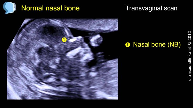 Contrast-enhanced examinations, 2 types of elastography, Fushion-technology for superimposing images in real time - all this is an innovative ultrasound diagnostic device PHILIPS EPIQ-7.
In the clinic on Rodionova, 199 doctors work on the VOLUSON E8 ultrasound machine - it supports innovative imaging technologies - HDlive Silhouette and HDlive Flow, which allow you to see the smallest details. The SonoRenderlive algorithm simplifies the workflow and gives you the ability to render surfaces by defining the transition area between tissue and fluid.
Some types of ultrasound require special preparation for the most accurate results.
Ultrasound diagnostics is available in each of the 4 branches of the Zdorovenok children's clinic.
In the children's clinic "Zdorovenok" experienced children's doctors of ultrasound diagnostics:
- Akatova Larisa Nikolaevna
- Baldina Natalia Nikolaevna
- Belousova Svetlana Viktorovna
- Burov Evgeny Valentinovich
- Ivantsova Elena Andreevna
- Maslenikova Elena Alexandrovna
- Odintsova Julia Evgenievna
- Smotrina Elena Viktorovna
Ultrasound examinations at Zdorovenok Clinic
Children's clinic "Zdorovenok" conducts all types of ultrasound examinations of children, including ultrasound of newborns, dopplerography and triplex scanning. Children's ultrasound in Nizhny Novgorod is carried out on a high-precision My Lab Class C device. This is the latest development of the European company Esaote, recommended specifically for examining children. The scanning depth of the sensors of the device is 25-30 cm, which allows diagnosing even deeply located organs, in particular the abdominal cavity and hip joints.
Neurosonography is an ultrasound examination of the brain in children under 1 year of age, which is performed through the fontanelle (cartilaginous connective tissue between the bones of the child's skull) to assess the state of the medulla, its anatomical structure, in order to identify or exclude brain developmental malformations, inflammatory and other changes. Neurosonography is recommended for all children aged 1 to 3 months.
Duplex scanning of cerebral vessels - assessment of functional parameters of blood flow, anatomical changes in the child's cerebral vessels, their patency and bends for the early detection of pathological changes in vessels, such as atherosclerotic plaques and other circulatory disorders.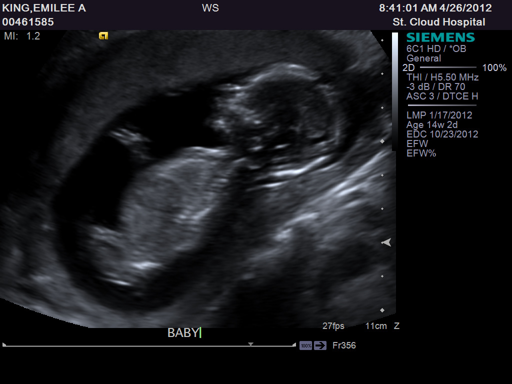 Dopplerography of vessels is used to identify the causes of headaches and dizziness in a child, memory loss, attention and other neurological diseases.
Echocardiography - ultrasound examination of the heart to assess the structure of the child's heart, its functional state and early detection of congenital and acquired malformations. It is also prescribed in the presence of heart murmurs and diseases of the thyroid gland
Ultrasound of the kidneys - performed to assess the state of the urinary excretory system in order to identify or exclude malformations in the child, since in most cases they are asymptomatic. Also, children's ultrasound of the kidneys is prescribed for suspected inflammatory processes in the kidneys (for example, pyelonephritis)
Ultrasound of the abdominal organs - assessment of the structure of the liver, its size, as well as the spleen, pancreas and gallbladder to detect abnormalities in case of suspected diseases of these organs.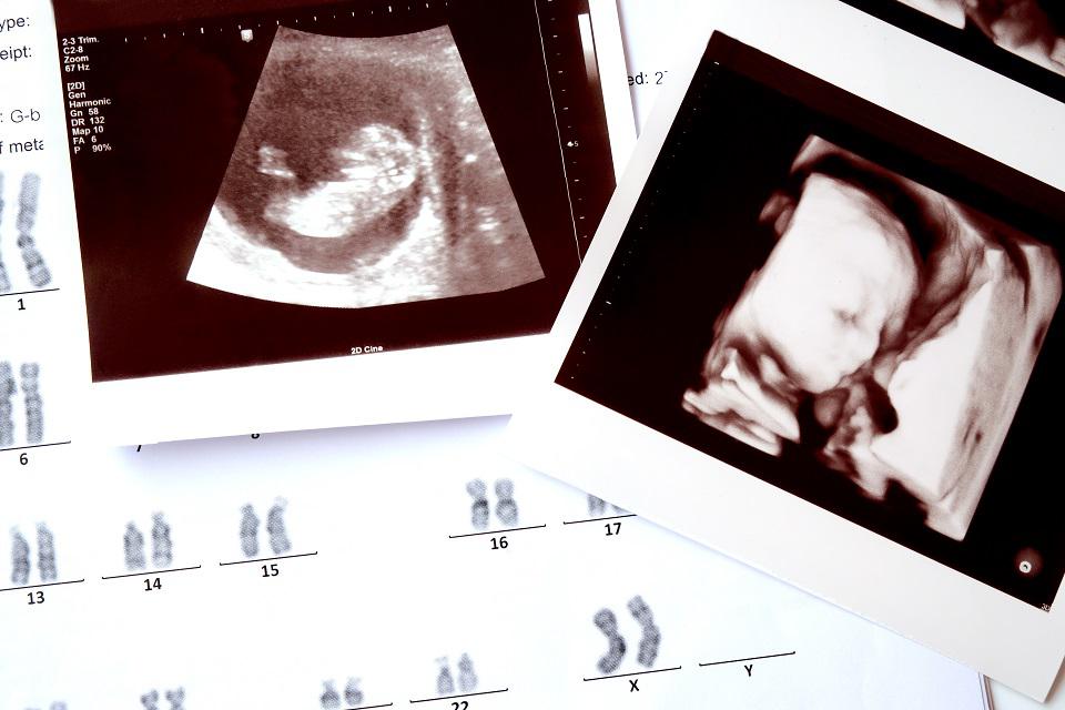 In infants, abdominal ultrasound allows early detection of signs of reactive pancreatitis (pancreatic changes) and effective treatment.
Ultrasound of the hip joints - the possibility of diagnosing underdevelopment or abnormal development of the hip joints in the form of congenital hip dislocation. Ultrasound of the hip joints should be performed in all children aged 1 to 3 months. The earlier the disease is detected, the greater the possibilities of its drug treatment without complications in the future.
Ultrasound of the pelvic organs in girls organs of the girl and early pathological processes for further treatment.
Ultrasound of the scrotum is performed for boys of any age and allows you to assess the location and shape of the testicles, the degree of their blood supply, their size compliance with the norm for age, the presence of congenital malformations of the reproductive system and the presence of additional formations (neoplasms, tumors) in the testicular tissue.
Preparing the child for examination
Some types of ultrasound examinations require special preparation for the most accurate results:
- Abdominal ultrasound.
It is important for parents to remember that an abdominal ultrasound is performed on an empty stomach, so both children and adults should not eat for at least six hours before the procedure. This examination is easier to carry out in the morning, before breakfast. If the examination is not carried out in the morning and the child wants to eat, then you can eat a piece of dried white bread and drink a mug of unsweetened tea.
The night before, the child can have dinner, but exclude gas-producing foods from the diet (black bread, dairy products, cabbage, apples, legumes). If the child is a newborn or infancy, then the study is carried out 3-4 hours after feeding (that is, before the next feeding).
- Ultrasound of the kidneys, adrenal glands, bladder, prostate, bladder, Ultrasound of the pelvic organs in girls - performed with a full bladder.
For other types of ultrasound such as:
- Ultrasound of the lymph nodes;
- Ultrasound of the hip joints;
- Neurosonography;
- Thyroid ultrasound;
- Ultrasound of the head, neck vessels;
- breast ultrasound;
- Ultrasound of the scrotum;
- Echocardiography with vascular Doppler.
Special preliminary preparation of children for the study is not required.
Regular ultrasound examinations at the Zdorovenok clinic is a great opportunity for parents to know EVERYTHING about their baby's health and control his development year after year! 8 (831) 430-01-30.
Ultrasound of the fetus in Nizhny Novgorod at the Tonus clinic
Ultrasound diagnostics is an integral element of monitoring the growth and development of the unborn baby and the health of the mother. This study is carried out both to establish the presence of pregnancy, and to monitor the growth and development of the fetus (ultrasound of the fetus), the formation of its anatomical structures. Fetal ultrasound is also a screening diagnosis, that is, it is aimed at early detection of developmental pathologies, threats of abortion and other complications. Therefore Ultrasound of the fetus must be done so that the expectant mother is sure that her unborn child will be born on time and healthy!
Ultrasound of the fetus during pregnancy is necessary at least 3 times.
Ultrasound of the fetus at different stages of pregnancy provides certain information about its growth and development at this stage, so ultrasound is performed several times during pregnancy.
What can you find out with a fetal ultrasound at different stages of pregnancy?
The pregnancy can be confirmed at 5 to 8 weeks. At such early stages of pregnancy (with its uncomplicated course), the diameter of the fetal egg and the coccyx - the parietal size of the embryo are measured. First 6 weeks, it is possible to register fetal heartbeats. After 7 weeks of pregnancy, ultrasound determines the motor activity of the fetus. These signs are an undoubted criterion for assessing the vital activity of the fetus during ultrasound.
The optimal time for the first fetal ultrasound is between 10 and 12 weeks of pregnancy.
An obligatory component of fetal ultrasound at this time is fetometry (determining the size of the fetus), which allows you to assess the compliance of the size of the fetus with the gestational age, clarify the gestational age and diagnose congenital malformations or fetal growth retardation. At this time, facial features of the future baby begin to form, which can be seen.
It is extremely important that at this time to do an ultrasound of the fetus, which allows you to identify signs that indicate chromosomal pathologies (Down syndrome, Turner syndrome, Patau syndrome, Turner syndrome, etc.). Markers of chromosomal abnormalities are necessarily included in the ultrasound protocol of the fetus at this time.
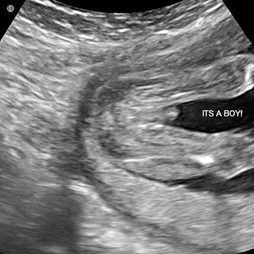
Ultrasound of the fetus at 20-24 weeks allows you to examine the fetus for malformations, assess the degree of development and functioning of internal organs, the musculoskeletal system, and the nervous system of the fetus. The nature of the amniotic fluid (their quantity and transparency) surrounding the baby is also assessed, the state of the placenta (its size and location), which unites the woman and the fetus into a single organism, the state of the umbilical cord is determined.
Fetal ultrasound at 30 to 34 weeks is a control study.
At this time, all systems and organs of the unborn child are sufficiently formed and actively functioning. An important component of fetal ultrasound at this time is the measurement of all fetometric indicators to exclude the syndrome of fetal growth retardation, which manifests itself after 30 weeks. Amniotic fluid, the state of blood flow between the circulatory systems of the mother and fetus are assessed. An ultrasound at this time gives an idea of how ready a woman is for childbirth.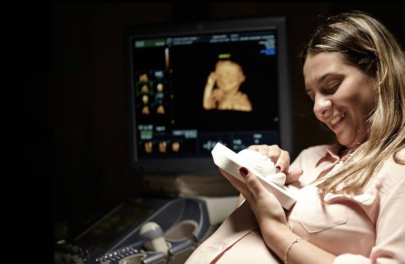 Sometimes it will be necessary to do an ultrasound of the fetus before childbirth, if the doctor needs to clarify the position of the fetus and its possible entanglement with the umbilical cord.
According to various indications (including the detected pathology of pregnancy or fetus) of the obstetrician-gynecologist leading your pregnancy, the frequency of fetal ultrasound can be increased.
Where can I get a fetal ultrasound?
Future parents for whom the question is relevant, where to do a fetal ultrasound? Ultrasound of the fetus in Nizhny Novgorod You can do it at the Tonus Medical Center. The clinic uses modern expert-class equipment that has a high resolution, which means it allows you to get a high-quality image (including in 3d mode, that is, a three-dimensional image in real time). In our clinic (in Nizhny Novgorod), fetal ultrasound is performed by competent and attentive specialists who will explain the results of the study to future parents in detail, talk and, of course, dispel fears and doubts.


