Displaced hips in babies
Developmental dysplasia of the hip
Developmental dysplasia of the hip (DDH) is a condition where the "ball and socket" joint of the hip does not properly form in babies and young children.
It's sometimes called congenital dislocation of the hip, or hip dysplasia.
The hip joint attaches the thigh bone (femur) to the pelvis. The top of the femur (femoral head) is rounded, like a ball, and sits inside the cup-shaped hip socket.
In DDH, the socket of the hip is too shallow and the femoral head is not held tightly in place, so the hip joint is loose. In severe cases, the femur can come out of the socket (dislocate).
DDH may affect 1 or both hips, but it's more common in the left hip. It's also more common in:
- girls
- firstborn children
- families where there have been childhood hip problems (parents, brothers or sisters)
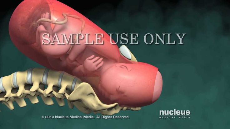
- babies born in the breech position (feet or bottom downwards) after 28 weeks of pregnancy
Without early treatment, DDH may lead to:
- problems moving around, for example a limp
- pain
- osteoarthritis of the hip and back
With early diagnosis and treatment, children are less likely to need surgery, and more likely to develop normally.
Diagnosing DDH
Your baby's hips will be checked as part of the newborn physical screening examination within 72 hours of being born, and again at 6 to 8 weeks of age.
The examination involves gently moving your baby's hip joints to check if there are any problems. It should not cause them any discomfort.
If a doctor, midwife or nurse thinks your baby's hip feels unstable, they should have an ultrasound scan of their hip between 4 and 6 weeks old.
Babies should also have an ultrasound scan of their hip between 4 and 6 weeks old if:
- there have been childhood hip problems in your family
- your baby was born in the breech position (feet or bottom downwards) after 28 weeks of pregnancy
If you have had twins or multiples and 1 of the babies was in the breech position, each baby should have an ultrasound scan of their hips by the time they're 4 to 6 weeks old.
Sometimes a baby's hip stabilises on its own before the scan is due, but they should still be checked to make sure.
Get help and support from the charity Steps if your baby's been diagnosed with DDH
Treating DDH
Pavlik harness
Babies diagnosed with DDH early in life are usually treated with a fabric splint called a Pavlik harness.
This secures both of your baby's hips in a stable position and allows them to develop normally.
Credit:
DR P. MARAZZI/SCIENCE PHOTO LIBRARY https://www.sciencephoto.com/media/729142/view
The harness needs to be worn constantly for 6 to 12 weeks and should not be removed by anyone except a health professional.
The harness may be adjusted during follow-up appointments. Your clinician will discuss your baby's progress with you.
Your hospital will provide detailed instructions on how to look after your baby while they're wearing a Pavlik harness.
This will include information on:
- how to change your baby's clothes without removing the harness – nappies can be worn normally
- cleaning the harness if it's soiled – it still should not be removed, but can be cleaned with detergent and an old toothbrush or nail brush
- positioning your baby while they sleep – they should be placed on their back and not on their side
- how to avoid skin irritation around the straps of the harness – you may be advised to wrap some soft, hygienic material around the bands
Eventually, you may be given advice on removing and replacing the harness for short periods of time until it can be permanently removed.
You'll be encouraged to allow your baby to move freely when the harness is off. Swimming is often recommended.
Surgery
Surgery may be needed if your baby is diagnosed with DDH after they're 6 months old, or if the Pavlik harness has not helped.
The most common surgery is called reduction. This involves placing the femoral head back into the hip socket.
Reduction surgery is done under general anaesthetic and may be done as either:
- closed reduction – the femoral head is placed in the hip socket without making any large cuts
- open reduction – a cut is made in the groin to allow the surgeon to place the femoral head into the hip socket
Your child may need to wear a cast for at least 12 weeks after the operation.
Their hip will be checked under general anaesthetic again after 6 weeks, to make sure it's stable and healing well.
After this investigation, your child will probably wear a cast for at least another 6 weeks to allow their hip to fully stabilise.
Some children may also require bone surgery (osteotomy) during an open reduction, or at a later date, to correct any bone deformities.
Late-stage signs of DDH
The newborn physical screening examination, and the infant screening examination at 6 to 8 weeks, aim to diagnose DDH early.
But sometimes hip problems can develop or show up after these checks.
It's important to contact a GP as soon as possible if you notice your child has developed any of the following symptoms:
- 1 leg cannot be moved out sideways as far as the other when you change their nappy
- 1 leg seems to be longer than the other
- 1 leg drags when they crawl
- a limp or "waddling" walk
Your child will be referred to an orthopaedic specialist in hospital for an ultrasound scan if your doctor thinks there's a problem with their hip.
Hip-healthy swaddling
A baby's hips are naturally more flexible for a short period after birth. But if your baby spends a lot of time tightly wrapped (swaddled) with their legs straight and pressed together, there's a risk this may affect their hip development.
Using hip-healthy swaddling techniques can reduce this risk. Make sure your baby is able to move their hips and knees freely to kick.
Find out more about hip-healthy swaddling on the International Hip Dysplasia Institute website
Page last reviewed: 08 August 2022
Next review due: 08 August 2025
Developmental Dislocation (Dysplasia) of the Hip (DDH) - OrthoInfo
The hip is a ball-and-socket joint. In a normal hip, the ball at the upper end of the thighbone (femur) fits firmly into the socket, which is part of the large pelvis bone. In babies and children with developmental dysplasia (dislocation) of the hip (DDH), the hip joint has not formed normally. The ball is loose in the socket and may be easy to dislocate.
Although DDH is most often present at birth, it may also develop during a child's first year of life. Recent research shows that babies whose legs are swaddled tightly with the hips and knees straight are at a notably higher risk for developing DDH after birth. Given the popularity of swaddling, it is important for parents to learn how to swaddle their infants safely, and to understand that when done improperly, swaddling may lead to problems like DDH.
In all cases of DDH, the socket (acetabulum) is shallow, meaning that the ball of the thighbone (femur) cannot firmly fit into the socket. Sometimes, the ligaments that help to hold the joint in place are stretched. The degree of hip looseness, or instability, varies among children with DDH.
- Dislocated. In the most severe cases of DDH, the head of the femur is completely out of the socket.
- Dislocatable. In these cases, the head of the femur lies within the acetabulum, but it can easily be pushed out of the socket during a physical examination.

- Subluxatable. In mild cases of DDH, the head of the femur is simply loose in the socket. During a physical examination, the bone can be moved within the socket, but it will not dislocate.
(Left) In a normal hip, the head of the femur fits firmly inside the hip socket. (Right) In severe cases of DDH, the thighbone is completely out of the hip socket (dislocated).
In the United States, approximately 1 to 2 babies per 1,000 are born with DDH. Pediatricians screen for DDH at a newborn's first examination and at every well-baby checkup thereafter.
DDH tends to run in families. It can be present in either hip and in any individual. It usually affects the left hip and is more common in:
- Girls
- Firstborn children
- Babies born in the breech position (especially with feet up by the shoulders). The American Academy of Pediatrics now recommends ultrasound DDH screening of all female breech babies.
- Family history of DDH (parents or siblings)
- Oligohydramnios (low levels of amniotic fluid)
Some babies born with a dislocated hip will show no outward signs.
Contact your pediatrician if your baby has:
- Legs of different lengths
- Uneven skin folds on the thigh
- Less mobility or flexibility on one side
- Limping, toe walking, or a waddling gait
To Top
In addition to visual clues, your child's doctor will perform a careful physical examination to check for DDH, such as listening and feeling for "clunks" as the hip is put in different positions. The doctor will use specific maneuvers to determine if the hip can be dislocated and/or put back into proper position.
During the exam, your child's doctor will maneuver your baby’s legs and hips in certain ways to detect hip instability.
Reproduced and adapted from JF Sarwak, ed: Essentials of Musculoskeletal Care, ed. 4. Rosemont, IL, American Academy of Orthopaedic Surgeons, 2010.
Newborns identified as at higher risk for DDH are often tested using ultrasound, which can create images of the hip bones. For older infants and children, X-rays of the hip may be taken to provide detailed pictures of the hip joint.
- When DDH is detected at birth, it can usually be corrected with the use of a harness or brace.
- If the hip is not dislocated at birth, the condition may not be noticed until the child begins walking. At this time, treatment is more complicated, with less predictable results.
Treatment methods depend on a child's age as well as the severity of the DDH.
Newborns. The baby may be placed in a soft positioning device, called a Pavlik harness, for 1 to 3 months to keep the thighbone in the socket. This special brace is designed to hold the hip in the proper position while allowing free movement of the legs and easy diaper care. The Pavlik harness helps tighten the ligaments around the hip joint and promotes normal hip socket formation.
Newborns may be placed in a Pavlik harness for 1 to 3 months to treat DDH.
Parents play an essential role in ensuring the harness is effective. Your doctor and healthcare team will teach you how to safely perform daily care tasks, such as diapering, bathing, feeding, and dressing. It is very important to attend all of your baby's scheduled clinic visits so the doctor can check the hip and the fit of the Pavlik harness.
1 month to 6 months. Similar to newborn treatment, a baby's thighbone is repositioned in the socket using a harness or similar device. This method is usually successful, even with hips that are initially dislocated.
How long the baby will require the harness varies. It is usually worn full-time for at least 6 weeks, and then part-time for an additional 6 weeks.
If the hip will not stay in position using a harness, your child's doctor may try an abduction brace made of firmer material that will keep your baby's legs in position.
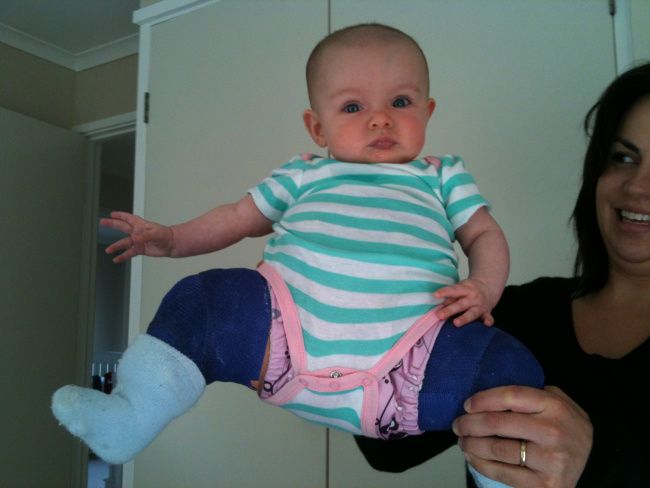
In some cases, a closed reduction procedure is required. Your child's doctor will gently move your baby's thighbone into proper position, then apply a body cast (spica cast) to hold the bones in place. This procedure is done while the baby is under anesthesia.
Caring for a baby in a spica cast requires specific instruction. Your child's doctor and healthcare team will teach you how to perform daily activities, maintain the cast, and identify any problems.
6 months to 2 years. Older babies are also treated with closed reduction and spica casting. Skin traction may be used for a few weeks prior to repositioning the thighbone. Skin traction prepares the soft tissues around the hip for the change in bone positioning. This may be done at home or in the hospital.
Surgical Treatment6 months to 2 years. If a closed reduction procedure is not successful at putting the thighbone in its proper position, open surgery is necessary. In this procedure, an incision is made at the baby's hip that allows the surgeon to clearly see the bones and soft tissues.
In some cases, the thighbone will be shortened to properly fit the bone into the socket. X-rays are taken during the operation to confirm that the bones are in position. Afterward, the child is placed in a spica cast to maintain the proper hip position.
Older than 2 years. In some children, the looseness worsens as the child grows and becomes more active. Open surgery is typically necessary to realign the hip. A spica cast is usually applied to maintain the hip in the socket.
RecoveryIn many children with DDH, a body cast and/or brace is required to keep the hip bone in the joint during healing. The cast may be needed for 2 to 3 months. Your child's doctor may change the cast during this time period.
X-rays and other regular follow-up monitoring are needed after DDH treatment until the child's growth is complete.
- Children treated with spica casting may have a delay in walking. However, when the cast is removed, walking development proceeds normally.
- The Pavlik harness and other positioning devices may cause skin irritation around the straps, and a difference in leg length may remain. Rarely, positioning in the Pavlik may also cause nerve compression in the leg, with loss of motion. The nerve almost always recovers if the harness is removed or adjusted.
- Growth disturbances of the upper thighbone are rare, but may occur due to a disturbance in the blood supply to the growth area in the thighbone.
- Even after proper treatment, a shallow hip socket may persist, and surgery may be necessary in early childhood to restore the normal anatomy of the hip joint.
If diagnosed early and treated successfully, children are able to develop a normal hip joint and should have no limitation in function. Left untreated, DDH can lead to pain and osteoarthritis by early adulthood. It may produce a difference in leg length or decreased agility.
Even with appropriate treatment, hip deformity and osteoarthritis may develop later in life. This is especially true when treatment begins after the age of 2.
To assist doctors in the management of pediatric developmental dysplasia of the hip, the American Academy of Orthopaedic Surgeons has conducted research to provide some useful guidelines. These are recommendations only and may not apply to every case. For more information: Pediatric Developmental Dysplasia of the Hip - Clinical Practice Guideline (CPG) | American Academy of Orthopaedic Surgeons (aaos.org)
To Top
Reviewed by members of
POSNA (Pediatric Orthopaedic Society of North America)
The Pediatric Orthopaedic Society of North America (POSNA) is a group of board eligible/board certified orthopaedic surgeons who have specialized training in the care of children's musculoskeletal health.
Learn more about this topic at POSNA's OrthoKids website:
Developmental Dysplasia of the Hip
Hip dysplasia in young children
At an orthopedist's appointment in the first month of a child's life, an unpleasant detail may turn out: the baby has immaturity of the pelvic bones.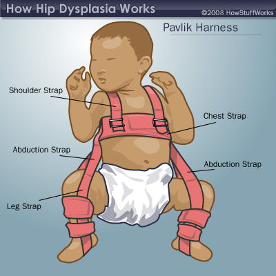 Most often, at the same time, the pediatrician pronounces the words “hip dysplasia”, which instantly frighten all young parents without exception. But being scared is not the right thing to do in this situation. You need to be patient and strictly follow the recommendations of the attending physician. Nevertheless, all doctors will be right when they tell you that joint dysplasia, which was left without the attention of parents and doctors in the first year of a baby’s life, can form the most severe inflammatory processes in a child by the age of two or three, painful hip dislocation and in the future - lameness for life.
Hip dysplasia is a congenital disorder of the formation of the joint that can cause dislocation or subluxation of the femoral head. In this condition, either underdevelopment of the joint, or its increased mobility in combination with connective tissue deficiency, can be observed. Predisposing factors are unfavorable heredity, gynecological diseases of the mother and pathology of pregnancy.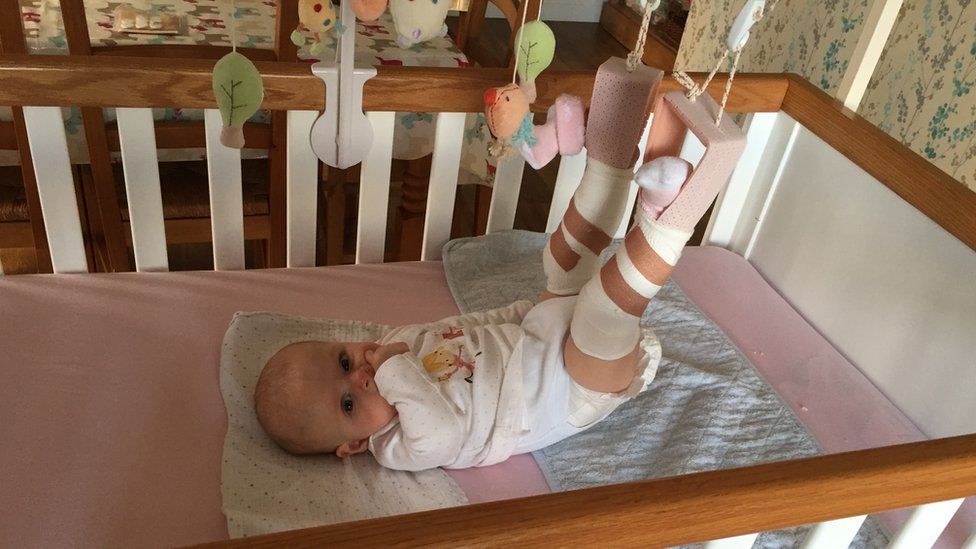 Hip dysplasia is 10 times more common in those children whose parents had signs of congenital hip dislocation and in those born with a breech presentation of the fetus, more often in the first birth. Often, dysplasia is detected during drug correction of pregnancy, during pregnancy complicated by toxicosis. The left hip joint is most often affected (60%), less often the right (20%) or both (20%). The relationship of morbidity with environmental problems has been noted. With untimely detection and lack of proper treatment, hip dysplasia can cause dysfunction of the lower limb and even disability. Therefore, this pathology must be identified and eliminated in the early period of the baby's life.
Hip dysplasia can present in a variety of ways. There are three main forms of dysplasia: acetabular dysplasia - acetabular dysplasia; dysplasia of the proximal femur; rotational dysplasia.
With timely detection and proper treatment, the prognosis is conditionally favorable.
Statistics say: up to 25% of newborn children have some form of hip dysplasia, in other words, they are born with subluxations.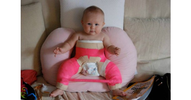 In most cases, under the constant supervision of an orthopedist, the joints “grow” on their own and return to the anatomical norm. For the rest, they just need a little help.
A preliminary diagnosis can be made even in the maternity hospital. In this case, you need to contact a pediatric orthopedist within 3 weeks, who will conduct the necessary examination and draw up a treatment regimen. In addition, to exclude this pathology, all children are examined at the age of 1-4 months. Particular attention is paid to children who are at risk. This group includes all patients with a history of maternal toxicosis during pregnancy, a large fetus, breech presentation, as well as those whose parents also suffer from dysplasia. If signs of pathology are detected, the child is sent for additional studies.
To clarify the diagnosis, methods such as radiography and ultrasonography are used. In young children, a significant part of the joint is formed by cartilage, which is not displayed on radiographs, so this method is not used until the age of 2-3 months.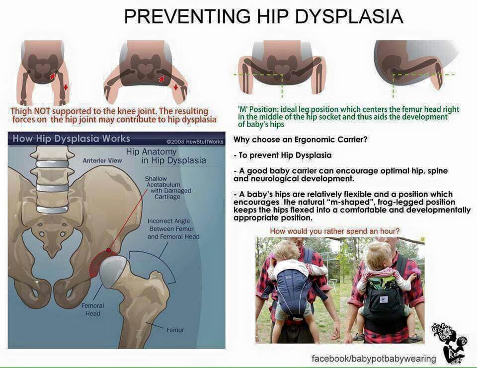 Ultrasound diagnostics is a good alternative to X-ray examination in children during the first months of life. This technique is practically safe and quite informative.
With dysplasia, the shape, relationship and size of the structures of the hip joint change significantly. The hip joint of a newborn, even in normal conditions, is an immature biomechanical structure. If the development of the joint is disturbed, the excessively elastic ligaments and the articular capsule are not able to hold the head of the femur in the articular cavity, it shifts upward and laterally (outwards). With certain movements, the femoral head can extend beyond the acetabulum. This condition of the joint is called "subluxation". In severe hip dysplasia, the head of the femur extends completely beyond the acetabulum, a condition called hip dislocation. Hip dysplasia can manifest itself not only as a violation of the acetabulum (acetabular dysplasia), but also as an abnormal development of the proximal femur.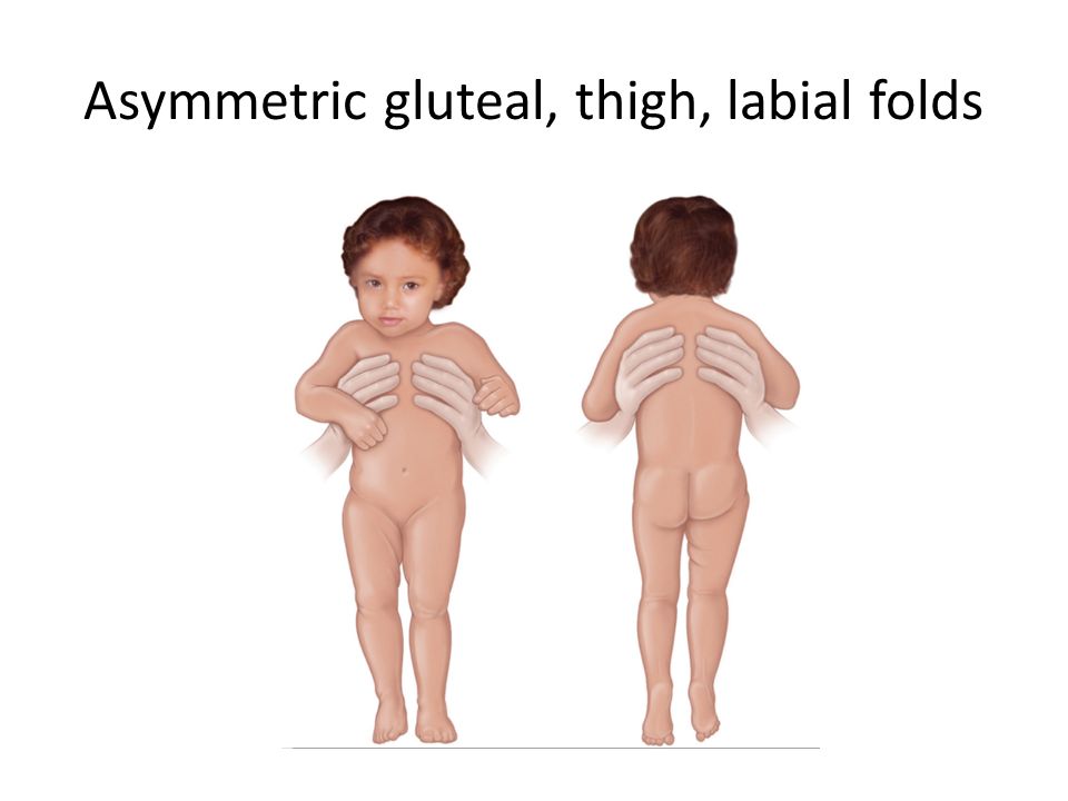
Hip dysplasia is suspected in the presence of shortened hip, asymmetric skin folds, limited hip abduction, and Marx-Ortolani slipping. Asymmetry of the inguinal, popliteal and gluteal skin folds is usually better detected in children older than 2-3 months. During the inspection, they pay attention to the difference in the level of location, shape and depth of the folds. It should be borne in mind that the presence or absence of this symptom is not enough to make a diagnosis. With bilateral dysplasia, the folds may be symmetrical. In addition, the symptom is absent in half of the children with unilateral pathology. The asymmetry of the inguinal folds in children from birth to 2 months is of little information, since it sometimes occurs even in healthy infants. But the most important sign indicating congenital dislocation of the hip is the “click” or Marx-Ortolani symptom. Slowly abduct the hips evenly to both sides. With normal relations in the joints, both hips in the position of extreme abduction almost touch the outer surfaces of the table plane. With dislocation, the femoral head slips into the acetabulum at the moment of abduction, which is accompanied by a characteristic push - the moment when the femoral head from the dislocation position is reduced into the acetabulum.
Another symptom that indicates the pathology of the joint is limitation of movement. In healthy newborns, the legs are retracted to a position of 80-90 ° and freely placed on the horizontal surface of the table. When the abduction is limited to 50-60°, there is reason to suspect a congenital pathology. In a healthy child of 7-8 months, each leg is retracted by 60-70°, in a baby with congenital dislocation - by 40-50°.
The main method of preventing hip dysplasia is wide swaddling. As soon as at 19In 1971, the national health program promoted wide swaddling; already a few years later, only 0.2% of children over the age of one year suffered from this disease. Orthopedic devices that securely fix the baby's legs in a bent and divorced form. These devices include all kinds of splints (a kind of spacers between the legs), plastic corsets and even plaster retainers. The most popular fixing device is the so-called Pavlik stirrups. Moreover, Pavlik here is not a boy who was the first to try a miracle unit on himself, but a talented Czech orthopedic doctor who came up with the idea of fixing a baby’s legs with a special harness. Massage and gymnastics. Your attending orthopedist will teach you specific exercises and techniques for daily massage and gymnastics, since the set of manipulations strictly depends on how the joint is under-formed. Use of carriers, slings, backpacks and car seats. But only those models that allow the baby to hold freely, legs wide apart. In the countries of Asia and Africa, where women have been carrying their babies on themselves since ancient times, tying them on their backs or on their stomachs (that is, the child spends all the time in a sitting position, with legs wide apart), there is no such phenomenon as hip dysplasia in children at all. exists.
Treatment should begin as soon as possible. Alas, the treatment of dysplasia is not a quick matter.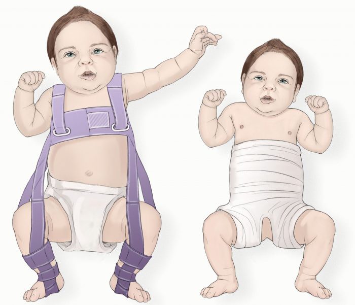 As a rule, it takes several months, sometimes - a year and a half. This is understandable: the hip joint cannot take the correct position and acquire reliable ligaments in a couple of days. But believe me, your efforts and patience are worth it! Of course, it’s not too pleasant to “hobble” your baby with orthopedic stirrups every day, and swaddle him with a pillow between his legs or “shackle” him in a plastic corset at night. But it’s better to be a little sad before he is even a year old, so that later you can see how famously he dances at his 17-18 years old at the prom. The opposite is true: to be touched by crooked legs now and do nothing, and then reap the terrible consequences of your carelessness ... Isn't it?
Radiologist of ME "3rd City Children's Clinical Clinic"
Krasovsky Vyacheslav Fedorovich
Pelvic tilt - treatment, symptoms, causes, diagnosis
The pelvis is one of the most important, although sometimes ignored, parts of the skeleton.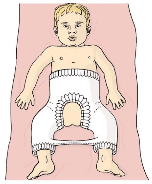
The pelvis is shaped like a basket with a tip and contains many vital organs, including the intestines and bladder. In addition, the pelvis is in the center of gravity of the skeleton. If the body is compared to a pencil balancing horizontally on a finger, its point of balance (center of gravity) will be the pelvis.
Therefore, it is obvious that the position of the pelvis greatly affects posture. This is the same as if the central block in the tower is displaced, in which case all blocks above the displacement are at risk of falling. And if you compare the central unit with a box, then the tilt can lead to the box falling out. Similar mechanisms take place when the pelvis is tilted, and the contents of the pelvis are shifted forward. As a result, there is a protruding abdomen and bulging of the buttocks. Since the pelvis is the junction of the upper and lower torso, it plays a key role in body movement and balance. The pelvic bones support the most important supporting part of the body - the spine.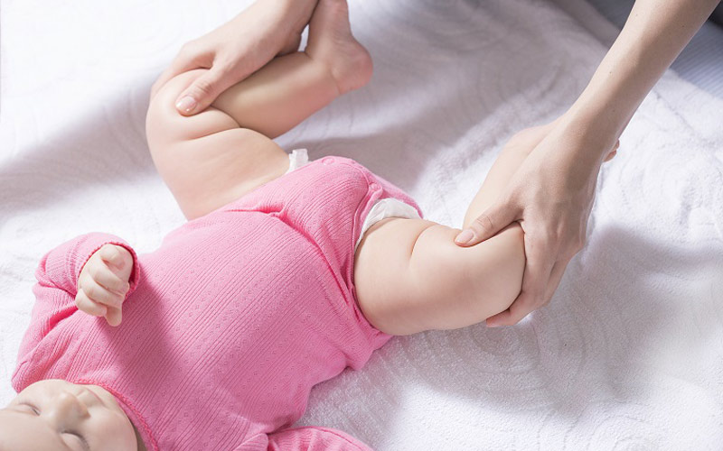 In addition, the pelvis allows the lower limbs and torso to move in a coordinated manner (in tandem). When the pelvis is in a normal position, various movements are possible, twisting, tilting and movement biomechanics are balanced and the distribution of load vectors is even. Displacement (skew) of the pelvis from normal positions causes dysfunctional disorders of the spine, as there is a change in the axis of distribution of loads during movement. For example, if there is an axle shift in a car, then the wheels wear out quickly. Something similar happens in the spine, there are leverage effects and excessive load on certain points, which lead to rapid wear of the structures of the spine. Therefore, often the main cause of pain in the back and neck is a change in the position of the pelvis (displacement, distortion). A change in position changes biomechanics, which can lead to degenerative changes in the spine, to disc herniation, scoliosis, osteoarthritis, spinal canal stenosis, sciatica, etc.
 Pelvic tilt also leads to pain and dysfunction in the neck, neck pain radiating to the shoulders, arms, contributes to the development of carpal tunnel syndrome and other problems in the extremities.
Causes of misalignment (displacement) of the pelvis
First of all, misalignment of the pelvis is caused by a normal muscle imbalance. Technology is developing very quickly and a sedentary lifestyle is one of the main reasons for the development of imbalance, because our body requires a certain amount of movement that it does not receive. Prolonged sitting and low physical activity are sufficient conditions for the development of muscle imbalance, leading to pelvic tilt and, as a result, the appearance of dysfunctional disorders in the spine and the occurrence of back pain.
Accidents and injuries are common causes of pelvic tilt , such as side impact, heavy lifting while twisting, falling to one side, carrying heavy loads from the side, such as carrying a child on the hip or carrying a heavy bag constantly on one shoulder. In women, the pelvis is less stable from birth than in men, since a certain flexibility and elasticity of the pelvic structures is necessary for the normal course of pregnancy and childbirth. Therefore, pregnancy is often the main cause of pelvic displacement in women.
Injury to the pelvic muscles is the most common cause of misalignment. Injured muscles tend to thicken and shift in order to protect the surrounding structures. If the muscles in the pelvic area, such as the sacrum, are damaged, then the tightening of the muscles will lead to an effect on the ligaments attached to the pelvis and joints. As a result, structures such as the sacroiliac joints will also have a certain disposition. Muscle compaction after damage persists until the muscle is fully restored and during this period of time the pelvis remains in an abnormal position.
Difference in leg length can also cause pelvic tilt and in such cases the tilt can be from right to left or vice versa. But the displacement can also be forward or backward, or it can be twisting of the pelvis.
Many conditions can lead to muscle spasms that cause pelvic twist. A disc herniation can cause muscle spasm of an adaptive nature and, in turn, antalgic scoliosis with functional pelvic tilt . Active people often experience tension in the calf muscles, which in turn creates tension around the pelvis. Surgery such as hip replacement can also cause the pelvis to reposition itself.
Since the pelvis is one of the most stressed areas of the body due to movement and weight support, movements that cause pain and stiffness are a clear indicator of pelvic alignment problems. Back pain in particular is a common indicator of pelvic tilt . In addition to participation in the movement in the pelvic cavity are: part of the digestive organs, nerves, blood vessels, reproductive organs. Therefore, in addition to back pain, there may be other symptoms, such as numbness, tingling, bladder and bowel problems, or reproductive problems. Most often, changes in the following muscles lead to pelvic disposition:
M. Psoas major (lumbar muscle) anatomically can lead to extension and flexion of the hip, which leads to an anterior displacement of the pelvis.
M.Quadriceps (quadriceps), especially the rectus muscle, can lead to hip flexion.
M.Lumbar erectors may cause lumbar extension.
M.Guadratus lumborum with bilateral compaction can cause an increase in lumbar extension.
M.Hip adductors (adductors of the thigh) can cause the pelvis to tilt forward as a result of internal rotation of the hip. This leads to shortening of the adductor muscles.
M. Gluteus maximus (gluteus maximus) is responsible for hip extension and is an antagonist of the psoas major muscle.
M.Hamstrings Muscle of the back of the thigh, this muscle can be hardened. The muscle can be weak, at the same time hardened due to the fact that it is a synergist of the gluteus maximus muscle and this can be of a compensatory nature. The deep muscles of the abdominal wall, including the transversus abdominis and internal obliques, may become tense due to weakening of the lumbar erectors muscles
Symptoms
Symptoms of pelvic misalignment can be moderate or severe and significantly affect the functionality of the body. With moderate misalignment, a person may feel unsteady when walking or frequent falls are possible.
The most common symptoms are pain:
- In the lower back (radiating to the leg)
- Pain in the thigh, sacroiliac joints or groin
- Pain in the knee, ankle or foot Achilles tendon
- Pain in shoulders, neck
If the pelvis is displaced for a long time, then the body will correct and compensate for the biomechanical disturbance and asymmetry and the corresponding adaptation of the muscles, tendons and ligaments will occur. Therefore, treatment may take some time. In addition, pelvic tilt can be difficult to correct, as a pathological stereotype of movements is formed over time. The longer the period of pelvic tilt, the longer it takes to restore normal muscle balance.
Diagnosis and treatment
Pelvic tilt is usually well diagnosed on physical examination of the patient. If it is necessary to diagnose changes in the spine or hip joints, instrumental examination methods are prescribed, such as radiography or MRI (CT).
