Can pap smears cause miscarriage
Pap smears are safe – before, during and after pregnancy | Women's Health | Your Pregnancy Matters
×
What can we help you find?Refine your search: Find a Doctor Search Conditions & Treatments Find a Location
Appointment New Patient Appointment
or Call214-645-8300
MedBlog
Women's Health; Your Pregnancy Matters
August 3, 2021
Women's Health; Your Pregnancy Matters
Yetunde Awosemusi, M. D. Obstetrics and Gynecology
Pregnancy induces changes in your body and your mind. Many mothers-to-be experience anxiety, which can lead some to second-guess their most routine medical procedures. While this mindset is understandable, one thing you can definitely cross off your “worry list” is the myth that Pap smears are not safe during pregnancy.
It is normal to experience some spotting after a Pap smear, even when you are pregnant. However, this natural response has caused some misconceptions that the Pap smear, a potentially life-saving procedure, isn’t safe during pregnancy.
The truth is that getting a Pap smear is safe for you and your baby; it will not cause a miscarriage.
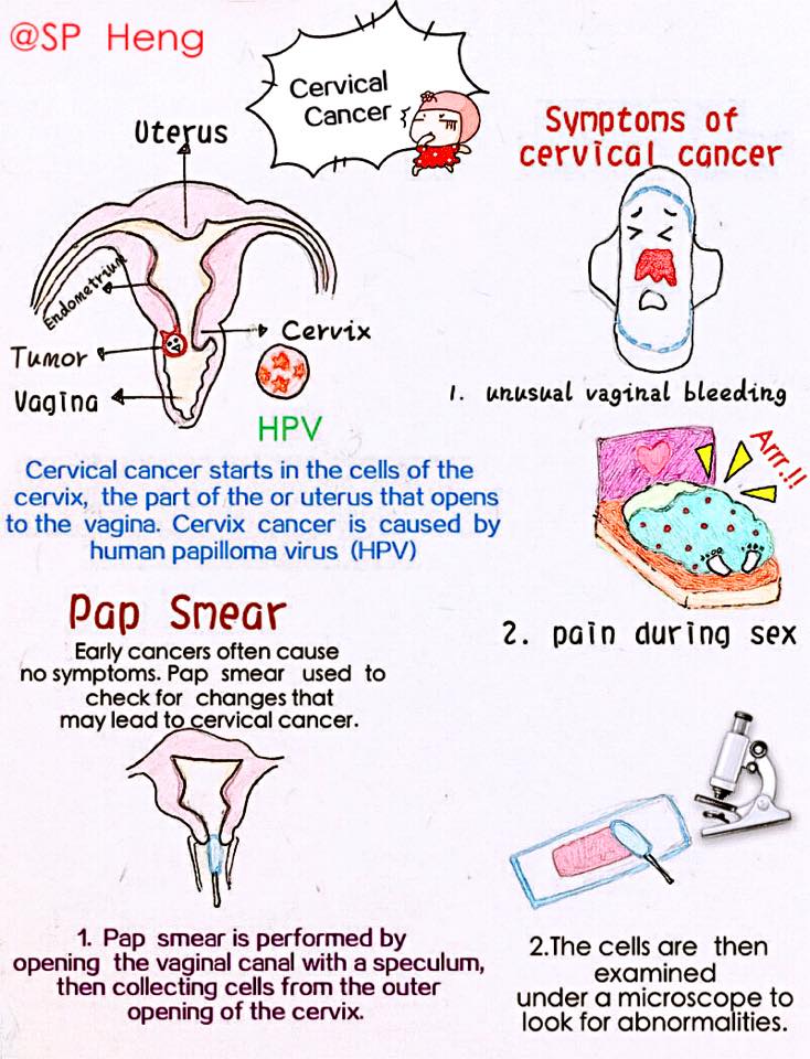
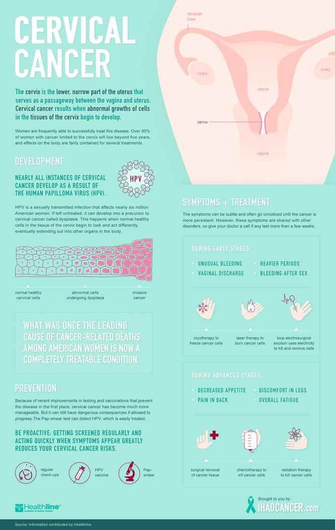
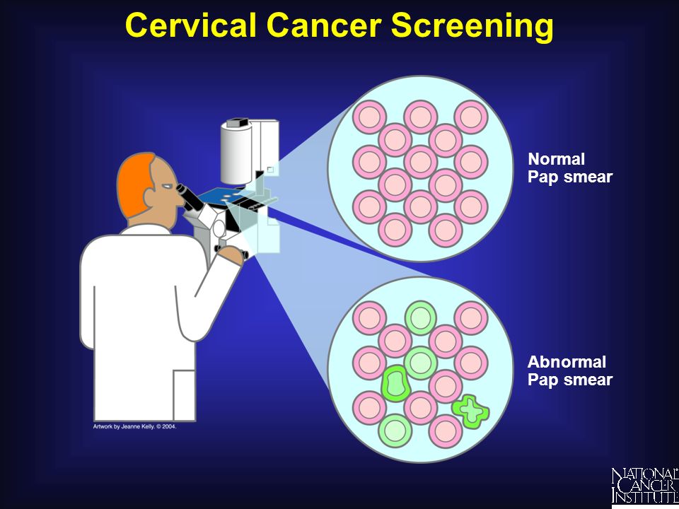
Whether you’re pregnant or not, the Pap smear is the gold standard for screening for cervical cancer. It allows us to collect cells from the surface of the cervix and examine them for abnormalities.
Cervical cancer and abnormal Pap smear results are almost always caused by the human papillomavirus (HPV), a sexually transmitted disease currently infecting around 80 million people in the United States. Though the virus is common, the likelihood of it causing cancer is much less common. In fact, most infections go away on their own. Cancer develops when the infection isn’t detected and managed appropriately, which is why Pap smears are so important.
Data from the Centers for Disease Control and Prevention (CDC) show that cervical cancer deaths have decreased over the last several decades as a result of increased Pap smears. If you follow up on your Pap smear results as recommended by your provider, we almost always will catch the cancer.
What’s even more encouraging is that the HPV vaccine, which the CDC recommends for all children ages 11 or 12, can prevent cervical cancer from developing. We still recommend routine Pap smears for people who receive the vaccine, but we are finding very low rates of abnormal Pap smears within this vaccinated population.
As more people recognize the importance of regular Pap smears, questions about the procedure continue to swirl. We’ve answered the most common ones below.
How often should I get a Pap smear?
We recommend a Pap smear every three years for women between the ages of 21 and 30. If your results are normal and you are negative for HPV, you can extend that to every five years from ages 31 to 65. Higher-risk populations, such as people who have an autoimmune disease, should discuss more frequent testing with their provider.
Do I need a Pap smear when I’m pregnant?
Reviewing your Pap smear history is an early, important step during prenatal care. If you received a normal test result within the last three years, you likely don’t need a Pap smear while pregnant.
We realize, however, that many newly pregnant people are seeing a doctor for the first time in quite a while – maybe their first time ever as an adult – and haven’t had a Pap smear before, or for several years. To support a healthy pregnancy, we want to rule out the possibility of cervical cancer development as soon as possible.
If I do need one, is it safe?
Pap smears while pregnant are safe for you and your baby. Any suggestion that it might cause a miscarriage is false. It is normal to experience minor bleeding after a Pap smear when you are pregnant because blood flow to the uterus increases, which causes the cervix to bleed more easily when touched.
Any type of bleeding is understandably alarming while pregnant, but in this case the blood is coming from the outside of your cervix – not inside the uterus, where the baby is safe and developing.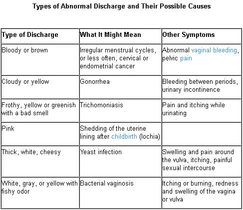
What happens if I have an abnormal Pap smear?
The next step may be to perform a colposcopy, which allows us to look more closely at the cervix and take a small tissue sample to test the abnormal cells for cancer. The colposcopy is safe during pregnancy – the only cells we touch are on the outside of the cervix, not the inside near your baby.
Generally, we divide these test results into what we call low-grade or high-grade “dysplasia” – the existence of abnormal cells:
- Low-grade dysplasia doesn’t usually progress to cancer, so we just watch it and will likely encourage you to come back in a year for another Pap smear.
- High-grade dysplasia has an increased chance of progressing to cancer, so we will likely recommend a loop electrosurgical excision procedure (LEEP) after delivery.
LEEP removes the outside portion of the cervix containing the abnormal cells, and it can increase the risk of pregnancy complications like preterm birth or losing the baby in the second trimester.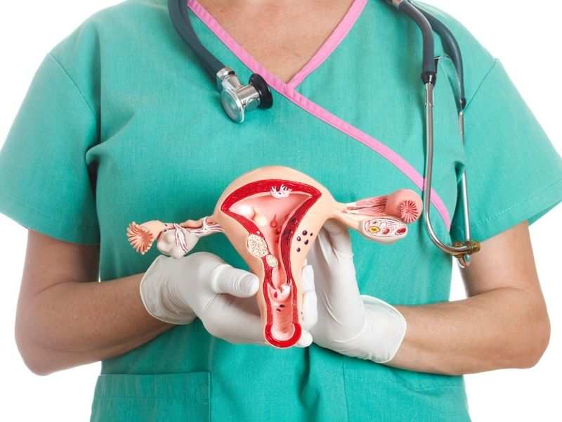 Because cervical cancer is a slow-growing disease, we can usually postpone the procedure until after delivery and watch the area closely, performing a colposcopy every trimester to monitor the dysplasia’s progress.
Pregnancy weakens the immune system, which can increase the likelihood of abnormal results, so sometimes the high-grade dysplasia resolves itself after pregnancy. If it does progress to cancer, we will work with you to develop a personalized treatment plan that incorporates your specific health conditions and needs. As an academic medical center, we can streamline your access to some of the best oncologists in the U.S., right here at UT Southwestern.
If you need a Pap smear, it is safe to get one during pregnancy. Along with the HPV vaccine, Pap smear is a vital tool that helps make cervical cancer preventable – and it will give you one less thing to worry about as you prepare for the healthy birth of your baby.
To visit with an Ob/Gyn or certified nurse-midwife, call 214-645-8300 or request an appointment online.
More in: Women's Health, Your Pregnancy Matters
Plastic Surgery; Women's Health
- Abby Culver, M.D.
- Christine Carman Stiles, M.D.
December 5, 2022
Women's Health
- Maude Carmel, M.
 D.
D.
October 17, 2022
Men's Health; Women's Health; Your Pregnancy Matters
- Yair Lotan, M.D.
September 6, 2022
Pediatrics; Women's Health
- Nirupama DeSilva, M.
 D.
D. - Jason Jarin, M.D.
August 31, 2022
Women's Health
- Abey Eapen, M.D., Ph.D.
August 18, 2022
Men's Health; Women's Health
July 26, 2022
Prevention; Women's Health
- Andrea Jochim, M.
 D., Ph.D.
July 25, 2022
Cancer; Discovery; Women's Health
- Kevin Albuquerque, M.D.
- Chika Nwachukwu, M.D., Ph.D.
May 18, 2022
Diet and Nutrition; Women's Health; Your Pregnancy Matters
- Robyn Horsager-Boehrer, M.
 D.
D. - Carrie McAdams, M.D., Ph.D.
May 17, 2022
More Articles
Risk of Miscarriage from a Pap Smear?
Home » Pregnancy Tests » Does taking a Pap smear in pregnancy increase the risk of miscarriage?
Pregnancy Tests
Sometimes when I see a patient for the first antenatal visit she is overdue due for her Pap smear.
She comments her general practitioner doctor was not prepared to take a smear as she is pregnant and was worried taking the smear may cause a miscarriage.
In the diagram adjacent the transformation zone is shown. The columnar skin is coloured red.
While talking a Pap smear will often result in minor bleeding for a short time, taking a Pap smear will not cause a miscarriage.
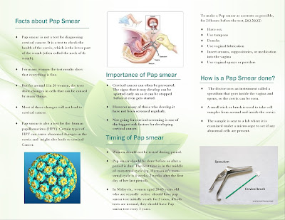
A Pap smear is taken from the uterine cervix where the type of cervix skin cell changes from one called ‘squamous’ to one called ‘columnar’. This part of the cervix is called the squamo-columnar junction of the cervix and also called the ‘transformation zone’ of the cervix. It is this area of the cervix that abnormal changes can occur that can develop into the commonest cervical cancer (squamous cell carcinoma). There needs to be both squamous cells and columnar cells in a smear specimen for it to be classified as ‘satisfactory’ quality.
In the diagram adjacent the transformation zone is shown. The columnar skin is coloured red.
In pregnancy the squamo-columnar junction often is more on the outside part of the cervix (called ‘ectocervix’). That means there are more columnar cells also on the outside part of the cervix. In contrast to squamous cells, columnar cells are more friable and bleed more easily when scraped. As well the cervix is more vascular in pregnancy and this increases the likelihood of it bleeding with superficial trauma.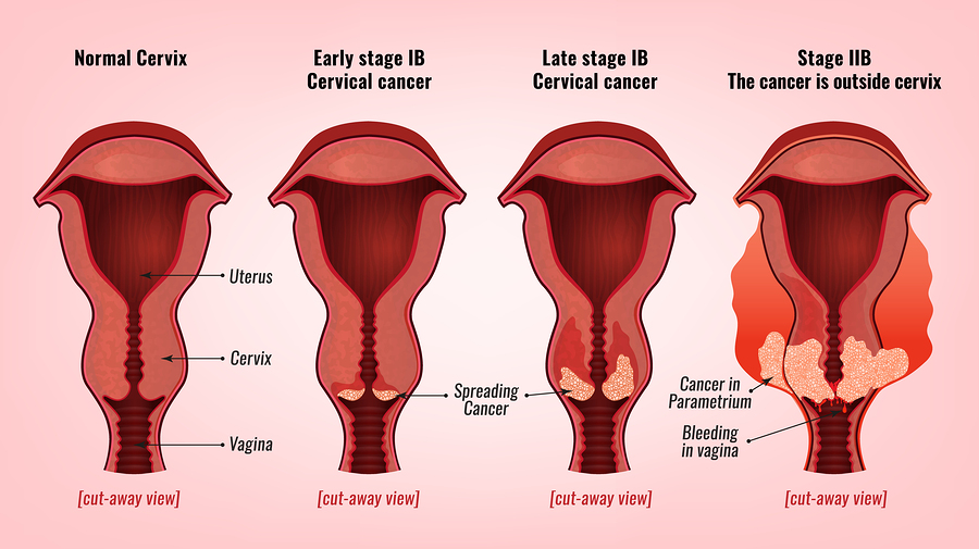
By the way it is because of these same changes (more columnar cells on the ectocervix) that pregnant women can have minor bleeding after sex (the penis superficially traumatising this part of the cervix). As well columnar cells are glandular cells and having more on the ectocervix is why a pregnant woman typically has more vaginal discharge.
It is because of these changes, and the greater tendency for the cervix to bleed when taking a smear that it is more difficult in pregnancy to get a good quality smear, without too much blood on the slide. If there is too much blood on the Pap smear slide the smear will not be of satisfactory quality for checking for abnormal cells.
A ‘cervix sampler’ is usually used to take smear. It scrapes the ectocervix except for its central three brushes which go into the cervix canal to a maximum depth of 0.7cm. The cervix sampler at the entrance of the cervical canal is shown in the diagram. The cervical canal typically is 3 – 4.5 cm long. So there is a considerable distance between where the smear is taken and where the baby is in the uterine cavity. This is shown in the diagram. Hence there is no risk scraping cells from the transformation zone will cause miscarriage.
I have had quite a number of women from whom I have taken a smear at the first antenatal visit which has had an abnormal result. If there are significant abnormal changes then colposcopy and colposcopically directed cervical biopsy is indicated. Taking the biopsies will result in cervical bleeding, but again this is not associated with miscarriage
As long as the abnormal cervical changes are less than cancer then there is no cervical treatment while pregnant, even if there are severe precancerous changes. Rather the cervix is reassessed postnatally. Then the cervix can be treated as necessary.
I will only take a Pap smear if indicated and agreed by the patient and if the patient in the first trimester of pregnancy. Otherwise I will leave it until her 6 week postnatal visit
For more information, have a chat with us on 02 9680 3004 or contact us today.
- Monday all day 9.00am to 4.30pm
- Tuesday all day 9.00am to 4.30pm
- Wednesday all day 9.00am to 4.30pm
- Thursday morning 9.00am to 12.30pm
- Thursday alternate afternoons 2.00pm to 4.30pm
- Friday alternate mornings 9.00am to 1.00pm
- Friday afternoon 2.00pm to 4.30pm
- Saturday mornings 9.30am to 12.00 midday*
*Saturday morning appointments are not available for initial antenatal visit.
What to do after a miscarriage
If a woman has a miscarriage, it is important to take competent measures to restore health. This will help to cope mentally and prepare the ground for a new pregnancy. According to medical statistics, 15-20% of pregnancies end in spontaneous termination for various reasons. The symptoms of what happened rarely go unnoticed, which makes it possible to diagnose the pathology in time, consult a gynecologist, undergo adequate treatment and plan the birth of a child for the future.
Specialists classify spontaneous abortion into two categories:
1. Termination of biochemical pregnancy - the embryo leaves the uterine cavity in the first or third weeks after conception. A woman during this period most often does not suspect that she is carrying a child. Pregnancy becomes known only when testing for the content of hCG in the urine and blood. The blood that has left the body is usually perceived as menstruation, which, for unknown reasons, began outside the scheduled time. Units who carefully monitor their health go to the doctor.
2. Spontaneous abortion or miscarriage in early pregnancy - up to 22 weeks, when the weight of the embryo does not reach 0.4 kg.
Medical therapy
Any method is useful to maintain pregnancy. A qualified doctor develops an individual treatment protocol based on the available diagnostic data. Drugs used may include:
- sedatives;
- restorative therapy;
- hormone stabilizing drugs;
- uterine antispasmodics;
- vitamin and mineral supplements.
The specialist eliminates the threat of miscarriage in the early stages, tells how to prevent a relapse. In the later stages, the cervix is fixed with a special suturing (usually for a period of 16-25 weeks, if there is an ICI).
If an attempt to stop a spontaneous abortion fails, the following treatment tactics are used:
- Waiting – an organism freed from an embryo does not require specialized treatment.
- Drug therapy - the patient is prescribed drugs that complete the removal of foreign tissues from the body. By causing severe spasms of the muscular walls of the uterus, the tablets provoke the expulsion of residues from the cavity.
- Surgery - is used in case of complications or inconvenient for the independent exit of the fetus, the bending of the uterus.
Curettage
Having symptoms of a miscarriage in early pregnancy and faced with the need for a curettage (gynecological cleaning), a woman worries about the state of her reproductive system.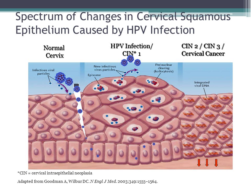 It is not worth doing this, the operation takes place in a gentle mode, with maximum delicacy in relation to the patient's childbearing ability. Curettage is performed when there is a risk of incomplete exit of the embryo from the uterine cavity and the development of infection in the pelvic organs due to the elements remaining in it. Ignoring the procedure can lead to blood poisoning and the formation of a pathology that prevents re-conception.
Vacuum aspiration, however, is performed more frequently, which is more gentle. The complex application of the method with hysteroscopy allows you to carefully examine the internal contents of the uterus in order to prevent poorly cleaned areas on the mucous membrane.
Preparation for gynecological cleaning (curettage)
Gynecological cleaning is performed for diagnostic and therapeutic purposes for various indications :
- after childbirth;
- in missed pregnancy, miscarriages;
- for menstrual irregularities;
- for accurate diagnosis of gynecological disorders.

Curettage is recommended a few days before the onset of menstruation. In this case, blood loss decreases and a favorable prognosis is given for rapid tissue recovery. The operation requires a preliminary examination, testing. This is :
- complete blood count;
- blood coagulation tests;
- smear for examination of the bacteriological environment;
- STI testing.
Before curettage, you stop taking any medications, dietary supplements that have not been discussed with a specialist. Even plant components that can affect blood clotting and provoke blood loss during surgery can be dangerous. Your healthcare provider should be made aware of the medications you are taking so that they know what risks may arise.
Rules for preparing for the procedure:
- refrain from sexual intercourse three days before the operation;
- avoid the use of intimate hygiene products (gels, creams, ointments, liquids), suppositories, tablets and vaginal sprays;
- Do not douche;
- Do not eat or drink 10 hours before surgery.
 This is necessary for high-quality anesthesia.
Cleaning
Curettage is carried out in a hospital, the woman is placed on the gynecological chair of the operating room. The doctor removes the upper layer of the mucous lining the uterine cavity from the inside. The exclusion of pain involves anesthesia. If there were signs of miscarriage in the early stages of pregnancy or at a later period, after which it spontaneously terminated, the dilated cervix allows for curettage without anesthesia. For anesthesia, intravenous administration of the drug is used, selected individually, taking into account the characteristics of the patient's body. A few seconds after the injection, the woman falls into a shallow sleep, the discomfort disappears, which makes the doctor's actions painless.
A dilator inserted into the cervix straightens the walls of the organ, facilitating access to the internal cavity. Holding the neck, the specialist inserts a rounded probe with a small diameter, after which he replaces it with a more voluminous analogue.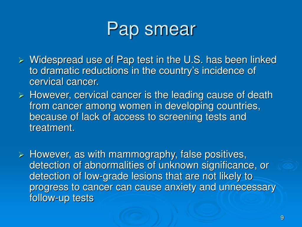 A special video camera attached to the end of the probe allows for hysteroscopy - examination of the cavity before curettage. Cleaning is done with a curette, shaped like a small spoon on a long handle. Carefully collected tissues are stored in a specialized sterile tube, which is later sent to the laboratory for histological examination.
The procedure rarely takes more than one hour, usually 20 minutes is enough for the doctor. Together with the cavity, the cervical canal is cleaned. Manipulations are called RDV - separate diagnostic curettage. Collected samples are placed separately. Histology is used to identify the structure of tissues in order to exclude the presence of atypical cells in them, indicating cancerous lesions, precancerous conditions. The study is carried out within two weeks, after receiving the results, the woman revisits the gynecologist for a follow-up examination.
Curettage is often carried out for diagnostic purposes to determine the symptoms of pathological conditions in the functioning of the organs of the reproductive system.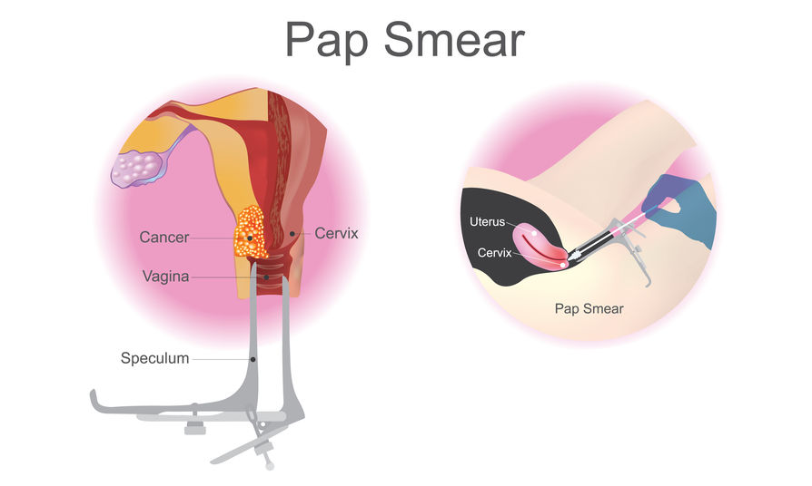 These can be:
- irregular cycle;
- voluminous discharge and painful menstruation;
- bleeding during menopause;
- difficulties in conceiving in the absence of visible causes of pathology;
- suggestion of developing uterine cancer.
Possible complications
Complications can occur, as after any surgical intervention. A serious consequence is the discovery of uterine bleeding. In order to prevent it, oxytocin is used - injections stimulate the cessation of abnormal blood flow. Oxytocin will help if the bleeding is due to insufficient contraction of the uterus. In violation of blood clotting, it is ineffective.
Another complication of is hematometra, when blood clots accumulate in the uterine cavity, which can cause an inflammatory process in the tissues. It is caused by a spasm of the cervix that occurred immediately after cleaning, which interferes with the evacuation of blood. Experts recommend the use of antispasmodics that relax the muscles of the organ and contribute to the normal outflow of blood. A woman should be alerted by pulling pains in the lower abdomen and a sharp cessation of discharge.
After cleansing, endometritis may occur when inflammation affects the lining of the uterus. A measure of therapy for a dangerous diagnosis is a course of antibiotics. Pain in the abdomen and a sharp increase in body temperature testify to the pathology. Any dangerous change in condition should be reported to the doctor immediately. In this case, countermeasures will be taken in a timely manner, which will eliminate the risks of developing more formidable complications.
How to behave after a miscarriage
A miscarriage that has occurred requires a certain tactic of behavior. Among the measures recommended by doctors:
- It is advisable to postpone a new pregnancy attempt for 3-6 months . Otherwise, the risk of repeating the undesirable development of events is high.
 If pregnancy occurs before the expiration date, there is no need to panic. The main thing is the supervision of a specialist.
 - If you are waiting for , ask for advice on effective contraception.
- Follow your doctor's advice .
- Pass the necessary examinations , take tests.
Ask about the effect of the drugs you take on the fetus if you become pregnant during therapy. Find out after what period of time you can fearlessly try to conceive a child.
How to detect genetic pathologies during repeated pregnancy
If a miscarriage of the first pregnancy occurs due to a genetic factor, it is especially scary to decide on a second one. But you should not be afraid of this, with a well-designed therapy, the chances of success are more than great. Diagnostic procedures today are highly accurate and allow you to identify pathology in the early stages. Examination in this case is mandatory, as well as the following:
- who are over 35;
- has screening changes;
- who had markers of chromosomal pathologies and malformations of the embryo;
- who already have children with chromosomal abnormalities.
Ultrasound diagnostics can detect malformations in 80-85% of cases. However, the technology is not impeccably reliable, as it misses pathologies in 20% of situations. Biochemical screening, invasive examinations have valid data. The latest version of the study allows you to identify up to 99% anomalies.
When planning a new pregnancy, it is imperative to visit a geneticist. Screening diagnostics for the detection of abnormal genes will help eliminate the risks of possible pathologies, the factor of heredity and genetic failure during conception. Sometimes the threat of miscarriage in the early stages exists in almost healthy carriers. The examination will allow you to find out about the anomaly in advance and undergo treatment.
What is a miscarriage like
A miscarriage that occurs is complete when all parts of the embryo come out of the uterine cavity together with membranes and amniotic fluid. If parts of the fetus remain in the uterus, they speak of an incomplete miscarriage, which occurs more often in the early stages of pregnancy. To neutralize the negative consequences, to prevent the development of an infectious process in the tissues, the product of conception is evacuated from the uterine cavity by the methods of medical interruption, gynecological curettage, and vacuum aspiration. Therapy may include the use of drugs aimed at contracting the uterus and pushing the contents out. Ultrasound examination is considered to be the control method of diagnostics.
Why the body rejects the embryo
The causes of miscarriage often lie in the presence of chromosomal abnormalities in the fetus. Among the factors that provoke rejection of embryos are also:
- Heredity and genetic failure at the stage of fertilization of the egg by the sperm.
- A non-viable fetus may appear as a result of various risk factors - environmental conditions, occupational hazards, viral illness of parents. It is impossible to neutralize these factors.
 The only way out of the situation is to reduce the likelihood of their manifestation by protecting the expectant mother from dangers during gestation.
 - Hormonal imbalance caused by disruption of the endocrine system. The situation can be affected by an insufficient amount of progesterone in the mother's body or an excess of testosterone. With early detection of a failure of the hormonal system, a woman undergoes specially organized therapy before pregnancy.
- The presence of tumors , neoplasms in the pelvic organs.
- Isthmic-cervical insufficiency when the isthmus and cervix dilate prematurely, unable to cope with the increasing pressure caused by the growing fetus in the body.
- There is a risk of miscarriage in the presence of anomalies in the work of the cardiovascular and renal systems.
- Drug addiction , alcohol addiction, substance abuse of mother and father.

- Depressive conditions , stress, nervous stress of a pregnant woman.
- Mechanical stress , blows, bruises, excessive physical labor of the future woman in labor.
- X-ray examination - radiation can cause miscarriage.
- Drug use . In the first trimester, you can not use potent medicinal formulas. Drugs can cause the development of defects in the embryo. Some decoctions of herbs are also contraindicated - parsley, tansy, cornflower, nettle, St. John's wort. It is forbidden to self-medicate. Each drug is agreed with the attending physician.
- Infectious and viral process in the body. Any sexually transmitted infection can provoke a miscarriage, which must be cured before pregnancy, otherwise there is a high risk of infection of the fetus in the womb. A great threat of miscarriage in the early stages exists due to viral infections and inflammation of the internal organs.
 A dangerous symptom is the high temperature of the mother, accompanied by intoxication of the body. At the stage of pregnancy planning, it is important to stop chronic diseases.
 - History of abortion , unsuccessful surgery, unprofessionalism of the doctor and unfortunate circumstances.
- Immunological factors .
The list of causes of miscarriage in the early stages of pregnancy and in the later period may be more extensive, in each case, doctors identify the pathology individually.
Isthmic-cervical insufficiency (ICI)
One of the most common causes of spontaneous miscarriage during pregnancy is CSI - dilatation of the cervix and isthmus of the uterus as a result of increasing pressure from the growing fetus. Pre-pregnancy manipulations with the uterus (cervical dilation due to abortion, childbirth or curettage) affect the condition of the muscle ring. Damaged areas are tightened by scar tissue that does not have elasticity, is not amenable to stretching and contraction. ICI also has a functional nature when there is a hormonal imbalance.
ICI occurs in the period from the 11th to the 27th week after conception, when the embryo begins to produce androgens in the mother's body with the launch of the adrenal glands. Taking into account the mother's hormones, their indicator can be exceeded - this softens the cervix, opens and shortens it. Harmful bacteria and microorganisms penetrate into the formed channel, infecting the fetal egg. The initial stages of ICI do not have obvious symptoms, since they do not entail the tone of the uterine muscles. With the loss of strength of the membranes, amniotic fluid pours out. There are no pain sensations.
If a woman has had a miscarriage that started with amniotic fluid, she should report it to her doctor when monitoring a subsequent pregnancy.
Treatment of isthmic-cervical insufficiency
Endocrine disorders are corrected by prescribing hormonal drugs. An assessment of the condition of the uterus is carried out by a doctor a couple of weeks after the start of taking medications. They say about positive dynamics when the opening is suspended and no further expansion of the neck is observed. In the absence of the planned effect, surgical intervention is prescribed. Similar measures are used for the traumatic nature of the neck deformity. You should not be afraid of the operation, the doctor acts delicately, without causing additional injuries to the patient, without causing discomfort to the baby growing in the womb. The procedure is most effective in the early stages of pregnancy. Suturing can significantly reduce the risk of infection of the embryo through the lower edge of the cavity.
Surgical intervention takes place in a hospital. Before the operation, the pregnant woman is examined. After the procedure, the vagina is sanitized, for which the suturing site is treated with chlorhexidine and furatsilin for three days. The patient needs to undergo a weekly follow-up examination with the attending physician, where he assesses the situation, making adjustments to the therapeutic protocol if necessary. The sutures are removed at the 38th week of pregnancy. During this time, the neck matures, preparing the birth canal for the passage of the fetus. Many women in labor worry that they will need a caesarean section if they have stitches, but this is not true. In most cases, women give birth on their own.
Immediate action is recommended if the amniotic sac prolapses (falls out) into the cervix between 16 and 24 weeks. The suturing of the neck obliges the woman to observe bed rest, strictly follow the daily routine, avoid physical exertion, and do not skip taking medications. In rare cases, complications occur. Among them, the eruption of sutures through the tissues, provoked by the frequent tension of the muscles of the uterus. To prevent tone, tocolytics are prescribed - medicines to prevent premature birth. The expectant mother should be prepared for frequent examinations and smears, which may be caused by the likelihood of accumulation of pathological microflora on the suture threads.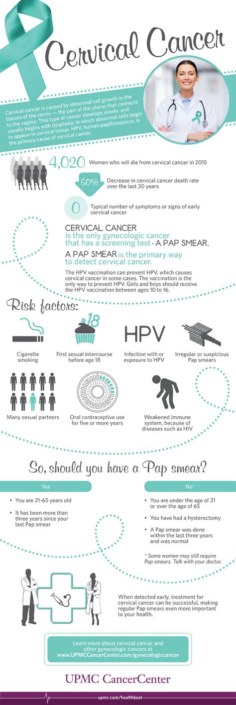
It is also important to conduct psychological therapy, where a woman is taught relaxation techniques. The behavior of the future mother is a decisive factor in the successful bearing of the fetus in case of pregnancy complications. Panic and fuss create an unfavorable prognosis in stabilizing the situation. If a spontaneous abortion occurred for reasons of ICI, when you can get pregnant after a miscarriage, the doctor will say. Ideally, the period should be at least two years. The specialist must also take measures to prevent a repeated situation of losing a child.
In addition to the suture, ICI correction is also carried out using an obstetric pessary. An alternative method is the imposition of a special ring of hypoallergenic materials on the cervix. Silicone is the most commonly used. The ring creates additional support, preventing the opening of the neck.
Uterine hypertonicity - risk prevention
Uterine contractions before natural delivery is called hypertonicity.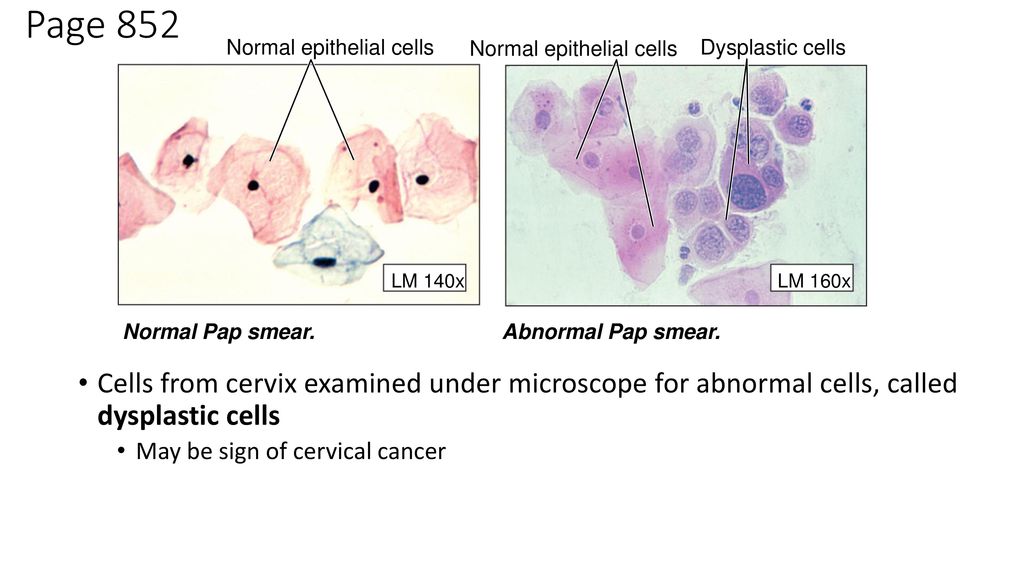 The condition is not an independent disease, it signals a malfunction in the body, often manifesting itself in the early stages of pregnancy. The causes of the pathological phenomenon are:
- Hormonal disorders caused by insufficient function of the placenta, ovaries, problems with the adrenal glands, causing imbalance.
- Genital infantilism organ defects.
- Neoplasms , tumors in the uterus that are not necessarily malignant (eg, fibroids).
- During pregnancy infectious processes, viral diseases.
- CCI - opening of the cervix under increasing pressure created by the growing embryo.
- Immunological problems .
- Chronic diseases of the body (cardiovascular disorders, renal insufficiency).
- Past miscarriages early pregnancy, symptoms of which may recur, induced abortions.

In addition to physiological causes, psychological factors are of no small importance. A woman who is in a depressed state can provoke hypertonicity in herself.
You can feel the tension of the muscles of the uterus on your own, without the help of a specialist. This is evidenced by the heaviness that appears in the lower abdomen, pulling pains in the lumbar region. Symptoms are similar to painful menstruation. Arising in the first trimester, the condition provokes spontaneous abortion, missed pregnancy, death of the fetal egg. In the subsequent period, premature birth due to hypertonicity is likely.
Why does the tension of the walls of the uterus cause irreversible consequences? The reason is the disturbed blood supply to the placental tissues, the occurrence of hypoxia of the embryo and the slowdown in the development of the emerging child. Following the contraction of the muscles of the uterus, the placenta does not contract, which causes its detachment and provoking the release of the fetal bladder.
Hypertonicity is diagnosed during a scheduled visit to a specialist. Stabilization of the situation requires the appointment of sedative drugs and antispasmodics. A strengthening effect is provided by therapy with the inclusion of vitamin B6, magnesium. In most cases, the measures taken are sufficient to neutralize the risks. Self-treatment, which can cause irreversible consequences, is strictly prohibited. With hypertonicity, the main rule for a pregnant woman is calmness and lack of physical activity. Some women who have had a successful delivery say they "didn't get up" during their entire pregnancy. With hypertonicity, sexual intercourse is also excluded.
If the threat cannot be neutralized, hospitalization is recommended. It is especially dangerous when severe cramping pain is complemented by spotting. To lie down "for preservation" is an adequate measure in the struggle for the birth of a healthy and strong baby. In the hospital walls, a pregnant woman is prescribed a vaginal examination, ultrasound.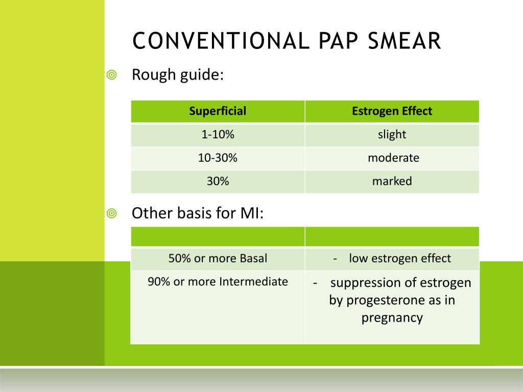 If necessary, a woman takes urine and blood tests, checks the hormonal background, and is examined for the presence of STIs.
At the onset of labor activity before the 34th week, the condition is tried to be stabilized with tocolytics. The most dangerous period is from the 25th to the 28th week, when the woman is recommended the maximum possible bed rest. After that, the fetus has every chance of survival. In order to quickly form the pulmonary system of the embryo, allowing it to survive with an early birth, hormones are prescribed.
Having an unfavorable prognosis for miscarriage and the threat of miscarriage, it is necessary to take up prevention at the stage of conception planning.
Stages of spontaneous abortion
There are certain signs that attract attention and divide the course of a miscarriage into specific stages:
- Threat - having noticed factors threatening pregnancy, you can take measures to restore the situation, normalize the mother's well-being.
- Start of abortion - at this stage, the doctor can apply life-saving manipulations and give recommendations to the pregnant woman.
- Miscarriage in progress – the condition is irreversible, it is impossible to stop the pathology. The death of the fetal egg begins, which leaves the uterine cavity.
- Completed abortion - the uterus gets rid of the residual tissues of the embryo, cleanses, restores its original parameters. It is important to prevent the remnants of foreign fibers inside, otherwise the organ becomes infected with decaying residues and toxins go into the bloodstream.
Symptoms of miscarriage - how not to miss the threat
If there is a threat of miscarriage in the early stages, the following symptoms may occur: The pain may be monotonous or come in waves.
 Bright scarlet blood can stand out, it is often confused with menstruation.
 A woman who does not know how an early miscarriage occurs should listen to her inner state.
Should alert:
- spasmodic pain impulses;
- Drawing pain in the lumbar region.
In the later stages, the above symptoms are added:
- liquid discharge from the vagina, which may indicate damage to the amniotic sac;
- pain when urinating;
- internal bleeding, which warns of a deterioration in the general condition, fainting, dizziness, pallor of the skin. All this is an indication for emergency hospitalization of a pregnant woman.
The beginning of an abortion is characterized by more pronounced symptoms of a miscarriage - contraction-like pain, severe dizziness, loss of strength. Instead of smearing discharges, clot-like ones appear, abundantly manifested during movement. Pregnancy can be saved if the area of detachment of the fetal egg is small and the fetal heartbeat is determined.
The third stage is useless for saving the fetus. There is girdle pain in the lower back and abdomen. Together with abundant blood loss, a fetal egg comes out of the uterus. Incomplete miscarriage requires curettage of the uterine cavity if parts of the embryo or membranes of the fetal egg remain in it, otherwise there is a high risk of complications that will endanger the life of the mother.
In rare cases, complications and serious health consequences can occur after a spontaneous abortion. But in the majority of situations, the body independently copes with what happened, expelling the parts remaining in the uterine cavity with a natural contraction of the muscles. An early spontaneous miscarriage does not always occur, a dangerous condition can also occur in the later stages. Some women try to provoke the release of the fetus with decoctions of herbs and medications.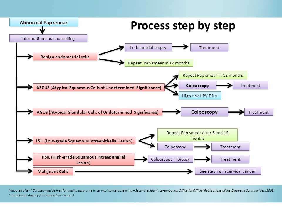 This is fraught with complications, including sepsis, dysfunction of the reproductive organs, after which pregnancy becomes impossible.
Methods of diagnosis
The symptoms of a threatened miscarriage at an early stage will be determined by a doctor during a visit to the antenatal clinic. The specialist will check the size of the uterus, determine the tone of its muscles, the condition of the cervix, and examine the discharge from the genital organs. A reliable method to identify the existing threat is transvaginal ultrasound diagnostics. The doctor draws attention to segmental muscle contractions of the uterus, detachment of the fetal egg. Genetic testing will help analyze the likely causes of a miscarriage. The patient's history is carefully collected.
Planning a new pregnancy
The medical community is unanimous in the issue of planning a new pregnancy after a spontaneous abortion. Conception is not recommended for at least 3-6 months. During this period, the woman's body will recover and gain strength to bear the fetus. Observation by a doctor, harmonization of hormonal levels, examination of parents to identify possible pathologies are important. In order not to become pregnant in the first months, it is recommended to use contraceptive methods prescribed by your doctor.
Examination after a miscarriage includes blood and urine tests, examination of the microflora of the vagina with a smear, detection of overt and latent genital infections, glucose and hormone tests, examination of partners for biological compatibility. Planning is an important step towards having a healthy baby. After the studies, the woman is prescribed strengthening therapy. It is important to completely reconsider eating habits, to exclude factors that are harmful to well-being. Vitamins, folic acid are used. Fast food, food containing carcinogens and preservatives are excluded from the diet. Subject to the rules recommended by the doctor, a successful pregnancy with a favorable outcome is likely.
It is not uncommon for a pregnancy test to show two lines after a miscarriage. This is due to the restructuring of the body, the organs of the reproductive system. It is important to report the incident to your doctor. The presence of remnants of embryonic tissue in the uterus can provoke a positive test result. In this case, immediate curettage is necessary, which neutralizes the risk of inflammation and infection. To accurately determine her condition, a woman needs to undergo an ultrasound diagnosis, take tests to determine hCG in the blood.
The question of whether it is possible to get pregnant after a miscarriage worries many parents. The answer is unequivocal - yes, if you follow the recommendations of experts, carefully plan a new conception, monitor your well-being and state of your health.
Components of success after a miscarriage
Spontaneous abortion can provoke not only the health of the patient, but failure to follow simple rules can be a threat. To reduce the risk of losing a child during pregnancy, you need to:
1. Keep calm – it is important for a mother to eliminate all factors that make her nervous from her life. Irritation is not the best way to normalize the condition. In order to stabilize the emotional background, rest is recommended, the use of soothing teas with the permission of the doctor. Good results are given by decoctions of chamomile, lemon balm, mint.
2. Avoid taking unnecessary medicines and preparations. But it is unacceptable to stop the therapy prescribed by the doctor on your own. Each step must be discussed with the gynecologist.
3. Eliminate harmful occupational factors. Work in the chemical industry and other hazardous facilities can create an undesirable background in the body, which prevents normal gestation. It is important to understand what is of great value to the mother - the birth of a healthy baby or a career factor. Many refuse to work to increase the chance of having a baby.
4. Eliminate bad habits. It is unacceptable for a woman who has experienced miscarriage to drink alcohol and smoke. It is forbidden to do this and the future father. This negatively affects the quality of spermatozoa, provokes difficulties with conception and risks of deviations in the development of the embryo.
5. Take vitamin complexes, specially designed to prepare the body for pregnancy, the formation of basic conditions for its favorable course.
6. Eat right. A complete, balanced diet works wonders. With a lack of weight, a nutritionist will develop an adequate diet for a woman with the inclusion of a large amount of protein foods rich in vitamins and trace elements of vegetables, fruits, and cereals. Recommended fats contained in fish, seeds, nuts, avocados, olives.
7. Get rid of extra pounds. Obesity adversely affects the development of pregnancy. Science has proven that enhanced nutrition during this period is not required. The main thing is its balance.
Infections during pregnancy
Infectious processes transferred before pregnancy develop immunity in the mother to similar agents of influence. Primary infection poses a great threat, so vaccination will be useful before planning conception. Perinatal diagnosis allows you to detect the infectious process at the initial stage and prevent its harmful effects. This is possible if the pregnant woman is registered from an early date.
Infection may develop due to an infection transmitted by airborne droplets. It is the most dangerous, since it is almost impossible to prevent it. This applies to mumps, measles, rubella. HIV and hepatitis infect the body through sexual contact, similar to chlamydia. Listeriosis is transmitted with poor-quality products. A pregnant woman can pass infections to a developing baby. Pathology is determined by profile tests of latent infection.
Routine pregnancy monitoring involves regular testing. Sexual infections are determined using a smear, ultrasound shows deviations in the development of the baby, and KGT is aimed at listening to the work of the fetal heart muscle. If there is a suspicion of a serious infection of the embryo, blood sampling from the umbilical cord and amniotic fluid analysis are practiced.
Infection of a child also depends on concomitant factors. The speed of diagnosis, the literacy of the treatment, the type of pathogen, the duration of the pregnancy are taken into account. The following infectious processes deserve special attention:
1. Viral etiology - a huge number of viruses poses a danger to a pregnant woman. The threat is genital herpes, rubella, infectious type erythema, cytomegalovirus, hepatitis B, measles, mumps, chickenpox.
2. Bacterial infections, detected during the analysis of biological materials (feces, urine, blood), examination of certain organs of the body. Active reproduction provokes a rapid growth in the number of bacteria in the vagina. Not all microorganisms pose a threat to the child. Dangerous candidiasis, streptococcus, chlamydia, bacterial vaginosis, cystitis.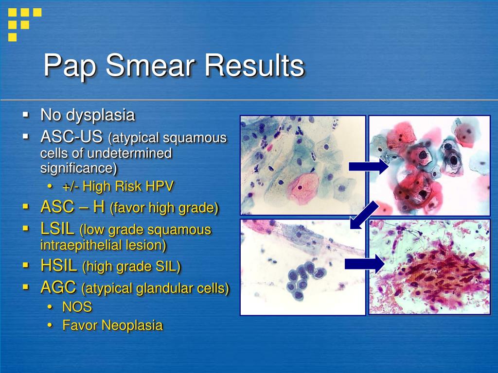
The successful course of pregnancy is threatened by intestinal infections, often activated in the summer. Their carriers can be animals and poorly processed food before consumption. Of particular danger are listeriosis, salmonellosis, toxoplasmosis.
Prevention of infections during pregnancy
Infection of the mother poses a threat to the life of the fetus. From the 3rd to the 12th week, the infected organism responds with a miscarriage or the formation of malformations of the child. From the 11th to the 25th - developmental delay. At a later date, organs are deformed and prerequisites for premature birth are created. In order to prevent intrauterine infection, it is recommended to apply a number of rules:
- be examined for the detection of STIs;
- examine blood, determine the presence of antibodies to infection carriers, pathogens;
- avoid contact with sick people, visits to crowded places where there is a possibility of infection by airborne droplets;
- screen pets for dangerous infections, treat them if necessary, or remove them from the home until the threat is eliminated;
- exclude fast food, store-bought semi-finished products from the diet, thoroughly heat treat meat, fish;
- remove from the diet sushi and other culinary delights purchased in restaurants, cafes;
- thoroughly wash hands, fruits, vegetables with special disinfectants that are not capable of harming the pregnant woman and the child;
- it is planned to visit a gynecologist, undergo examinations recommended by a doctor, take tests, take vitamins;
- register at the first sign of pregnancy;
- prepare for conception, cure infections, vaccinate.
It is also important for the child's father to follow most of the recommended rules. If only the mother undergoes treatment, a relapse is likely during sexual intercourse, neutralizing the beneficial effect of therapy.
A woman who has had a miscarriage in the past should be alert to any deviations from the norm in her state of health. It is important to pay attention to ailments, pain, weakness, dizziness. Accounting for an early consultation will create conditions for the bearing of the fetus and the birth of a child. There is no need to be afraid that a miscarriage will forever deprive the joy of motherhood.
Having completed a course of examinations, passing tests and following the measures prescribed by the doctor to treat imbalances in the body, you will create all the conditions for a favorable pregnancy outcome. Tune in to the positive, protect yourself from worries, worries, stress. Feel the support of loved ones, hope for the best! Get advice from good specialists to rule out any unfavorable prognosis before conception or take steps to neutralize them. Health and prosperity to you, your families and loved ones!
Flora smear during pregnancy - when and why to take it
- When to take a flora smear
- What a flora smear shows
- Flora smear preparation rules
The flora smear is the most common test prescribed by an obstetrician-gynecologist. To conduct this study, the doctor, while examining a woman in a gynecological chair, takes the contents of the vagina from the posterior fornix (this is the space that is located between the back wall of the vagina and the cervix), the cervical canal and the discharged urethra, applies the material to the glass slide and directs him to the lab.
A smear examination for flora in the laboratory is carried out by a doctor of laboratory diagnostics under a microscope. This study allows you to determine the nature of the microflora (types of microorganisms) of the vagina, cervical canal and urethra, to identify the inflammatory process in the genitals of a woman, in some cases it also allows you to determine the causative agent of this inflammatory process (for example, gonococcus, Trichomonas).
When to take a smear for flora
It is mandatory for all pregnant women to take a smear twice - when registering and at 30 weeks of pregnancy, often another smear for flora is taken at 36-37 weeks to assess the state of the vaginal microflora before childbirth. During these periods, the analysis is given even in cases where the patient is not bothered by anything. This is carried out in order to identify a hidden inflammatory process that can lead to serious complications during pregnancy. During pregnancy, due to changes in hormonal levels and a decrease in immunity, exacerbation of chronic infections, as well as candidiasis (thrush), is much more likely. Any inflammatory process in the vagina during pregnancy can lead to serious complications of pregnancy - premature rupture of amniotic fluid, premature birth, oligohydramnios or polyhydramnios, intrauterine growth retardation and others.
If a pregnant woman has complaints - the appearance of copious discharge from the genital tract, itching, burning or discomfort in the genital area, a swab for flora is also taken.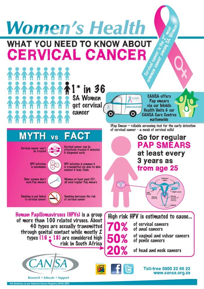 In some pathological conditions, for example, in the presence of past miscarriages associated with infectious complications of pregnancy, cervical incompetence, a smear for flora is taken once a month, and after 30 weeks once every two weeks. Smear sampling is an absolutely safe procedure and does not lead to any complications, therefore it can be performed at any stage of pregnancy.
Find out more about the services:
- Tests for pregnant women
What a flora smear shows
A flora smear is evaluated according to the following indicators:
Epithelium
Squamous epithelium is the cells of the surface layer of the mucous membrane of the vagina and cervix. The presence of a large amount of squamous epithelium in a smear may indicate an inflammatory process. The absence of epithelium in the smear indicates a violation of the hormonal background.
Leukocytes
These are blood cells involved in the destruction of pathogenic bacteria. Leukocytes are able to actively penetrate through the wall of blood vessels into the tissues of the body and participate in the fight against infectious agents. Normally, no more than 10 leukocytes are present in a smear for flora from the vagina, no more than 15 leukocytes per field of view from the cervical canal, and up to 2 leukocytes per field of view from the urethra. An increase in the content of leukocytes in a smear is a sign of inflammation, while the higher the content of leukocytes in a smear, the more pronounced the inflammatory process.
Erythrocytes
These are red blood cells. Normally, single erythrocytes (1-2 in the field of view) can be found in a flora smear. An increase in the number of red blood cells indicates the presence of a chronic inflammatory process, and also be a sign of injury or occult bleeding, for example, in the presence of cervical ectopia (the so-called erosion, when the vaginal part of the cervix is covered with a cylindrical epithelium normally lining the inside of the cervix).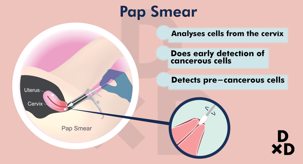
Slime
Normally, there is no mucus in the urethra, a moderate amount of mucus is detected in the vagina, and there may be a large amount of mucus in the cervix. An increase in the amount of mucus may be a sign of an inflammatory process, but this criterion does not have great diagnostic value, and doctors rarely rely on it when making a diagnosis.
Bacteria
Normally, flora should not be detected in the urethra, rod flora is detected in a moderate amount in the vagina and cervix. Rod flora is most often lactobacilli, which are 95% are normal vaginal biocenosis. Lactobacilli actively colonize the vagina and create an acidic environment in it, thereby preventing the growth and reproduction of pathogenic bacteria.
In addition to lactobacilli, other rod bacteria, such as E. coli, bacteroids, and various cocci, may also be present in the vagina. These are bacteria that, under microscopy, have the shape of balls. This group of bacteria includes streptococci, staphylococci, enterococci.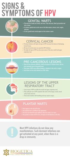 In a small amount, they are normally present in the vagina. If their number increases sharply against the background of the death of normal lactobacilli, this can lead to the development of an inflammatory process. Unfortunately, according to the results of a routine smear on the flora, it is impossible to determine which specific bacteria and in what quantity are present in the vagina. Therefore, with a pronounced inflammatory process, as well as when a large amount of coccal flora is found in a smear on the flora, the doctor prescribes an additional analysis to make the correct diagnosis - sowing on the flora with the determination of sensitivity to antibiotics.
Opportunistic flora
These are micro-organisms that live in the human body in small numbers without causing harm, but under certain conditions can lead to an inflammatory process. Such microorganisms found in a smear on the flora include fungi of the genus Candida and gardnerella.
Gardnerella ("key cells")
Gardnerella and other bacteria living in anoxic conditions (so-called anaerobic bacteria) normally live in the vagina in small numbers, without causing symptoms of an inflammatory process. With a decrease in local immunity, which is quite common during pregnancy, there is an increase in the proportion of these bacteria in the vaginal microflora, a disease occurs - bacterial vaginosis (vaginal dysbiosis). At the same time, “key” cells are found in a smear on the flora - these are cells of the vaginal mucosa, covered with gardnerella and other anaerobic bacteria. The gardnerella themselves are not visible in a normal unstained smear. They can only be detected by staining smears with special dyes.
Mushrooms
Microorganisms of the genus Candida are part of the normal microflora of the mouth, vagina and colon of most healthy people. Normally, the number of these microorganisms is small and they do not cause an inflammatory process. Normally, in some women, a small amount of spores of the fungus may be detected in a vaginal smear. In the absence of an inflammatory reaction and complaints of the patient, the treatment of this condition is not carried out. The detection of a large number of spores or mycelium of a yeast-like fungus of the genus Candida in a smear on the flora makes it possible to diagnose candidiasis (or thrush)
Pathogenic flora
There are microorganisms that should not normally be present in the vagina of a healthy woman, and the detection of which in a flora smear indicates the presence of a serious sexually transmitted disease. Of these infections in the smear, Trichomonas and gonococci are most often detected.
Trichomonas
These are the simplest microorganisms that have a flagellum and are capable of movement. Detection in a smear on the flora of Trichomonas indicates the presence of a sexually transmitted disease - trichomoniasis. Trichomoniasis in a pregnant woman increases the risk of preterm birth, premature rupture of amniotic fluid, intrauterine growth retardation. In addition, there is a risk of infection of the baby when passing through the birth canal, therefore, if Trichomonas is found in a smear, antibacterial treatment is mandatory during pregnancy.
Gonococci
These are bacteria that look like double bean-shaped balls in the smear, adjacent to each other with a concave side. Detection of gonococci in a smear allows the doctor to make a diagnosis - gonorrhea. This is a sexually transmitted disease, which must also be cured during pregnancy. The inflammatory process caused by gonococcus significantly complicates the course of pregnancy, can lead to miscarriage, premature birth, premature rupture of amniotic fluid, infection of the placenta and membranes, and in addition, when the baby passes through the birth canal, the eyes of the newborn are affected by gonococcus.


