Baby soft spots location
5 Warning Signs From Your Baby’s Soft Spot – Cleveland Clinic
When you’re a new parent, you learn that you need to protect that soft spot, or fontanelle, on your baby’s head. It rarely requires much attention, but it reminds you just how fragile your infant is.
That’s why, if something changes — is the soft spot sunken in a little? — you may worry there’s something wrong.
A change in the fontanelle isn’t always a major problem, but it can sometimes reveal internal issues, says Violette Recinos, MD, Section Head of Pediatric Neurosurgery at Cleveland Clinic.
“It is a good indicator of the baby’s potential hydration status and brain status,” she says. “It’s like an automatic pressure sensor.”
What is the fontanelle?
The fontanelle is the space between different plates of a baby’s skull that will eventually come together. This aspect of an infant’s skull structure typically allows for easy delivery through the birth canal and for rapid head growth during the first year of life, Dr. Recinos says.
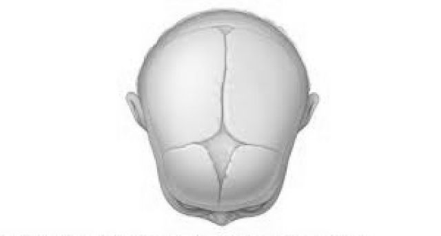
There are actually two soft spots — one at the back of the head and another on top. The posterior one closes within a few months of birth, while the top fontanelle typically remains until just past a child’s first birthday.
Dr. Recinos explains what changes in the fontanelle can tell you about your infant’s health.
Sunken in soft spot
This is often a sign of dehydration, she says. It may occur if your child is sick and not getting enough fluids.
What you should do: See your pediatrician if the sunken appearance persists and you can’t get your baby to take in more fluids.
Advertising Policy
Swollen soft spot
After a fall, a swollen soft spot (particularly if it’s accompanied by vomiting) is sometimes a sign of head trauma.
What you should do: Seek medical treatment right away.
Bulging soft spot
Fluid buildup (hydrocephalus) can cause rapid head growth and can make the soft spot look “full,” Dr. Recinos says.
A bulging fontanelle also might signal internal bleeding or a tumor or mass causing pressure in the head.
What you should do: If their soft spot is bulging, that’s a reason to seek care from your pediatrician, she says.
Seek emergency care if your infant exhibits fatigue, vomiting or unusual mental status along with the fontanelle’s fullness. Treatment for these conditions may include surgery to insert a shunt that relieves fluid buildup or to remove any underlying mass, Dr. Recinos says.
Disappearing soft spot
Most of the time the soft spot is obvious, particularly on a newborn. But at times it can seem to disappear quickly.
This may scare parents, but it typically means it’s just a “quiet fontanelle,” not that it has fused together prematurely, Dr. Recinos says.
Advertising Policy
As long as your child’s head is growing normally, all is probably well, she says. But your pediatrician may suggest an imaging test to make sure the fontanelle is still open.
Occasionally, though, the skull bones do close earlier than normal on one side, causing craniosynostosis. Depending on which bones fused, the baby may develop an abnormal head shape. For example, sagittal craniosynostosis, the most common form, results in a longer head that is shaped somewhat like a football.
What you should do: Children with craniosynostosis may need surgery to open the fused bones and reshape the skull. In some cases, the child will wear a helmet afterward until the site heals and the head shape normalizes.
Soft spot that doesn’t close
If the soft spot stays big or doesn’t close after about a year, it is sometimes a sign of a genetic condition such as congenital hypothyroidism.
What you should do: Talk to your doctor about treatment options.
The bottom line? If you have any questions or concerns about your baby’s soft spot, it’s a good idea to check in with your pediatrician to make sure all is well.
Baby Soft Spot: How to Care for Your Baby’s Head
As you spend hours gazing at your newborn and stroking his face, you’ll no doubt notice a couple of soft spots on his head. These soft spots, called fontanelles, are perfectly normal and actually play an important role in your baby’s development.
Learn more about these soft spots, including how to protect them, when the bones of the skull will harden, and which warning signs to look out for by reading on.
What Are Baby Soft Spots and Where Are They?
All babies are born with two soft spots (fontanelles) on their heads: The larger soft spot (anterior fontanelle) is toward the front of the head, and the smaller soft spot (posterior fontanelle) is toward the back.
These softer areas are made up of immature skull bones that are still forming and expanding as your baby’s brain grows.
Why Do Babies Have Soft Spots?
The soft spots on babies' heads have two main functions:
They make it possible for the bony plates of the skull to compress and overlap as the head passes through the narrow birth canal during a vaginal delivery
They allow a baby’s skull to expand, making room for the rapid brain growth that happens in the first year.

related baby tool
Keep an eye on your baby’s average growth by tracking height, weight, and head circumference with our simple tool.
Fill out your baby's details*:
What is your child*
Boy Girl
This is a mandatory field.
Age (between 0 and 24 months)
This is a mandatory field.
Weight (lbs.)
This is a mandatory field.
Height (in.)
This is a mandatory field.
Head circumference (in.)
This is a mandatory field.
*Input details of your baby’s last measurements. **Source: World Health Organization
When Does a Baby's Soft Spot Close or Go Away?
In your baby’s first few months, both soft spots should be open and flat. At about 2 to 3 months of age the soft spot at the back of your baby’s head may close. The soft spot at the front may close around the time your toddler turns 18 months old.
What Happens If You Touch the Soft Spot on Your Baby's Head?
As long as you touch your baby’s soft spots gently — for example, when you’re holding your baby and supporting his head and neck or when you’re washing your baby’s hair — you shouldn’t be afraid of hurting him.
There is a thick and durable membrane just under your baby’s scalp that protects her brain, so gently touching the fontanelles won’t hurt her.
To help ensure your baby’s head is protected, it’s a good idea to remind friends, family members, and caregivers to be careful and gentle with your baby’s head.
What Does It Mean When a Baby's Soft Spot Is Pulsating?
Sometimes it may appear that your baby’s soft spot is pulsating. This is completely normal — blood is pulsing through your baby’s body, and this movement can sometimes be visible where the soft spot is. There’s no need to worry if you see your baby’s soft spot pulsing.
What Causes a Sunken Soft Spot on Your Baby’s Head?
A sunken soft spot may be due to dehydration, which can happen if your baby does not get enough breast milk or formula. Your baby may also be more likely to be dehydrated if she has a fever, has been vomiting, or has diarrhea.
Beyond a sunken soft spot, these are some of the other signs of dehydration:
Fewer wet diapers
Sunken eyes
A dry mouth
Cool skin
Drowsiness
Irritability.
Contact your baby’s healthcare provider right away if you’re concerned your newborn may be dehydrated.
Keep in mind, a sunken soft spot can sometimes occur in babies who are not dehydrated. It’s safest for your baby’s healthcare provider to make a diagnosis.
What Should You Do If Your Baby Hits His Soft Spot?
Contact your baby’s healthcare provider if your baby hits his soft spot.
If you notice swelling/bulging of the soft spot and/or bruising around her eyes or behind her ears, it may be due to a concussion. Call 911 immediately.
Other signs of a head injury or trauma requiring immediate medical attention include:
Nonstop crying
Your baby being unwilling to feed
Vomiting
Seizures
Discharge or blood from ears or nose
Difficulty waking after sleep.
When Should You Be Concerned About Your Baby's Soft Spot?
The lack of soft spots on your baby’s head may be a sign of very rare condition called craniosynostosis, a birth defect in which your baby’s skull bones fuse together earlier than normal, resulting in a misshapen head. Contact your baby’s healthcare provider if
your baby seems to lack soft spots
there are raised, firm edges where the skull plates meet
your baby’s skull shape seems misshapen and is not growing over time.
The Bottom Line
Although it might seem a little odd that your baby would have soft spots on her head, they actually serve two important purposes: to make it easier for your baby to pass through the birth canal during a vaginal delivery, and to ensure your baby’s skull can expand to make room for her growing brain.
By around 18 months, your baby’s fontanelles will have closed. In the meantime, be gentle with your baby’s head when holding her.
If your baby accidentally bumps or hits a soft spot, or if you’re concerned that one of the soft spots may be sunken or injured, contact your child’s healthcare provider right away.
When it comes to the shape of your little one’s head, if you’ve noticed flatter spots, it could be because your little one is spending too much time lying on his back looking the same way. Prolonged pressure on the softer skull bones can flatten out the area. Find out more about flat head syndrome and what you can do to treat or prevent it.
Correct posture for children at a desk
Incorrect posture, stoop - the scourge of all schoolchildren. Problems with the spine lead to rapid fatigue, respiratory failure, the development of scoliosis, and the appearance of headaches. Also, the curvature of the spine leads to deformation of the internal organs and the appearance of an intervertebral hernia.
To avoid this, it is necessary to form the correct posture in the child from the age of 5. The first thing to do is to choose the right chair for the shift table.
Organization of the child's workplace
The kid has been preparing for school for 5 years. It is from this age that he should begin to explain how to properly sit at the shift table. A growing desk and chair will help your child get ready for school. Make sure that there is a palm-width distance between the child's body and the edge of the desk.
When working at a laptop, the distance from the eyes to the screen should be at least 50 cm, while the gaze should be directed to the center of the monitor or to a height of 2/3 of its height.
Correct position of the child at the desk
In many schools, the same type of desks are still installed, which are not designed for the age difference among schoolchildren. An excellent solution would be a growing desk. Plus it is that it easily adapts to the characteristics of your baby.
It is also important to observe the following points:
- the knees of the child sitting at the desk must be bent and form a right angle;
- the neck must be straight and stretched forward;
- elbows must be on the tabletop;
- back should be at a 90 degree angle;
- the back must not touch the back of the chair;
- from the table to the eyes must be at least 30-35 cm.
Chairs and armchairs
Children's computer chair for a shift table can either have armrests or be without them. On sale you can find both plastic and soft, wooden models. There are also specimens with a footrest, which is adjusted to the height of the child.
However, it is worth considering that for a long time on a chair that is not adjustable in height, and the back cannot be adjusted, you will not sit. After a couple of hours, the spine will begin to get tired, and the muscles will numb. The baby will start fidgeting. He will try to lean on his arm / armrest, which will negatively affect his posture.
Therefore, it is important that the seat and back are adjustable. As a rule, these are orthopedic models - they are specially designed to correct and support the lumbar spine. The back will help reduce the load on the spine and prevent the development of many diseases.
Child seats for shift tables are easy to use. They provide prof. actions that will help protect the child from the development of a number of diseases. As a rule, in orthopedic chairs, you can adjust the inclination of the backrest, as well as the height of the seat. As a rule, chairs are supplemented with headrests, wheels and armrests. There are models with a stand for the baby.
What is the best chair or chair for a student?
Chair or chair? What is the difference? As a rule, chairs do not rotate on their axis, which cannot be said about armchairs. On the one hand, it is convenient when the chair rotates - it will be easier to deliver the items you need during the game / training, being on the sidelines. On the other hand, it can also distract the child.
It is much more convenient to move a chair from corner to corner than a chair. The chairs have wheels, but the chairs don't.
These items differ in size. Chairs are more compact than armchairs.
If you are planning to organize a workspace for your child, a growing desk for a student will be a win-win solution. By choosing it, you do not have to change the shift table to a new one every couple of years. After all, the height of the growing desk can be adjusted to the height of the child.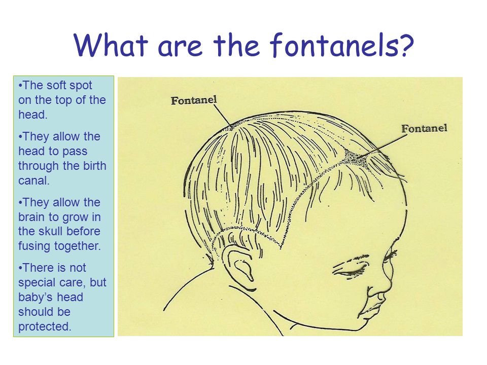
Pediatric neurologist. First visit | Children's neurologist
Your baby is one month old. It's time to visit a pediatric neurologist to make sure that the baby is all right. And if something is wrong, identify the pathology at the very beginning, prevent the disease from developing and moving into a more severe stage.
The fact is that the development of a young child is a very sensitive sign of the state of the organism. It depends both on hereditary characteristics and on a complex set of social conditions and requires the dynamic supervision of doctors.
Do not forget to show your baby to a pediatric neurologist within the prescribed timeframes - 1, 3, 6, 12 months!
If you invite a specialist to your home, then you must consider the following:
- the examination of the child should be carried out on a changing table or other soft, but not sagging surface;
- the temperature in the room should be around 25 ° C, the lighting should be bright but not annoying, the environment should be calm, if possible, eliminate distractions;
- inspection is preferably carried out 1.
 5-2 hours after feeding.
To determine the level of development of the child and exclude neurological pathology, it is important to evaluate the formed reactions to light, sound, motor and psycho-emotional activity of the newborn and his appearance. First of all, a pediatric neurologist, when examining a one-month-old baby, will pay attention to the shape and size of the skull, facial expression, posture, type of skin.
At the time of birth, when the child passes through the birth canal, the bones of the skull overlap each other. Features of the course of the birth process affect the change in the shape of the skull. With a complicated birth act, a sharp finding of the bones of the skull on top of each other may occur, and this will lead to its deformation, which will persist for quite a long time, and a pediatric neurologist has to deal with this.
A change in the shape of the skull can be expressed in the preservation of swelling of the soft tissues of the head in the place where the child moved forward along the birth canal. The swelling disappears within the first 2-3 days. Cephalhematoma (bleeding under the periosteum) also changes the shape of the skull. It resolves more slowly than swelling, and this process requires the supervision of specialists (neurologist, surgeon). The change in the shape of the skull is also associated with age-related features. In a newborn, the skull is elongated in the anterior-posterior direction, and after a few months the transverse size of the skull will increase, and its shape will change.
Some change in the shape and size of the skull can also occur during normal development in premature babies, or when the child is often laid on the same side, or when the child is lying on the back for a long time. All these features, according to pediatric neurologists, in themselves do not carry a negative value for the development of the child, if other provoking factors do not arise against their background.
Fontanelles
Examination of the fontanel by a pediatric neurologist is an almost obligatory procedure. Fontanelles are located in the area of convergence of the bones of the skull. Anterior, large, fontanel is located between the frontal and parietal bones. At birth, it measures from 2.5 to 3.5 cm, then gradually decreases by 6 months and closes at 8-16 months. The back, small, fontanel is located between the parietal and occipital bones. It is small and closes by 2-3 months.
In pathological processes accompanied by an increase in intracranial pressure, the fontanelles close later, and sometimes they open again. The small size of the anterior fontanel may be a variant of the norm if they are not accompanied by a decrease in the circumference of the skull, the rate of its growth, and a delay in psychomotor development.
The above signs do not limit the whole variety of possible deviations in a young child. However, it should be borne in mind that any unusual variant of the child's appearance requires careful examination and monitoring of its growth and development, which is well known to pediatric neurologists.
How a newborn's head grows is an important point that a pediatric neurologist pays attention to.
A newborn has an average head circumference of 35.5 cm (the normal range is 33.0-37.5 cm). The most intensive increase in head circumference in full-term children is observed in the first 3 months - on average, it is 1.5 cm for each month. Then the growth decreases slightly, and by the year the child's head circumference is on average 46.6 cm (normal limits 44.9- 48.9 cm).
The head circumference of a premature baby increases faster than that of a full-term baby, and the increase is most pronounced during the period of active weight gain, and by the end of the 1st year of life it reaches normal values. The exception is very premature babies.
The pediatric neurologist warns that it should always be borne in mind that even in the normal development of a child, there may be physiological deviations from the average values, which are often associated with constitutional features or environmental influences.
Right from birth, you can independently control the growth of the child's head circumference, which is one of the main indicators of normal and pathological conditions. To do this, measure the circumference of the child's head weekly and record the figures obtained in a specially wound notebook. When measuring, place the centimeter tape along the most protruding points of the skull (frontal and occipital tubercles). It is desirable that the measurements were carried out by the same person in order to avoid subjective errors.
In addition to the increase in head circumference, specialists - pediatric neurologists emphasize - it is possible to control the increase in chest circumference, which is one of the general anthropometric indicators of a child's development. To do this, measure your chest circumference weekly on the same day you measure your head circumference. Place the measuring tape at the level of the baby's nipple line.
Carrying out these simple measurements, you will help the pediatric neurologist to make an objective picture of the development of the child, and you yourself can be calm, eliminating the possibility of developing serious diseases (normally, the monthly increase in head circumference for the first three months in a full-term baby should not exceed 2 cm per month; up to a year, the circumference of the chest is approximately 1 cm larger than the circumference of the child's head).
Of course, not every deviation from the norm clearly indicates a pathological condition of the child. Clinical observations show that there are many factors that influence the shape and size of an infant's head.
Of course, even a small increase or decrease in the circumference of the skull in a newborn compared to the age norm can be considered a risk factor for the development of hydrocephalus or microcephaly, but you should not panic when you just find that the baby's head is slightly larger or smaller than normal : this circumstance should, first of all, become a signal for the need for additional examinations, primarily by a highly qualified pediatric neurologist to exclude pathological conditions.
An absolutely safe and reliable method is neurosonography (ultrasound examination of the brain through the large fontanel). This study will help not only to see changes in the structure of the brain and signs of increased intracranial pressure, but also to assess blood flow through the main vessels of the brain.