Chromosome testing at 12 weeks
First trimester screening - Mayo Clinic
Overview
Nuchal translucency measurement
Nuchal translucency measurement
First trimester screening includes an ultrasound exam to measure the size of the clear space in the tissue at the back of a baby's neck (nuchal translucency). In Down syndrome, the nuchal translucency measurement is abnormally large — as shown on the left in the ultrasound image of an 11-week fetus. For comparison, the ultrasound image on the right shows an 11-week fetus with a normal nuchal translucency measurement.
First trimester screening is a prenatal test that offers early information about a baby's risk of certain chromosomal conditions, specifically, Down syndrome (trisomy 21) and extra sequences of chromosome 18 (trisomy 18).
First trimester screening, also called the first trimester combined test, has two steps:
- A blood test to measure levels of two pregnancy-specific substances in the mother's blood — pregnancy-associated plasma protein-A (PAPP-A) and human chorionic gonadotropin (HCG)
- An ultrasound exam to measure the size of the clear space in the tissue at the back of the baby's neck (nuchal translucency)
Typically, first trimester screening is done between weeks 11 and 14 of pregnancy.
Using your age and the results of the blood test and the ultrasound, your health care provider can gauge your risk of carrying a baby with Down syndrome or trisomy 18.
If results show that your risk level is moderate or high, you might choose to follow first trimester screening with another test that's more definitive.
Products & Services
- Book: Mayo Clinic Guide to a Healthy Pregnancy
- Book: Obstetricks
Why it's done
First trimester screening is done to evaluate your risk of carrying a baby with Down syndrome. The test also provides information about the risk of trisomy 18.
Down syndrome causes lifelong impairments in mental and social development, as well as various physical concerns. Trisomy 18 causes more severe delays and is often fatal by age 1.
First trimester screening doesn't evaluate the risk of neural tube defects, such as spina bifida.
Because first trimester screening can be done earlier than most other prenatal screening tests, you'll have the results early in your pregnancy. This will give you more time to make decisions about further diagnostic tests, the course of the pregnancy, medical treatment and management during and after delivery. If your baby has a higher risk of Down syndrome, you'll also have more time to prepare for the possibility of caring for a child who has special needs.
Other screening tests can be done later in pregnancy. An example is the quad screen, a blood test that's typically done between weeks 15 and 20 of pregnancy. The quad screen can evaluate your risk of carrying a baby with Down syndrome or trisomy 18, as well as neural tube defects, such as spina bifida. Some health care providers choose to combine the results of first trimester screening with the quad screen. This is called integrated screening. This can improve the detection rate of Down syndrome.
First trimester screening is optional. Test results indicate only whether you have an increased risk of carrying a baby with Down syndrome or trisomy 18, not whether your baby actually has one of these conditions.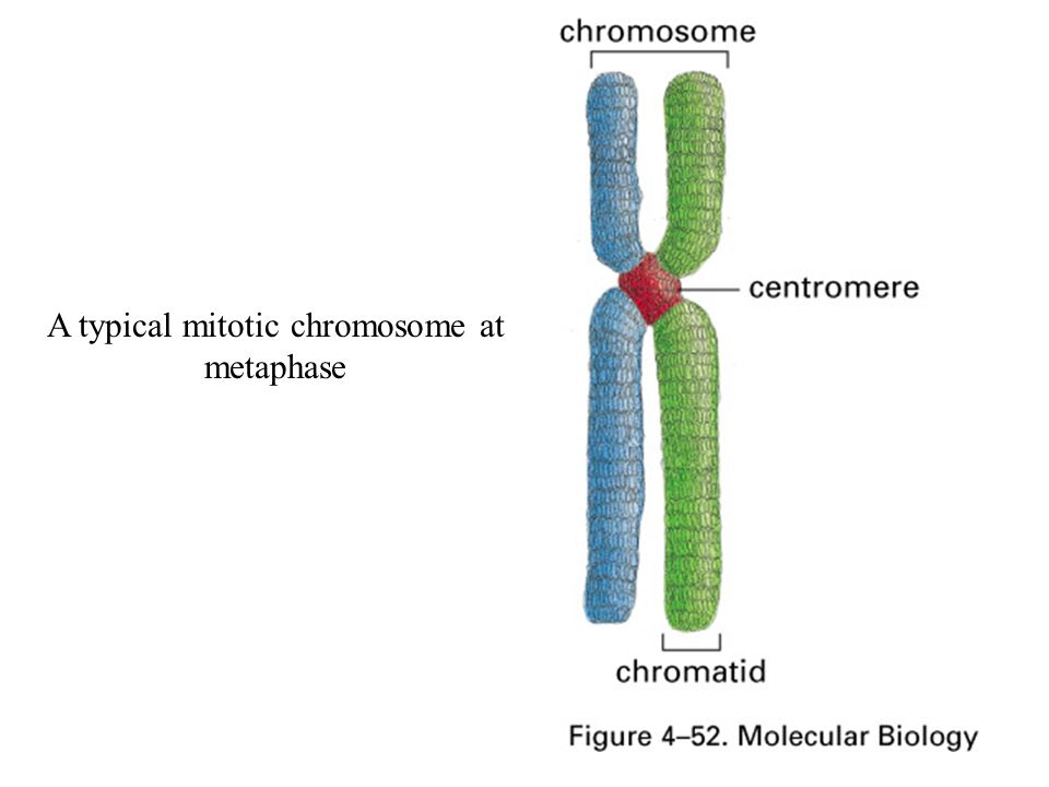
Before the screening, think about what the results will mean to you. Consider whether the screening will be worth any anxiety it might cause, or whether you'll manage your pregnancy differently depending on the results. You might also consider what level of risk would be enough for you to choose a more invasive follow-up test.
More Information
- Prenatal testing: Quick guide to common tests
Request an Appointment at Mayo Clinic
Risks
First trimester screening is a routine prenatal screening test. The screening poses no risk of miscarriage or other pregnancy complications.
How you prepare
You don't need to do anything special to prepare for first trimester screening. You can eat and drink normally before both the blood test and the ultrasound exam.
What you can expect
First trimester screening includes a blood draw and an ultrasound exam.
During the blood test, a member of your health care team takes a sample of blood by inserting a needle into a vein in your arm. The blood sample is sent to a lab for analysis. You can return to your usual activities immediately.
For the ultrasound exam, you'll lie on your back on an exam table. Your health care provider or an ultrasound technician will place a transducer — a small plastic device that sends and receives sound waves — over your abdomen. The reflected sound waves will be digitally converted into images on a monitor. Your health care provider or the technician will use these images to measure the size of the clear space in the tissue at the back of your baby's neck.
The ultrasound doesn't hurt, and you can return to your usual activities immediately.
Results
Your health care provider will use your age and the results of the blood test and ultrasound exam to gauge your risk of carrying a baby with Down syndrome or trisomy 18. Other factors — such as a prior Down syndrome pregnancy — also might affect your risk.
First trimester screening results are given as positive or negative and also as a probability, such as a 1 in 250 risk of carrying a baby with Down syndrome.
First trimester screening correctly identifies about 85 percent of women who are carrying a baby with Down syndrome. About 5 percent of women have a false-positive result, meaning that the test result is positive but the baby doesn't actually have Down syndrome.
When you consider your test results, remember that first trimester screening indicates only your overall risk of carrying a baby with Down syndrome or trisomy 18. A low-risk result doesn't guarantee that your baby won't have one of these conditions. Likewise, a high-risk result doesn't guarantee that your baby will be born with one of these conditions.
If you have a positive test result, your health care provider and a genetics professional will discuss your options, including additional testing. For example:
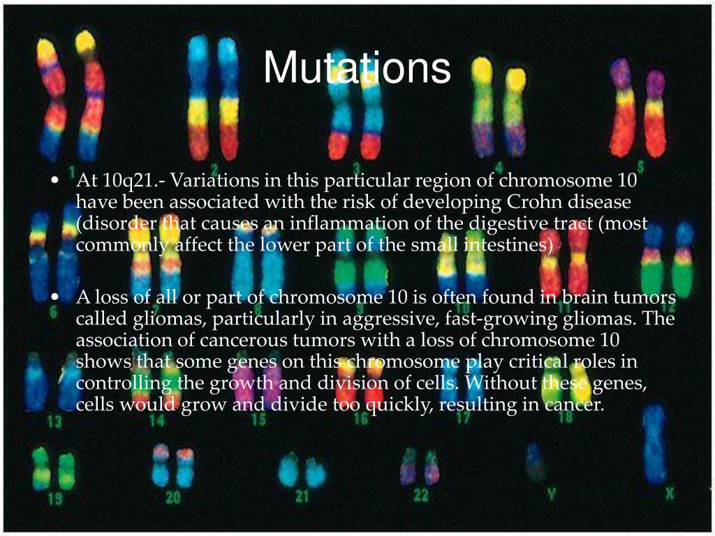
- Prenatal cell-free DNA (cfDNA) screening. This is a sophisticated blood test that examines fetal DNA in the maternal bloodstream to determine whether your baby is at risk of Down syndrome, extra sequences of chromosome 13 (trisomy 13) or extra sequences of chromosome 18 (trisomy 18).
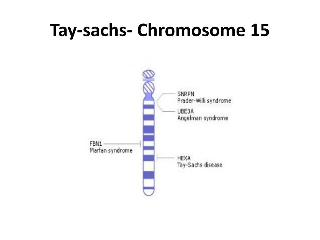 Some forms of cfDNA screening also screen for other chromosome problems and provide information about fetal sex. A normal result might eliminate the need for a more invasive prenatal diagnostic test.
 - Chorionic villus sampling (CVS). CVS can be used to diagnose chromosomal conditions, such as Down syndrome. During CVS, which is usually done during the first trimester, a sample of tissue from the placenta is removed for testing. CVS poses a small risk of miscarriage.
- Amniocentesis. Amniocentesis can be used to diagnose both chromosomal conditions, such as Down syndrome, and neural tube defects, such as spina bifida. During amniocentesis, which is usually done during the second trimester, a sample of amniotic fluid is removed from the uterus for testing. Like CVS, amniocentesis poses a small risk of miscarriage.
Your health care provider or a genetic counselor will help you understand your test results and what the results mean for your pregnancy.
By Mayo Clinic Staff
Related
Products & Services
Prenatal Genetic Screening Tests | ACOG
Amniocentesis: A procedure in which amniotic fluid and cells are taken from the uterus for testing. The procedure uses a needle to withdraw fluid and cells from the sac that holds the fetus.
Aneuploidy: Having an abnormal number of chromosomes. Types include trisomy, in which there is an extra chromosome, or monosomy, in which a chromosome is missing. Aneuploidy can affect any chromosome, including the sex chromosomes. Down syndrome (trisomy 21) is a common aneuploidy. Others are Patau syndrome (trisomy 13) and Edwards syndrome (trisomy 18).
Carrier Screening: A test done on a person without signs or symptoms to find out whether he or she carries a gene for a genetic disorder.
Cell-Free DNA: DNA from the placenta that moves freely in a pregnant woman’s blood. Analysis of this DNA can be done as a noninvasive prenatal screening test.
Cells: The smallest units of a structure in the body. Cells are the building blocks for all parts of the body.
Chorionic Villus Sampling (CVS): A procedure in which a small sample of cells is taken from the placenta and tested.
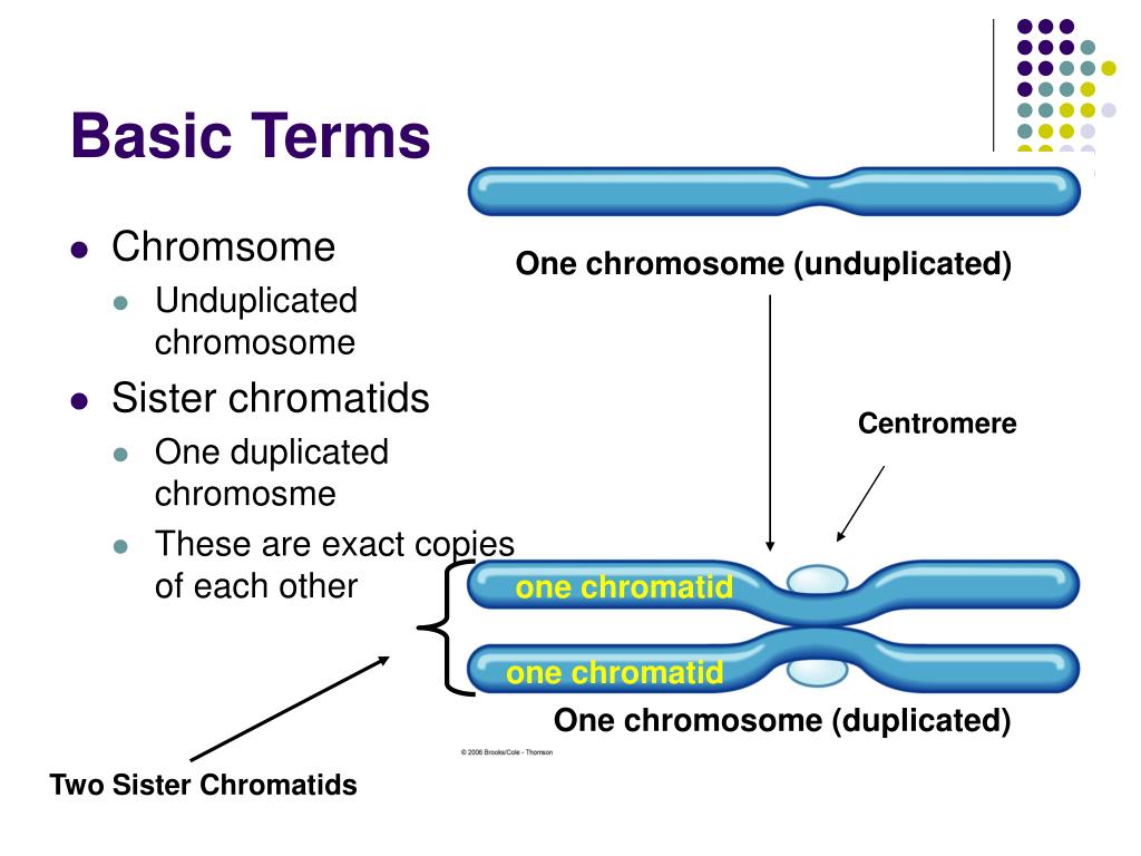
Chromosomes: Structures that are located inside each cell in the body. They contain the genes that determine a person’s physical makeup.
Cystic Fibrosis (CF): An inherited disorder that causes problems with breathing and digestion.
Diagnostic Tests: Tests that look for a disease or cause of a disease.
DNA: The genetic material that is passed down from parent to child. DNA is packaged in structures called chromosomes.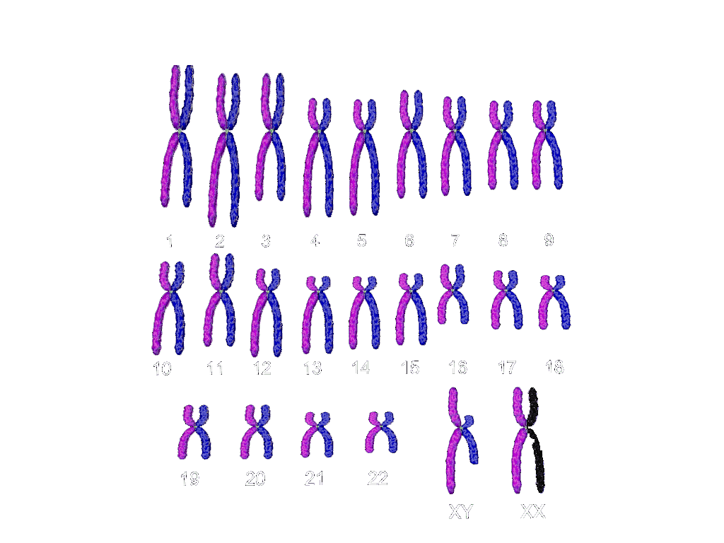
Down Syndrome (Trisomy 21): A genetic disorder that causes abnormal features of the face and body, medical problems such as heart defects, and mental disability. Most cases of Down syndrome are caused by an extra chromosome 21 (trisomy 21).
Edwards Syndrome (Trisomy 18): A genetic condition that causes serious problems. It causes a small head, heart defects, and deafness.
Fetus: The stage of human development beyond 8 completed weeks after fertilization.
Genes: Segments of DNA that contain instructions for the development of a person’s physical traits and control of the processes in the body. The gene is the basic unit of heredity and can be passed from parent to child.
Genetic Counselor: A health care professional with special training in genetics who can provide expert advice about genetic disorders and prenatal testing.
Genetic Disorders: Disorders caused by a change in genes or chromosomes.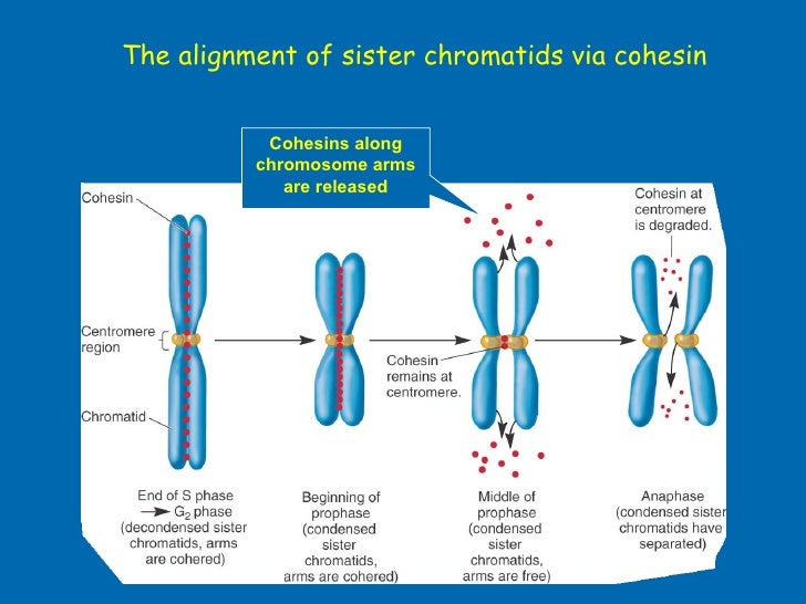
Inherited Disorders: Disorders caused by a change in a gene that can be passed from parents to children.
Monosomy: A condition in which there is a missing chromosome.
Mutations: Changes in genes that can be passed from parent to child.
Neural Tube Defects (NTDs): Birth defects that result from a problem in development of the brain, spinal cord, or their coverings.
Nuchal Translucency Screening: A test to screen for certain birth defects, such as Down syndrome, Edwards syndrome, or heart defects. The screening uses ultrasound to measure fluid at the back of the fetus’s neck.
Obstetrician: A doctor who cares for women during pregnancy and their labor.
Patau Syndrome (Trisomy 13): A genetic condition that causes serious problems. It involves the heart and brain, cleft lip and palate, and extra fingers and toes.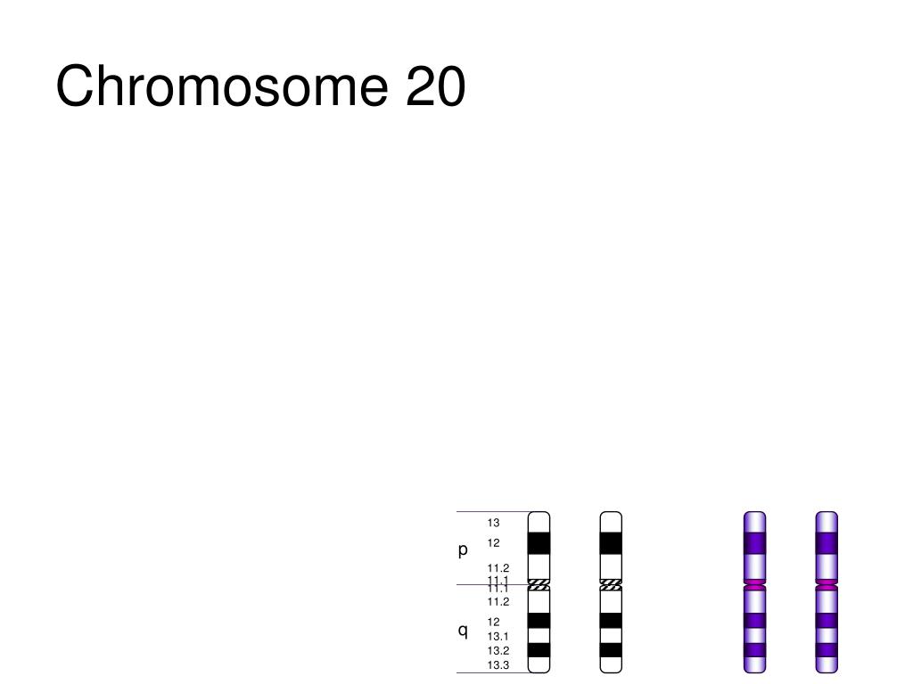
Placenta: An organ that provides nutrients to and takes waste away from the fetus.
Screening Tests: Tests that look for possible signs of disease in people who do not have signs or symptoms.
Sex Chromosomes: The chromosomes that determine a person’s sex. In humans, there are two sex chromosomes, X and Y. Females have two X chromosomes and males have an X and a Y chromosome.
Sickle Cell Disease: An inherited disorder in which red blood cells have a crescent shape, which causes chronic anemia and episodes of pain.
Tay–Sachs Disease: An inherited disorder that causes mental disability, blindness, seizures, and death, usually by age 5.
Trimester: A 3-month time in pregnancy. It can be first, second, or third.
Trisomy: A condition in which there is an extra chromosome.
Ultrasound Exams: Tests in which sound waves are used to examine inner parts of the body. During pregnancy, ultrasound can be used to check the fetus.
Frequently asked questions about biochemical screening of pregnant women
What tests are performed on pregnant women?
Currently, two main types of tests are recommended for pregnant women in St. Petersburg:
- analysis for PAPP-A and beta-hCG in the period of 9-13 weeks
- analysis for AFP and hCG.
Should I do a “triple test”?
Some private laboratories use the so-called “triple test”, using kits that, in addition to AFP and hCG, have added the determination of the concentration of another hormone - unconjugated estriol. According to modern data, its assessment for clarifying the risks of fetal chromosomal pathology has too little weight and is highly dependent on many other factors of the woman's condition, which is not significant for calculating risks for fetal chromosomal pathology. If the results of the “double” test reveal an increased risk for you, it is better to contact a geneticist and understand the situation, taking into account a professional clinical assessment of the test results.
How important is it to accurately indicate the weight of a pregnant woman when taking blood for “fetal proteins”?
Each woman's weight must be recorded on the examination referral. In the absence of such information, the risk can be calculated "according to the average" weight of pregnant women in this period - 60 kg.
All women are different - there are pregnant women weighing 45 and 145 kg. For a more accurate assessment of the results, an amendment is introduced in accordance with the "weight category". But absolute accuracy is not required here - individual grams will not change the calculations. An individual approach is important. Therefore, we always measure a woman's weight before taking tests.
I took a blood test for fetal proteins, as the doctor prescribed them immediately after taking them, now I'm worried, so I had a hearty breakfast in the morning. Will this affect the outcome of the study? Can I retake blood on an empty stomach?
Don't worry! Unlike most "adult" tests that are sensitive to food intake, the measurement of the amount of any proteins entering the mother's blood from the fetus is not dependent on the time of the meal.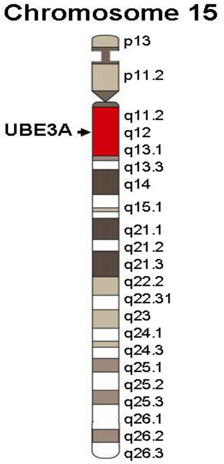 The most important thing here is to know exactly the gestational age established by ultrasound. It makes no sense to retake the analysis. And it is very important for pregnant women to eat breakfast and continue to eat more often during the day than usual.
In clinics where an individual approach to the examination of pregnant women is possible, and of course, in our Center for Fetal Medicine, blood for screening tests can be donated throughout the working day.
Which test is better - PAPP-A and beta-hCG or AFP and hCG?
At present, the first one has the absolute advantage. It has been proven that it is more specific for assessing risks for chromosomal pathology, including Down syndrome. Its important advantage is that it is carried out in the first trimester, you can donate blood from the age of 9weeks of pregnancy (determined by the size of the fetus on ultrasound). The most optimal terms for this analysis are 9-12 weeks. A study period of up to almost 14 (13 weeks 6 days) is allowed, but the reliability of the risk assessment will be lower.
If you have completed a full first trimester test, performed an ultrasound and received a conclusion from a geneticist that the fetus is at low risk for chromosomal abnormalities, testing for AFP and hCG is not worth it.
In special cases, after the first screening, a test for AFP and hCG is prescribed as an additional test on the recommendation of a geneticist.
If you missed the first screening test, then, of course, you need to donate blood at least for the second one within 15 to 18 weeks.
I would like to emphasize that, on the recommendation of international experts in prenatal diagnosis, analysis for PAPP-A and beta-hCG in a period of 9-12 weeks is recommended for all pregnant women at any age.
I went through the IVF procedure and used a donor cell, because my cells do not mature at the age of 46. Now 12 weeks. How do I correctly pass a biochemical test?
You urgently need to donate blood for PAPP-A and beta-hCG and perform an ultrasound. From your age, the IVF procedure itself does not change the amount of proteins. And the risk "by proteins" will be assessed depending on certain concentrations.
But the computer program calculates the "combined" risks - by proteins, by ultrasound and by the age of the woman, more precisely, by the "age of the egg." Accordingly, for this analysis, the age of the egg donor must be indicated in the calculation referral. If you do not know it exactly, you can calculate by low age risk, since all donors have age restrictions for participating in the IVF program. If you have already been calculated according to your age, don't worry, we can reassess the risks taking into account real data and issue you a Medical Genetic Conclusion based on the results of prenatal studies.
Doctors of the Fetal Medicine Center are one of the leading specialists in prenatal diagnostics, candidates of medical sciences, doctors of the highest categories with a narrow specialization and extensive experience in prenatal medicine.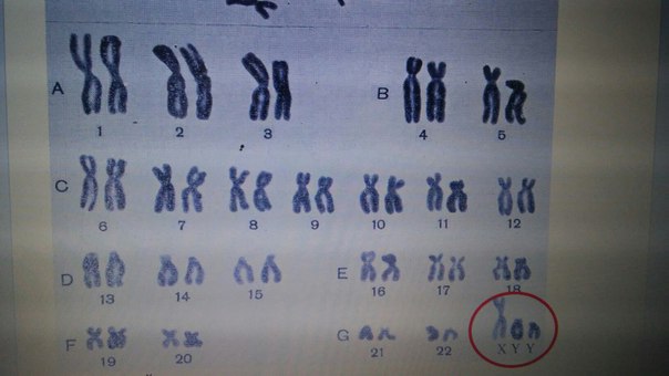
All ultrasound examinations in the center are carried out according to the international standards FMF (Fetal Medicine Foundation) and ISUOG (International Society for Ultrasound in Obstetrics and Gynecology).
Ultrasound doctors have international certificates from the Fetal Medicine Foundation (Fundamental Medicine, UK), which are are confirmed annually.
We take care of the most complex cases and, if necessary, it is possible to consult with specialists from King's College Hospital, King's College Hospital (London, UK).
The pride of our Centers - modern and high-tech medical equipment from General Electric: expert-class ultrasonic devices Voluson E8 / E10
The capabilities of these devices allow us to talk about a new level of information content.
To make an appointment and get an expert opinion of our ultrasound specialists, you can call the single contact center +7 (812) 458-00-00
assessment of the karyotype / genotype of the developing fetus - reliable confirmation or confirmation of chromosomal abnormalities at the earliest possible time - is of critical importance when planning further pregnancy management tactics.
There are a number of requirements for examination methods and algorithms, among which the main ones are the following:
- High accuracy - any analysis for chromosomal pathologies must have a high degree of reliability, otherwise it simply loses its meaning. The lower limit of accuracy for screening is 85-90%, and over 99% accuracy for the rest.
- Low cost - the lower the cost of the study, the better. With a minimum level of expenditure, there is the possibility of full coverage of pregnant women. At a higher cost, it is important that as many people as possible can afford the study.
- Technical simplicity and the level of medical support - certain technical support and doctors are required to conduct any study. Methods in which extremely expensive equipment or exceptional specialists are needed, even with excellent reliability of the results, will not be available for objective reasons.
- Maximum safety for the health of the pregnant woman, the developing fetus and the normal course of the pregnancy itself.

Based on these basic principles, in today's medical practice, the following test options for fetal chromosomal pathologies are used.
Traditional screening of pregnant women - blood markers + ultrasound
Relative accessibility, lack of special requirements for medical equipment and specialists (possibility of carrying out in almost any medical institution of the corresponding profile), low cost. This makes it possible to ensure the mass character of the type of screening, that is, a comprehensive survey.
The actual disadvantage is the low level of confidence, which is in the range of 85%. It is for this reason that screening during pregnancy is performed repeatedly - three times, at a certain time. Obtaining results regarding possible fetal chromosomal abnormalities is an estimated value with the need to clarify by other, more accurate methods.
Additionally, screening has a lot of limitations that reduce its reliability below the minimum limit in the case of: multiple pregnancy, IVF pregnancy, donation of reproductive material, etc.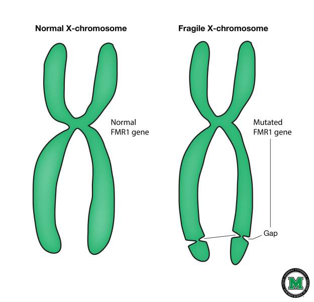
Invasive procedures for taking fetal biomaterial for analysis
The main ones are chorionic villus biopsy, amniocentesis with amniotic fluid sampling, blood sampling from umbilical cord vessels. The genetic material of the fetus is taken with the help of invasion, that is, penetration through the fetal membranes. The method is 99.9% reliable, however, it also has its drawbacks.
Relatively high cost, technical complexity and the need for special equipment - over time, the relevance of these factors will gradually decrease, the methods will become more accessible and cheaper. The only significant factor was and remains some risk for the mother, the fetus and the course of pregnancy, up to its termination. That is why invasive research methods have been and are used to a limited extent in the presence of strong indications, when there are simply no other possibilities to verify the fetal karyotype.
Non-invasive test for fetal chromosomal pathologies - Prenetix
The non-invasive prenatal test (NIPT) has already established itself as a screening for the most common chromosomal pathologies. Here are its main features:
-
The blood sample for examination is taken from the peripheral vein of the mother
-
The method is accurate, as fetal DNA fragments are examined
-
Technically available - delivery of biomaterial is possible throughout the country, and direct genetic testing is carried out in the laboratory where blood is delivered
-
The cost of the test is quite low and fixed
-
Informative for twin pregnancy, after IVF, surrogacy and reproductive donation.
First Expert Level Screening
First Expert Level Screening is state-of-the-art screening for the genetic health of an unborn baby with a high degree of certainty not previously available. Screening includes:
- Ultrasound by specially trained and licensed ultrasound screening specialists in the first trimester
- Determination of maternal serum markers (pregnancy-associated plasma protein A (PAPP-A) and free beta subunit of human chorionic gonadotropin)
- Non-invasive prenatal test Prenetix.


