Abdominal muscle separation post pregnancy
Diastasis Recti (Abdominal Separation): Symptoms & Treatment
Overview
Diastasis recti happens when a person's abdomen stretches during pregnancy and creates a gap in the abdominal muscles.What is diastasis recti?
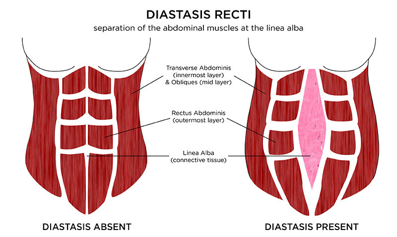
Diastasis recti (diastasis rectus abdominis or diastasis) is the separation of the rectus abdominis muscles during and after pregnancy. The rectus abdominis runs vertically along the front of your stomach. It's frequently referred to as someone's "six-pack abs." It's divided into left and right sides by a band of tissue called the linea alba that runs down the middle. As your uterus expands during pregnancy, the abdominals are stretched and the linea alba thins and pulls apart. This band of tissue gets wider as it's pushed outward.
Once you deliver your baby, the linea alba can heal and come back together. It's highly elastic and retracts backs (like a rubber band). When the tissue loses its elasticity from being overstretched, the gap in the abdominals will not close as much as it should. This is diastasis recti.
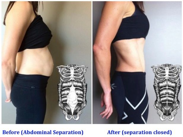
If you have diastasis, your belly may appear to stick out just above or below the belly button, making you appear pregnant months or years after giving birth.
Why does diastasis recti happen?
Pregnancy puts a lot of pressure on your abdomen (abs). The abdomen is made up of left and right ab muscles and a thin band of connective tissue (linea alba) in between. They are pushed outward and stretched to make room for the growing baby. Diastasis recti occurs when the linea alba is overstretched and doesn't come back together. The left and right sides of the abdominals stay separated. It's also referred to as an "ab gap" or abdominal separation.
Who gets diastasis recti?
Diastasis recti is most common in pregnant and postpartum women (it can also be seen in men and infants). Diastasis recti usually develops in the third trimester. There is increased pressure on the abdominal wall because the baby is growing quickly during this time. Most people don't notice diastasis recti until the postpartum period.
How common is diastasis recti?
Diastasis recti is extremely common in those who are pregnant and during the postpartum period. It affects 60% of people. It usually resolves itself within eight weeks of delivery. About 40% of those who have diastasis recti still have it by six months postpartum.
Symptoms and Causes
What are the symptoms of diastasis recti?
Most people don't notice signs of diastasis recti until they are postpartum. You can have diastasis recti during pregnancy, but it's hard to distinguish because your abdomen is stretched.
Common signs of diastasis recti during the postpartum period are:
- A visible bulge or "pooch" that protrudes just above or below the belly button.
- Softness or jelly-like feeling around your belly button.
- Coning or doming when you contract your ab muscles.
- Difficulty lifting objects, walking or performing everyday tasks.
- Pain during sex.
- Pelvic or hip pain.
- Low back pain.
- Poor posture.
- Urine leaking when you sneeze or cough.
- Constipation.
- Feeling weak in your abdominals.
What does diastasis recti feel like?
Diastasis recti is not painful. You may feel pain associated with some of the side effects of diastasis, but the ab separation itself doesn't hurt. You may feel weakness in your core when doing once easy tasks, like lifting a laundry basket. Some people feel a jelly-like texture in the space between the left and right abdominals when contracting the ab muscles.
How do I know if I have diastasis recti?
There are some common signs that can signal you have diastasis recti. One of the most common signs of diastasis recti is a bulge in your midsection that doesn't go away, even after exercising or losing weight gained during pregnancy. Another sign is that your belly cones or domes when you lean back on a chair or get up out of bed. You can check for diastasis recti on your own, but it is always a good idea to speak with your healthcare provider about your symptoms.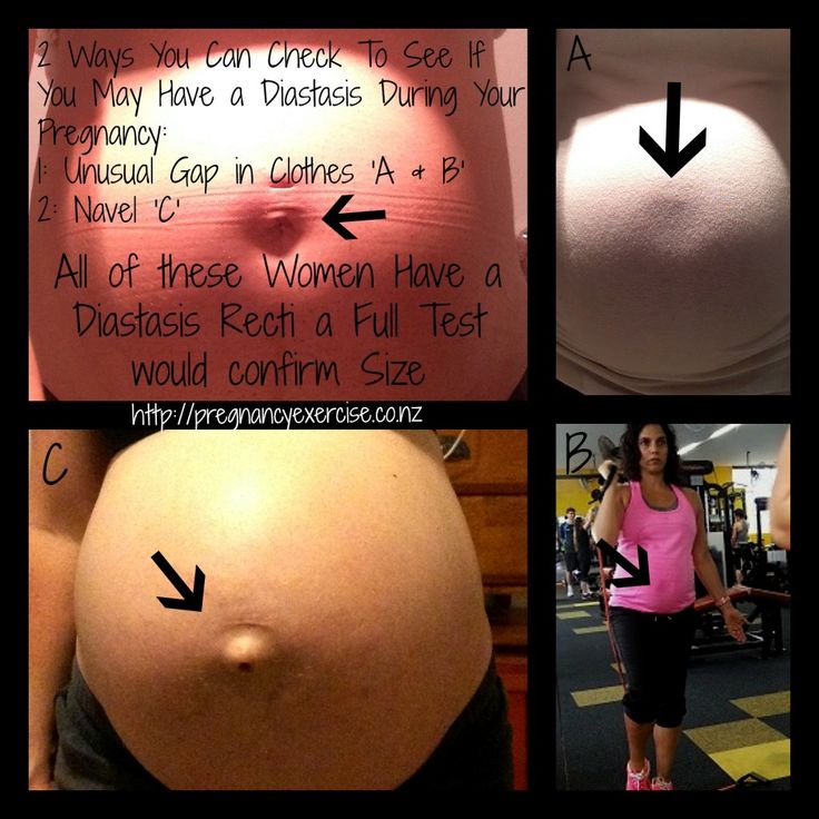
What are the risk factors for developing diastasis recti?
Several factors can increase your risk for developing diastasis recti:
- Having multiple pregnancies (especially back-to-back).
- Being over 35 years old.
- Having multiples (such as twins or triplets).
- Having a heavy or big baby.
- Being extremely petite.
- Vaginal delivery. Pushing can increase abdominal pressure.
Diagnosis and Tests
How is diastasis recti diagnosed?
Your healthcare provider will evaluate if diastasis is present, where it's located and how severe it is. Diastasis recti can occur above the belly button, below the belly button and at the belly button.
Your provider will use their hands and fingers to feel the abdominal area for gaps and muscle tone. Some providers may use ultrasound, measuring tape or a tool called a caliper for a more accurate measurement. This exam typically occurs at your postpartum appointment before being cleared for exercise.
An abdominal gap wider than 2 centimeters is considered diastasis recti. Diastasis recti is also measured in finger widths, for example, two or three fingers' separation.
Your healthcare provider may recommend movements for diastasis recti or they may refer you to a specialist for additional treatment.
How do I test myself for diastasis recti?
You can test yourself for diastasis recti:
- Lie on your back with your knees bent and feet flat on the floor.
- Lift your shoulders slightly off the ground, keeping one hand behind your head for support. Almost like you are doing a sit-up. Look down at your belly.
- Move your other hand above your belly button area, palms down and fingers towards your toes.
- Use your fingers to feel for a gap between the abs. See how many fingers can fit in the gap between your right and left abdominals.
If you feel a gap of two or more finger widths, discuss your concerns with your healthcare provider. They should confirm diastasis recti with a proper diagnosis and recommend appropriate care.
Management and Treatment
How can I fix diastasis recti?
To fix diastasis recti, you'll need to perform gentle movements that engage the abdominal muscles. Before starting an exercise program, be sure it's safe for diastasis recti. Work with a fitness professional or physical therapist who has experience with diastasis recti. They can create a treatment plan to make sure you are performing the movements correctly and progressing to more challenging movements at the right time.
Certain movements will make abdominal separation worse. During the postpartum period, there are some modifications you should make:
- Avoid lifting anything heavier than your baby.
- Roll onto your side when getting out of bed or sitting up. Use your arms to push yourself up.
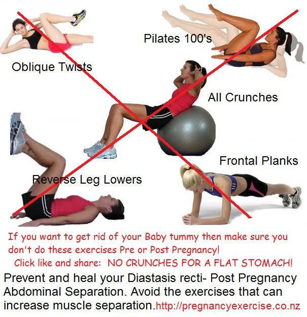
- Skip activities and movements that push your abdominals outward (like crunches and sit-ups).
Some people use binding devices (elastic belly bands) to help hold their belly in and support the lower back.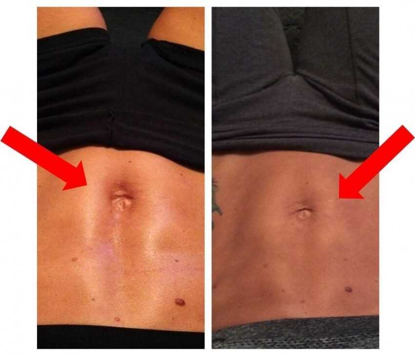 Wearing binders can't heal diastasis recti and will not strengthen your core muscles. It can be a good reminder of your diastasis recti and promote good posture.
Can you fix diastasis recti without surgery?
Yes, it's possible to fix diastasis recti without surgery. Surgery is rarely performed to fix diastasis recti. Healthcare providers will recommend physical therapy or at-home exercises to help heal diastasis before surgical methods. Surgery is performed in cases of hernia (when an organ pushes through the linea alba) or if a woman wants diastasis recti surgery (a tummy tuck).
What are the best exercises for diastasis recti?
The best exercises for diastasis recti are those that engage the deep abdominals. Most diastasis recti exercises involve deep breathing and slow, controlled movements. Unfortunately, many of the most common ab exercises (like crunches) can worsen your diastasis. Before starting abdominal exercises, ask your healthcare provider to check you for diastasis recti.
What movements make diastasis recti worse?
Any movement that bulges the abdominal wall forward can cause more damage to your diastasis recti. Everyday movements like getting out of bed or up off a chair can worsen diastasis. Try to be mindful about how you are using your abdominals as you go about your day.
These exercise movements should be avoided if you have diastasis recti:
- Crunches or sit-ups of any kind.
- Planks or push-ups (unless using modifications).
- Downward dog, boat pose and other yoga poses.
- Double leg lifts, scissors and other Pilates moves.
- Any exercise that causes your abdominals to bulge, cone or dome.
Prevention
How do I prevent diastasis recti?
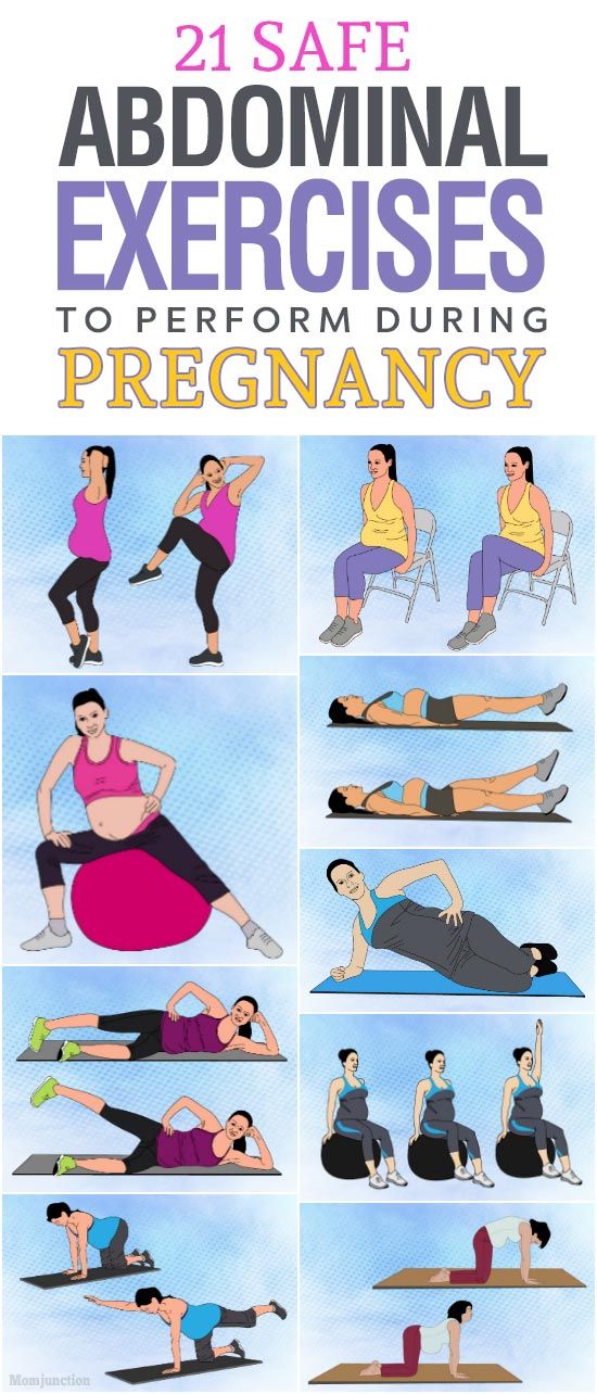
Some abdominal separation is normal and expected with pregnancy. There are some things you can do to lower your risk for developing diastasis recti:
- Healthy weight gain during pregnancy: Exercising and eating healthy foods to keep weight gain within a healthy range.
- Proper posture and deep breathing: Stand up straight with your shoulders back. Take deep breaths that allow your ribs to expand and not just your belly.
- Safe core exercises: Avoid exercises like sit-ups and crunches that put pressure on your abdominals after 12 weeks of pregnancy and postpartum.
- Don't strain while lifting: Certain day-to-day activities like lifting grocery bags or your children can put undue strain on your abdominals.
- Log roll when getting out of bed: If you're pregnant or postpartum, roll to one side and use your arms to push up out of bed.
Outlook / Prognosis
How long will it take to heal my diastasis recti?
The amount of time it takes to heal diastasis recti depends on the amount of ab separation and how consistent you are with strengthening exercises. After several weeks postpartum, this gap will start to close as your muscles regain strength.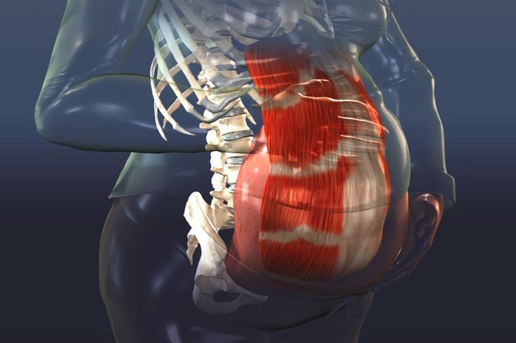 If you're making modifications to your lifestyle and performing exercises with good form, you're more likely to notice progress.
Can I get diastasis recti again?
Yes, you can heal your diastasis recti and get it again. Your risk for diastasis recti increases the more times you are pregnant. Think of the linea alba as a rubber band that is continuously stretched. Over time, the rubber band will lose its elasticity. The linea alba may not regain its original shape or form after being stretched through multiple pregnancies.
Is it too late to fix my diastasis recti?
It's never too late to repair your diastasis recti. With the proper exercises, you can fix your ab separation years after you've delivered your last baby.
Are there complications from diastasis recti?
If left untreated or in severe cases of diastasis recti, complications can include:
- Umbilical hernia.
- Increase in back pain.
- Pain during sex.
- Urinary incontinence.
- Pelvic and hip pain.
Living With
When should I see my healthcare provider?
Diastasis recti is a common and easily treated condition. If you have more than a two-finger gap between your abdominals or are experiencing pain, contact your healthcare provider for a diagnosis. They may want you to see a physical therapist or pelvic floor specialist to help strengthen your abdominal muscles.
A note from Cleveland Clinic:
Diastasis recti can make you appear pregnant years after your last baby. Discuss your concerns with your healthcare provider so they can diagnose and treat you. Getting treatment can help you feel more confident in your body and correct any pain you are experiencing.
How to Improve Diastasis Recti After Baby
Women often struggle to realize the flat and toned midsections that they enjoyed pre-baby won't always bounce right back post-delivery, and diastasis recti may be the culprit. Diastasis recti, more commonly known as abdominal separation, leaves many women looking pregnant months, even years, after delivery.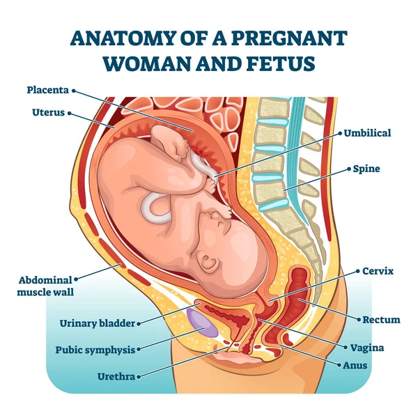 But this condition is more common than you may think.
If you’ve recently given birth and are looking to strengthen and tone your abdomen, or you’re wondering how to get rid of your “mommy tummy,” here are some key facts and helpful tips for you to keep in mind.
What is Diastasis Recti?
Diastasis recti, or abdominal separation, is a fairly common condition experienced by women during and after pregnancy, in which the right and left halves of the rectus abdominis muscle spread apart at the body’s midline.
In addition to reducing the integrity and functional strength of the abdominal wall, diastasis recti can also lead to pelvic girdle instability, causing women to experience pelvic and back pain, sacroiliac (SI) joint irritation, and restricted mobility.
Hi, I'm Cheryl.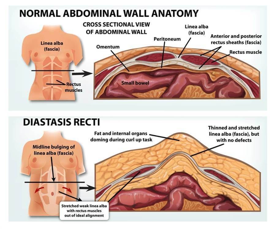 I work at Moreland OB-GYN and today I'm here to talk about diastasis recti. What is diastasis recti? It is the separation of the left and right abdominal muscles that happens during pregnancy.
Why does it happen? The connective tissue between your right and left abdominal muscle group is called the linea alba. The hormonal changes that happen during pregnancy soften connective tissue and break down the linea alba causing a protrusion of the abdominal area or a little pooch belly. Will you develop a diastasis recti? Some women will and some women won't. Women who are short or petite in nature, if you have had a separation of muscles in previous pregnancies, you're most likely to develop it again, or if you've had previous medical conditions. If you're a previous exerciser and have strong muscles, that will help you to prevent any weakness as baby grows. It's also encouraged to perform the "log roll" maneuver in bed before getting up. I hope you have found this video and instructions useful.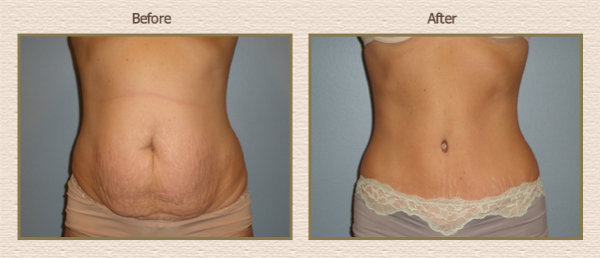 It is our mission at Moreland OB-GYN to lead you to better health.
What Causes Diastasis Recti?
During pregnancy, widening and thinning of the midline abdominal tissue occurs in response to the force of the growing uterus pushing against the abdominal wall. This, combined with pregnancy hormones that soften connective tissue, creates a small amount of widening of the midline and is normal.
Diastasis recti occurs in approximately 30-percent of all pregnancies and can develop anytime in the latter half of pregnancy. However, the condition is most commonly seen after pregnancy when the abdominal wall is lax, and the thinner midline tissue no longer provides adequate support for the torso and internal organs.
Those at the greatest risk of diastasis recti include:
- Women who are expecting more than one baby
- Petite women
- Women with a visible sway back and those with poor abdominal muscle tone
- Women who are considered obese
However, genetics also plays a significant role in determining whether your abdominal muscles will separate. For most women, it’s simply how their bodies respond to pregnancy.
How Do I Know If I Have Diastasis Recti?
A simple self-test will help you to determine if you have diastasis recti. Follow these steps:
- Lie on your back with your knees bent, and place the soles of your feet on the floor.
- Place one hand behind your head, and your other hand on your abdomen, with your fingertips across your midline, parallel with your waistline, at the level of your belly button.
- With your abdominal wall relaxed, gently press your fingertips into your abdomen.
- Roll your upper body off the floor into a “crunch,” making sure that your rib cage moves closer to your pelvis.
- Move your fingertips back and forth across your midline, feeling for the right and left sides of your rectus abdominis muscle. Test for separation at, above, and below your belly button.
If you feel a gap of more than two-and-a-half finger-widths when your rectus abdominis is fully contracted, the gap doesn’t shrink as your contract your abdominal wall, or you can see a small mound of tissue protruding along the length of your midline, you most likely have diastasis recti.
Are There Any At-Home Treatments for Diastasis Recti?
The good news is that the vast majority of women can close their midlines and flatten their abdominal walls with proper rehabilitation exercises.
Transverse Abdominis Activation
While lying on your back with your knees bent, draw your belly button in towards your spine. Contract your stomach muscles (as if coughing or sneezing) and hold this position. When this becomes easy and pain-free, move on to the next exercise.
- Repeat: 10 Times
- Hold: 3 Seconds
- Complete: 3 Sets
- Perform: 2 Times a day
Brace Heel Slides
While lying on your back with your knees bent, perform the transverse abdominis contraction as outlined in the first exercise. Hold this position with your stomach and slowly slide your heel forward on the floor or bed and then slide it back. Use your stomach muscles to keep your spine from moving. When this becomes easy and pain-free, move on to the next exercise.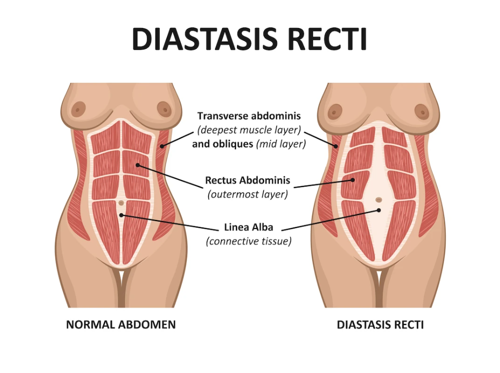
- Repeat: 10 Times
- Complete: 3 Sets
- Perform: 2 Times a day
Brace Marching
While lying on your back with your knees bent, perform the transverse abdominis contraction as outlined in the first exercise. Slowly raise up one foot a few inches and then set it back down. Next, perform on your other leg. Use your stomach muscles to keep your spine from moving. When this becomes easy and pain-free, move on to the next exercise.
- Repeat: 10 Times
- Complete: 3 Sets
- Perform: 2 Times a day
Brace Single Knee Extension
While lying on your back with knees bent, perform the transverse abdominis contraction as outlined in the first exercise. Slowly straighten out one knee while keeping the leg off the ground. Hold as indicated, then return to original position. Next, perform on the other leg. Keep your stomach muscles contracted.
- Repeat: 10 Times
- Complete: 3 Sets
- Perform: 1 Time a day
When Do I Need to See My Doctor for Diastasis Recti?
In extreme cases of diastasis recti, abdominal tissue may tear, which may cause internal organs to poke out of the opening, also known as a hernia.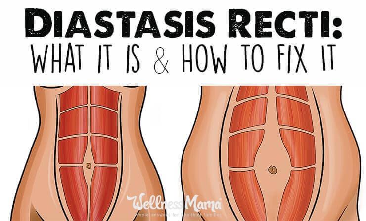 In severe cases, the protruding organ may become strangulated, causing the blood supply to get cut off, which may lead to infection and tissue death. If you suspect you have a hernia, we encourage you to see your doctor for a physical exam or other diagnostic tests.
Why Choose Moreland OB-GYN?
We understand how important it is for women to “get back to normal” as soon as possible after pregnancy, and while many are comfortable diving back into their previous exercise routines, some women may need a little guidance. We’re happy to answer any questions that you have and deliver a personalized care plan tailored specifically to your needs.
At Moreland OB-GYN, we specialize in women’s health care and prioritizing the needs of our patients at all ages and stages of life. We hope you’ll connect with us to answer your questions and we hope you’ll turn to our experts as a trusted source for information.
Diastasis of the abdominal muscles after childbirth in women in Moscow
Diastasis is a divergence to the sides of the rectus abdominis muscles.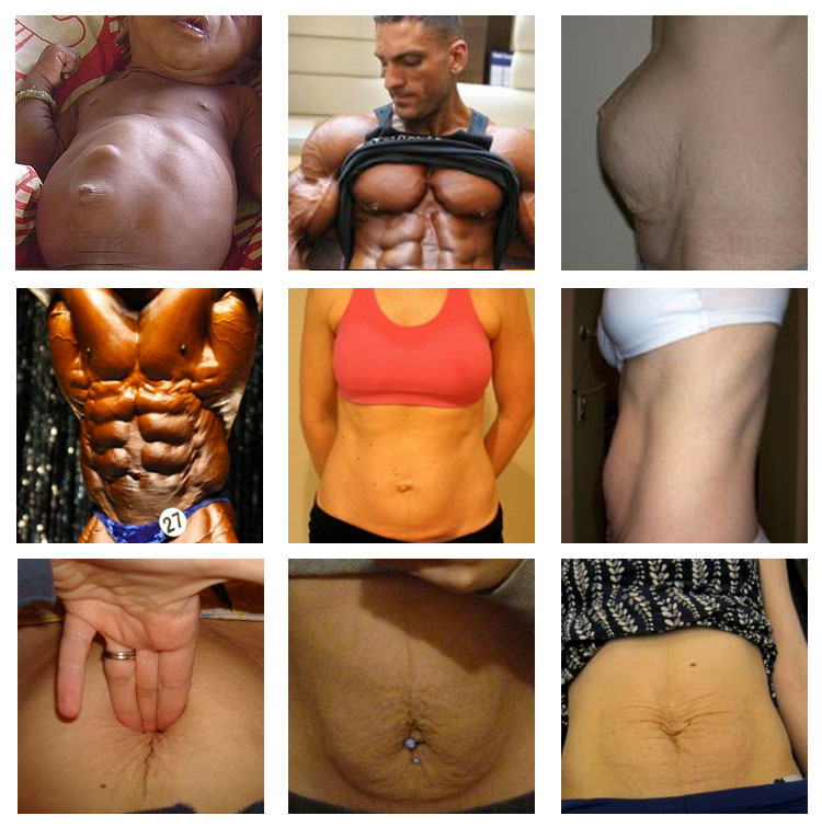 At the same time, the white line of the abdomen becomes thinner and stretched - a connective tissue structure that connects the muscles that form the very desired "press cubes".
If its width is more than 20 mm, we are talking about the presence of diastasis of the rectus muscles.
Diastasis itself is not a disease, but rather a physiological condition. This is not a hernia: there is no defect in the white line, so there is no risk of infringement of internal organs. At the same time, the expansion and thinning of the white line increases the potential risk of developing true hernias.
Causes of diastasis
Most often, diastasis appears in the third trimester of pregnancy. This is one of the manifestations of physiological changes in the connective tissue of a woman during this period under the influence of hormones - the anterior abdominal wall is stretched due to connective tissue structures (and primarily the white line) - otherwise the uterus with a growing child would simply not fit in the abdominal cavity. In this case, the center of gravity of the body shifts anteriorly, the bend of the spine increases, the pressure on the anterior abdominal wall increases. The second reason for the appearance and increase in diastasis is an increase in pressure in the abdominal cavity, including during certain physical exercises.
In the postpartum period, the distance between the muscles decreases, but this process takes about a year. A survey of 300 women conducted by our Norwegian colleagues showed that 6 weeks after birth, diastasis persists in 60% of women, and after 12 months - only in 33%. That is why we do not recommend considering the issue of surgical treatment of diastasis earlier than a year after childbirth.
Classification of diastasis
There are several classifications of diastasis. Back at 1990 Ranney proposed to define diastasis as small with a width of up to 3 cm, moderate - 3-5 cm and large - if the distance between the rectus muscles is 5 cm or more.
Today, we most often use the classification of Reinpold et al.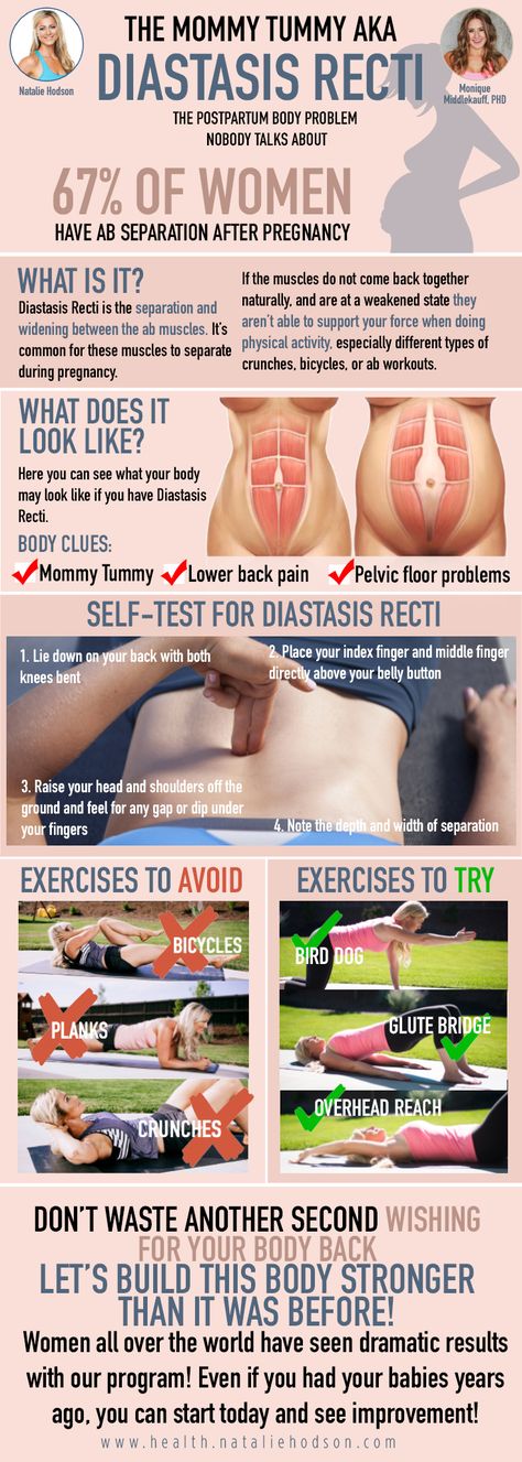 (2019), which is similar to the hernia classification of the European Society of Herniology. We also note the number of pregnancies, skin condition, clinical symptoms and a number of other parameters that help us to carefully describe the situation in each case and choose the best way to correct diastasis.
Symptoms of diastasis
Often diastasis is visible to the naked eye. For its clearer definition, we ask patients in the supine position to raise their heads and shoulders. At the same time, the rectus abdominis muscles contract and a protrusion becomes visible, or vice versa, a deepening along a line running from the sternum to the pubis. Palpation allows you to more accurately identify the presence and degree of diastasis.
Diastasis can also be manifested by clinical symptoms. The loss of a strong “foothold” for the muscles of the anterior abdominal wall leads to a redistribution of the static load, which in turn can lead to pelvic and lumbar pain, and in some, fortunately rare, cases, dysfunction of the pelvic organs.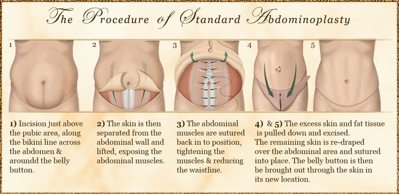
Diastasis diagnostics
We routinely use ultrasound to accurately determine the presence, length and width of diastasis, white line defects and hernias. Ultrasound is included in the consultation and is performed by the surgeon, who determines the tactics of treatment. This approach, in our opinion, is optimal and allows you to obtain reliable additional information that affects the adoption of a tactical decision.
In some cases, usually with severe diastasis and the presence of a hernia, we perform computed tomography, which gives us an understanding of the state of the anterior abdominal wall and allows us to determine and model the optimal method for diastasis plasty.
How can diastasis be treated?
As we said above, diastasis is not a disease, but rather a postpartum condition leading to an aesthetic and functional defect of the anterior abdominal wall. Accordingly, we prefer to replace "treatment" with "correction".
A properly selected set of exercises can often correct external manifestations.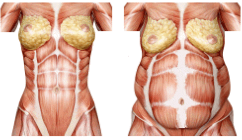 It must be remembered that not all exercises can be performed with diastasis. All types of exercises that can contribute to an increase in diastasis are prohibited - these are all basic exercises with weights (deadlift, squats), many exercises for the abdominal muscles - twisting, hanging leg raises, etc. The classic "bar" is also not recommended for patients with diastasis.
What exercises do we recommend for diastasis? Anything that strengthens the “deep core muscles”: side plank, tummy tuck, cat, Kegel exercises.
They cannot reduce the width of the white line (we remember that this is not a muscle, but a connective tissue that cannot be “pumped up”), but they can often improve the appearance of the abdomen and the quality of life of its owner, as a recent study by Thabet and Alshehri showed, although the level The evidence for this thesis is low. However, we recommend a training program for at least 6 months before considering diastasis surgery.
NB! This does not apply to situations where diastasis is combined with hernias - umbilical or white line. It is impossible to eliminate hernias with physical exercises, we immediately move on to the surgical treatment option (here, it is the treatment).
Diastasis surgical treatment
Surgery for diastasis is a rather extensive topic: there are several options for operations: laparoscopic (at least three fundamentally different methods) and open (also at least three), with or without implants, various suture materials, etc. We will devote a separate article to these features, and now we will only briefly dwell on the indications for surgical correction:
- Any diastasis associated with a hernia of the anterior abdominal wall or severe diastasis with a cosmetic defect and/or clinical symptoms
- Childbirth more than 12 months ago
- No effect after 6 months of training
- Not planning another pregnancy
If these four conditions are met, we can plan the surgical treatment of diastasis.
Prevention of diastasis
To date, there are practically no scientifically based measures for the prevention of diastasis. You can reduce pressure on the anterior abdominal wall by following these 5 principles:
- Do not slouch
- Before sitting or getting out of bed, roll onto your side to engage your lateral abdominal muscles when you get up
- Avoid heavy lifting during pregnancy, and if you have to, use proper lifting technique with a straight back
- A special supporting corset takes on part of the load, and in addition, physically pulls the rectus abdominis muscles together
- Don't forget about regular exercise during pregnancy to strengthen your core and pelvic floor muscles.
We will be happy to answer any questions on the topic of rectus diastasis by email [email protected] or +7(995)500-0324 (WhatsApp, Telegram).
Diastasis of the rectus abdominis muscles and diastasis of the womb. Solvable problems of pregnancy. Interview with Doctor of Medical Sciences, Professor M.A. Chechnyova
— What is muscle diastasis and what is pubic diastasis?
— Pregnancy is an amazing and wonderful time, but it is also a period of additional loads, which undoubtedly becomes a test of strength for the female body.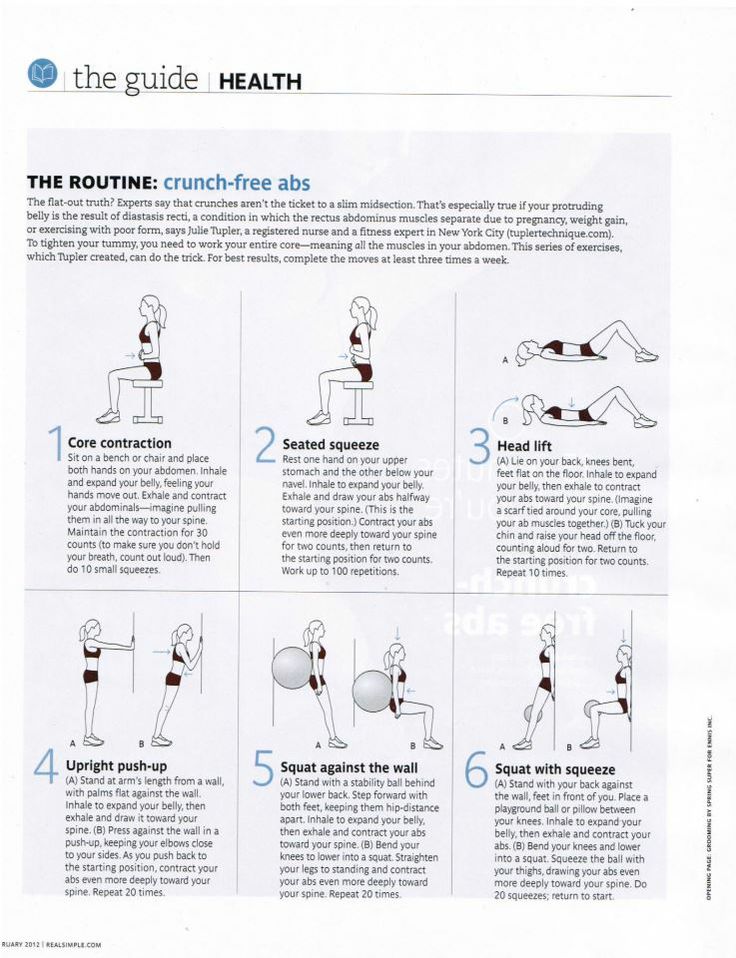
The previously existing everyday point of view that pregnancy rejuvenates and gives strength is not confirmed by anything. During the bearing of a child, significant additional loads are placed on the mother's body, which often lead to the manifestation of problems that were invisible before pregnancy.
Diastasis of the rectus abdominis muscles is a divergence of the inner edges of the muscles along the white line of the abdomen (connective tissue structure) at a distance of more than 27 mm. Pubic diastasis is one of the manifestations of pregnancy-associated pelvic girdle pain. This pathology affects the entire pelvic ring, sacroiliac joints and symphysis. And they certainly have common causes for the appearance.
The formation of such problems is facilitated by a decrease in the strength of collagen in the connective tissue. One of the reasons is an innate predisposition, the so-called connective tissue dysplasia, when the tissues are very elastic, extensible.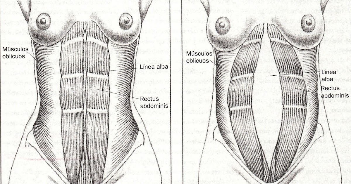 During pregnancy, the body of a woman increases the production of the hormone relaxin, which reduces the synthesis of collagen and enhances its breakdown. This is provided by nature to create maximum elasticity of the birth canal. However, other structures, such as the anterior abdominal wall and the pubic symphysis, also fall under the action of relaxin.
— How does diastasis of the muscles and diastasis of the pubis affect pregnancy and childbirth?
— The divergence of the rectus abdominis muscles is observed in approximately 40% of pregnant women. During pregnancy, it does not give serious complications that threaten the life of the mother or the condition of the fetus. However, the inferiority of the work of the rectus abdominis muscles forces the redistribution of the load on the back muscles, which can lead to lumbar-pelvic pain and, accordingly, discomfort in the back. During childbirth, the abdominal muscles are involved in attempts, and the violation of their anatomy and function can affect the birth act.
With diastasis of the pubis, things are more complicated. As already mentioned, this is only one of the manifestations of a violation of the structure and function of the pubic joint (symphysiopathy) during pregnancy. It occurs in about 50% of pregnant women in varying degrees of severity: in 25% of cases it leads to restriction of the mobility of the pregnant woman, in 8% - to severe disorders up to disability.
With symphysiopathy, the ligaments of the pubic articulation and cartilage that connect the pubic bones suffer. All this leads to severe pain in the pubic joint, pelvic bones, lower back, as well as to a violation of gait and the inability to stand up or lie down without outside help. Women with pelvic girdle pain syndrome experience significant levels of discomfort, disability, and depression, with associated social and economic problems. These include impaired sexual activity during pregnancy, chronic pain syndrome, risk of venous thromboembolism due to prolonged immobility, and even seeking early induction of labor or caesarean section to stop pain.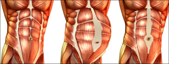
During childbirth, such a patient may have a rupture of the pubic symphysis, may require surgery to restore it.
— How to prevent the development of muscle and pelvic diastasis during pregnancy and childbirth? What factors increase the likelihood of its development?
- There is no recipe that will be one hundred percent. There is a wonderful term in the medical literature called "lifestyle modification". Whatever diseases we study, be it symphysiopathy, diabetes mellitus or preeclampsia, the risk group for pathology is always overweight women. You need to prepare for pregnancy, you need to be in good physical shape. During pregnancy, weight gain should be monitored. The recommendation to "eat for two" is not just wrong, but extremely harmful. Pregnant women should maintain reasonable physical activity. Weak and flabby abdominal muscles, combined with the large size of the fetus, undoubtedly increase the risk of diastasis.
The risk factors for symphysiopathy in numerous studies are hard physical labor and previous injuries of the pelvic bones.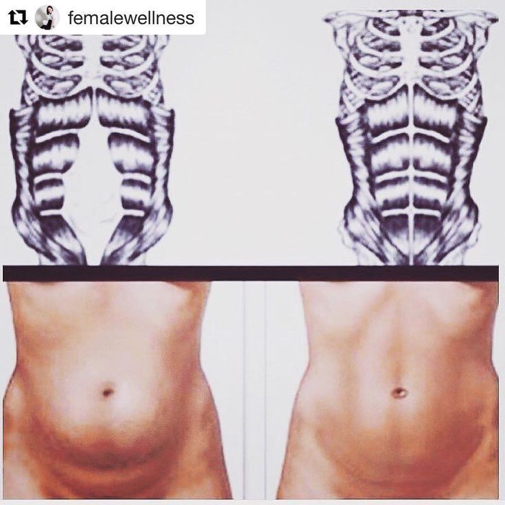 Factors such as time elapsed from previous pregnancies, smoking, use of hormonal contraception, epidural anesthesia, mother's ethnicity, number of previous pregnancies, bone density, weight and gestational age of the fetus (post-term fetus) are not associated with an increased risk of symphysiopathy.
— How to diagnose diastasis recti and diastasis pubis?
— In most cases, diastasis rectus abdominis can be diagnosed clinically. It happens that inspection, palpation and simple measurements are enough.
In the standing position, you can see the divergence of the muscles when the woman does not have subcutaneous fat. In this case, diastasis is defined as a vertical defect between the rectus muscles.
With tension of the abdominal press, a longitudinal protrusion is observed in the diastasis zone. Such a protrusion is especially noticeable if the patient in the supine position is asked to raise her head and legs. If necessary, you can measure the width of the defect simply with a ruler.
Ultrasound may be the most accurate diagnostic method. With ultrasound, the inner edges of the rectus muscles are clearly visible and the distance between them at different levels can be measured.
Computed tomography is used in the diagnosis of diastasis extremely rarely, mainly in scientific research.
For the diagnosis of symphysiopathy and diastasis pubis there is no one test as a "gold standard".
The first place, of course, is the questioning and examination of the patient. We pay attention to the gait of the pregnant woman, to how she sits down, lies down and how she gets up. Symphysiopathy is characterized by a “duck gait”, when a pregnant woman rolls from foot to foot. On palpation in the area of the womb, pain and swelling are noted. The so-called pain provocative tests are used, for example, a mat-test (pulling up an imaginary rug, mat with your foot towards you).
The following questionnaires are used to assess quality of life, pain and disability: Health-Related Quality of Life (HRQL), Oswestry Disability Index (ODI), Disability Rating Index (DRI), Edinburgh Postpartum Depression Scale (EPDS), Pregnancy Mobility Index (PMI), and Pelvic Ring Score (PGQ).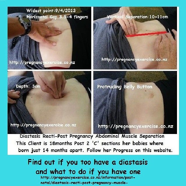
Of the instrumental methods, ultrasound is the most widely used, less often computed or magnetic resonance imaging. Ultrasound allows you to assess the condition of the ligaments of the pubic joint and the interpubic disc, the severity of the changes and the risk of natural childbirth.
— What is the treatment for diastasis recti or pubis?
— Primary prevention: when planning and during pregnancy, it is necessary to strengthen all muscle groups of the pelvic girdle, as well as the pelvic diaphragm.
More often, diastasis of the rectus muscles disappears on its own during the first months after childbirth. Special physical exercises to correct the work of muscles, to tone them and restore their basic functions should be performed under the guidance of a competent instructor. There are types of physical exercises that can, on the contrary, worsen the situation with diastasis of the rectus abdominis muscles. In some cases, when there is no effect from physiotherapy exercises, it is necessary to resort to surgical correction of the defect.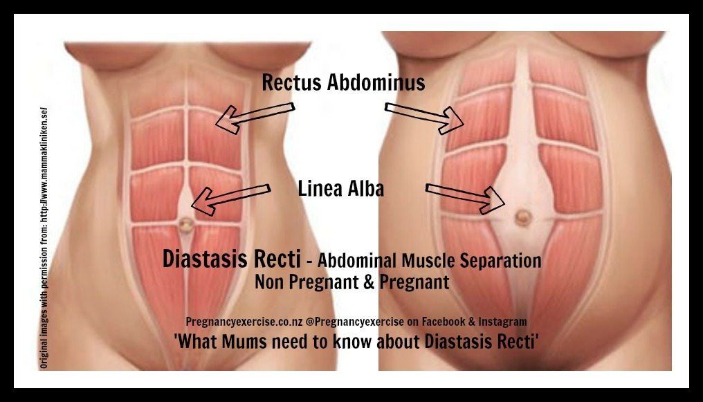 Currently, both endoscopic and open surgery are practiced. The choice of method depends on the size and localization of the defect.
In case of symphysiopathy, therapeutic exercises reduce back and pelvic pain. Acupuncture and wearing a pelvic bandage have a positive effect on symphysiopathy.
Initial treatment for pubic symphysis should be conservative even if symptoms are severe. Treatment includes bed rest and the use of a pelvic brace or corset that tightens the pelvis. Early appointment of physiotherapy with dosed therapeutic exercises will help to avoid complications associated with prolonged immobilization. Walking should be done with assistive devices such as walkers.
In most cases (up to 93%), the symptoms of dysfunction of the pelvic ring, including the pubic joint, progressively subside and completely disappear six months after birth. In other cases, it persists, becoming chronic. However, if the diastasis exceeds 40 mm, then surgical treatment may be required.