20 week ultrasound brain abnormalities
MRI helps diagnose fetal brain abnormality after 20 week scan, study finds
Intended for healthcare professionals
Toggle navigation
The BMJ logo
Site map
Search
Toggle top menu
- News & Views
- MRI helps diagnose...
- MRI helps diagnose fetal brain abnormality after 20 week scan, study finds
Research News BMJ 2016; 355 doi: https://doi. org/10.1136/bmj.i6730 (Published 15 December 2016) Cite this as: BMJ 2016;355:i6730
- Article
- Related content
- Metrics
- Responses
- Peer review
- Susan Mayor
- London
Carrying out a magnetic resonance imaging (MRI) scan helps to diagnose fetal brain abnormalities more accurately in women whose 20 week pregnancy ultrasound scan has shown a potential problem, a prospective study has found.1
The study included 570 women aged over 16 whose ultrasound scan at around 20 weeks of pregnancy suggested a brain abnormality in their developing fetus. This is relatively common, occurring in around three in 1000 pregnancies.
The women in the study had an in utero MRI scan within 14 …
View Full Text
Log in
Log in using your username and password
Log in through your institution
Subscribe from £164 *
Subscribe and get access to all BMJ articles, and much more.
Subscribe
* For online subscription
Access this article for 1 day for:
£30 / $37 / €33 (excludes VAT)
You can download a PDF version for your personal record.
Article tools
PDF0 responses
Email to a friend
Thank you for your interest in spreading the word about The BMJ.
NOTE: We only request your email address so that the person you are recommending the page to knows that you wanted them to see it, and that it is not junk mail. We do not capture any email address.
Username *
Your Email *
Send To *
You are going to email the following MRI helps diagnose fetal brain abnormality after 20 week scan, study finds
Your Personal Message
CAPTCHA
This question is for testing whether or not you are a human visitor and to prevent automated spam submissions.
- UK jobs
- International jobs
This week's poll
Back to top
Additional MRI After 20-Week Scan Could Improve Fetal Brain Abnormality Diagnosis
If you enjoy this content, please share it with a colleague
News | Magnetic Resonance Imaging (MRI) | January 05, 2017
U.K. study finds that adding an MRI scan increased accuracy of diagnosis in nearly all cases compared to ultrasound alone; second scan corrected diagnosis in a quarter of cases
January 5, 2017 — An extra scan using magnetic resonance imaging (MRI) could help more accurately detect brain abnormalities and give more certainty for parents whose mid-pregnancy ultrasound scan showed a potential problem, according to a study published in The Lancet.
The study involved 570 women whose mid-pregnancy ultrasound scan revealed a potential brain abnormality in the fetus. Within two weeks of their first scan they were given an extra scan using MRI, which increased accuracy of the diagnosis meaning almost all scans (93 percent) were correct. This extra information helped doctors give a more accurate diagnosis and advice to parents.
In comparison, the mid-pregnancy scan alone was correct in two-thirds of cases (68 percent). The accuracy of the scans was confirmed by scanning babies after birth or by autopsy for miscarriages and terminations.
The mid-pregnancy scan is an ultrasound scan given between 18 and 21 weeks of pregnancy to detect major physical abnormalities such as spina bifida, cleft lip, and heart and brain abnormalities. If a problem is found, women are referred for more tests and in some cases are offered a termination or counselling. Brain abnormalities occur in three in every 1,000 pregnancies and in some cases cause miscarriage or stillbirth.
"This study is the first of its kind and has shown that adding an MRI scan when a problem is detected provides additional information to support parents making decisions about their pregnancy." said lead author Prof. Paul Griffiths from The University of Sheffield, U.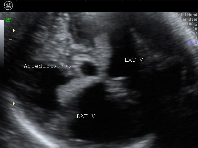 K. "Based on our findings we propose that an MRI scan should be given in any pregnancy where the fetus may have a suspected brain abnormality."
In the study, doctors analyzing the MRI and ultrasound scans were asked to rate how confident they were of their diagnosis. The study also analyzed how the MRI result changed the prognosis and advice given by doctors.
Overall MRI scans were accurate in almost all cases (93 percent) compared with around two-thirds (68 percent) for ultrasound. They also corrected the diagnosis from ultrasound in a quarter of cases (25 percent, 144 of 570).
The MRI gave extra information about the brain abnormality in half of cases (49 percent, 387 of 783). For example, in one case the MRI confirmed the ultrasound diagnosis and identified an additional abnormality. This information allowed doctors to give parents a more certain outlook, halving (55 percent) the number of cases in the 'unknown' prognosis group and increasing the number of pregnancies predicted to be 'normal' (135 percent), 'favorable' (18 percent) or 'poor' (56 percent).
Doctors agreed that the extra MRI scan changed the outlook for the pregnancy in at least a fifth of cases (20 percent, 157 of 783). And as a result this changed how the pregnancy was managed in one in three cases (34 percent, 269 of 783), with terminations offered in an extra 11 percent of cases (84 of 783) after the MRI and parents seeking counselling in 15 percent more cases (115 of 783).
Doctors using MRI were more confident of their diagnosis, saying they had 'high confidence' in 95 percent of cases (544 of 570), compared with 82 percent (465 of 570 cases) for doctors using ultrasound. In addition, 95 percent (257 of 270) of mothers said that they would have an MRI scan if a future pregnancy showed a brain abnormality and around 80percent (227 of 277) said that the information from the scan helped them better understand their baby's condition.
The researchers note that there will be further analysis of the trial's results, including a health economics analysis, to confirm if the extra scan should be used routinely. Within the study, two fetuses (of 570, less than 1 percent) were diagnosed correctly by the ultrasound and incorrectly by the MRI. Only cases identified by the ultrasound scan were given the extra MRI, so any cases where an ultrasound did not identify an abnormality would not have been included in this study.
Writing in a linked comment Prof. Rod Scott, University of Vermont, said, "Accurate diagnosis of significant brain abnormalities has important therapeutic implications. Consequently, it is essential that tools used for prenatal diagnosis are rigorously evaluated... This trial strongly supports the view that iuMRI is an excellent technique, and it should be incorporated into clinical practice as soon as possible."
The study was funded by the National Institute for Health Research Health Technology Assessment programme. It was conducted by scientists from University of Sheffield, Newcastle University, University of Birmingham, Birmingham Women's Foundation Trust and Leeds Teaching Hospitals NHS Trust.
For more information: www.thelancet.com
If you enjoy this content, please share it with a colleague
News | Prostate Cancer
Metastasis-Directed Radiation Therapy Plus Hormone Therapy Improves Progression-Free Survival for Men with Advanced Prostate Cancer
October 27, 2022 — Researchers from The University of Texas MD Anderson Cancer Center demonstrated that adding ...
October 27, 2022
News | X-Ray
How Many Bees Can You Fit in an X-ray Machine? That's Not a Joke
October 27, 2022 — Researchers at CU Boulder have, for the first time, used X-ray computed tomography (also known as a ...
October 27, 2022
News | Prostate Cancer
Lower Prostate Cancer Screening Rates Associated with Subsequent Increase in Advanced Cancers
October 24, 2022 — In the face of conflicting evidence over the risks and benefits of routine prostate cancer screenings .. .
.
October 24, 2022
News | Radiation Therapy
Radiation Therapy for High-risk, Asymptomatic Bone Metastases May Prevent Pain and Prolong Life
October 23, 2022 — Treating high-risk, asymptomatic bone metastases with radiation may reduce painful complications and ...
October 23, 2022
News | Prostate Cancer
Shortened Course of Radiation Therapy Safe and Effective for Men with High-risk Prostate Cancer
October 23, 2022 — A new randomized study confirms that men with high-risk prostate cancer can be treated with five ...
October 23, 2022
News | Radiation Therapy
MRIdian Clinical Studies and Initial MRIdian A3i Clinical Experience to be Highlighted at ASTRO 2022
October 21, 2022 — ViewRay, Inc. (NASDAQ: VRAY) today announced that the company's MRIdian MRI-Guided Radiation Therapy ...
October 21, 2022
News | Clinical Case Studies
Sexual Side Effects of Cancer Treatment Often Unaddressed with Female Patients
October 21, 2022 — A new study finds that sexual side effects of cancer treatment are discussed far less frequently with . ..
..
October 21, 2022
News | Magnetic Resonance Imaging (MRI)
Philips Advances MR Radiotherapy Imaging and Simulation for Head and Neck Cancers
October 20, 2022 — Royal Philips, a global leader in health technology, announced two new advances in MR-only workflows ...
October 20, 2022
News | Women's Health
Black Women, Breast Cancer and Clinical Trials
October 4, 2022 — For Black women, breast cancer is the most commonly diagnosed cancer and as of 2019 has surpassed lung ...
October 04, 2022
Feature | Radiology Business
Top 10: September's Most-read Content
Here is a recap of what ITN viewers found most interesting during the month of September: 1. Lasting Lung Damage Seen in ...
October 03, 2022
Subscribe Now
fetal ultrasound at 20 weeks
- Fetal development by week
- Our apparatus
- other types.
 ..
.. - Pregnancy management
- Fetometry data at various times
The cost of ultrasound in the second trimester in the period from 14 to 26 weeks is 550 hryvnia. The price includes prenatal screening, biometric protocols, 3D/4D visualization. The cost of comprehensive prenatal screening according to PRISCA (ultrasound + free estriol + alpha-fetoprotein + beta-hCG with the calculation of the individual risk of chromosomal pathologies (for example, Down syndrome or Edwards syndrome) and developmental defects (for example, neural tube defect) - 1060 hryvnia.
Ultrasound of the fetus at 20 weeks of pregnancy is performed routinely as part of prenatal diagnostics. During this period, an obstetrician-gynecologist conducting ultrasound during pregnancy can accurately determine the sex of the child, you yourself can verify this!
So, if you have a girl, her uterus is already formed, the ovaries contain seven million primitive eggs.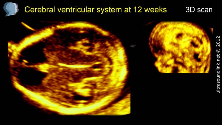 By the time of birth, there are 2 million of them in the norm. The eggs are exposed to negative environmental influences. For example, if you are taking certain medications, your daughter's egg count may drop dramatically and she may experience premature menopause or, God forbid, infertility. Therefore, taking any medication during pregnancy should be balanced and prescribed by a doctor if absolutely necessary. Your daughter is developing a vagina
If you have a boy, his testicles begin their journey from the abdomen, where they formed, to the scrotum.
Your child is actively growing. With ultrasound of the fetus at 20 weeks of gestation, its length is 19-20 cm, and its weight is about 400 grams. At the same time, the fetus still has enough space in its home, it moves freely, turns, which can be seen with an ultrasound of the fetus at 20 weeks of gestation. In most cases, at 20 weeks pregnant, you already feel the acrobat inside of you!
3d 4d ultrasound of the fetus during pregnancy will be informative, on which facial structures, back, head, etc. are available for viewing.
Conventional ultrasound measures the basic dimensions of the fetus to diagnose normal development.
Fetometry (fetal size) with ultrasound of the fetus at 20 weeks of pregnancy is normal
- BPD (biparietal size is 43-53 mm.
- LZ (frontal-occipital size) 56-68 mm.
- OG (fetal head circumference) 154-186 mm.
- OB (fetal abdominal circumference) is 102 mm 124 -164 mm.
Normal measurements of long bones on fetal ultrasound at 20 weeks gestation
- Femur 29-37 mm
- Humerus 26-34 mm
- Forearm bones 22-29 mm
- Lower leg bones 26-34 mm
Ultrasound of the fetus at 20 weeks of pregnancy must study the internal organs. The kidneys of the fetus are visible, expansion of the pelvis (pyeloectasia, hydronephrosis) in the fetus, stomach, gallbladder, bladder, intestines, liver, lungs, heart are excluded. In the study of the heart with ultrasound of the fetus at 20 weeks of gestation, the correct position of this organ, the symmetry of the chambers of the heart, the integrity of the partitions, and the correct exit of the main vessels are determined. If there is a discrepancy with the norm, the pregnant woman is sent for a specialized ultrasound of the fetal heart, since heart defects are among the most common fetal malformations. Diagnosis of heart disease is important for determining prognosis for life after childbirth. Sometimes surgery is required in the first hours of a child's life.
You may notice that your nails and hair are better. This happens under the influence of pregnancy hormones. A little later, when the consumption of calcium and other microelements and vitamins becomes greater due to the intensive growth of the fetus and with insufficient intake of these substances from food, you can note the opposite process. To prevent this from happening, eat at least 300 grams of hard low-fat cheese, cottage cheese, drink milk. Your doctor may recommend multivitamin supplements if needed.
You continue to experience heartburn symptoms and a feeling of heaviness in the abdomen. All the same hormones of pregnancy are guilty of this. Try to drink a glass of water every hour. This will relieve the symptoms. You also need to stop eating large portions. It is better to eat more often, but not much.
As your child grows, so does the amount of vaginal discharge. This is necessary to protect the birth tract from infection and ensures a normal balance of bacteria. Try not to be tempted to wear panty liners to prevent inflammation and thrush.
You may notice headaches more often after provocations, which include: bright, flickering lights, stuffy rooms and enclosed spaces, hot clothes, perfume smells. Try to avoid these irritants.
Weakness and dizziness are associated with increased blood volume in your body. Hot places should be avoided, such as staying near the stove for a long time while cooking. You need to get out into the fresh air more often.
Cramping of the calf muscles can be associated with a lack of calcium and magnesium, as well as with spasm of peripheral vessels. To reduce such manifestations, it is also important to drink enough (less than 1 glass per hour), eat foods containing calcium and magnesium.
Slight swelling of the legs and feet leads to the fact that you are forced to wear more comfortable, wider shoes. It's time to ditch your heels if you haven't already. Ankle ligaments become stretched under the influence of pregnancy hormones, it is much easier to injure them if you twist your leg. Be careful!
If severe swelling occurs, contact your doctor immediately!
The protruding navel on your tummy is due to the fact that the growing uterus is pressing on it from the inside. If you don't like your new look - don't worry - the belly button will return to its original place after childbirth as the stomach shrinks.
Continue monitoring uterine tone. When conducting fetal ultrasound at 20 weeks of pregnancy , it is possible to determine if there are segmental contractions of the myometrium (ultrasound of the uterus). You are already able to assess the tone of the entire uterus on your own by feeling pressure and tension in the abdomen below the navel. In such cases, it is necessary to lie on the left side, try to relax. In the absence of pain, such spasms are regarded as a variant of the norm. Be sure to tell your doctor about them at your next appointment.
read more: 21st week of pregnancy
- Hydrotubation (echohydrotubation): study of the patency of the fallopian tubes (ultrasonic hysterosalpingoscopy)
- Transvaginal
- Ovaries
- Uterus
- Mammary glands
Female ultrasound
- Duplex Scan
- Cerebral vessels
- Neck vessels (duplex angioscanning of the main arteries of the head)
- Veins of the lower extremities
Vascular ultrasound
- Transrectal (trusion): prostate
- Scrotum (testicles)
- Vessels of the penis
Male ultrasound
- Appendicitis
- Abdomen
- Gallbladder
- Stomach
- Intestines
- Bladder
- Soft tissue
- Pancreas
- Liver
- Kidney
- Joints
- Thyroid
- Echocardiography (ultrasound of the heart)
Organ ultrasound
- Varicose veins: ultrasound diagnosis of varicose veins
- Hypertension: Ultrasound diagnosis of hypertension
- Thrombosis: ultrasound diagnosis of vein thrombosis
- Ultrasound diagnosis of chronic pancreatitis
- for kidney stones
- for cholecystitis
Ultrasound diagnostics of diseases
- Hip joints in newborns (if hip dysplasia is suspected)
Pediatric ultrasound
If vascular plexus cysts (CSS) are found during ultrasound examination
fetal brain.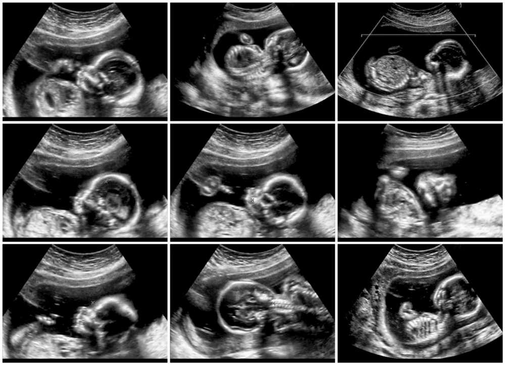 This is a complex structure, and the presence of both choroid plexuses confirms that both halves are developing in the brain. The choroid plexus produces fluid that nourishes the brain and spinal cord.
Occasionally, fluid forms within the choroid plexuses, which look like "cysts" on ultrasound.
Choroid plexus cysts can sometimes be found on ultrasound at 18-22 weeks of gestation. The presence of cysts does not affect the development and function of the brain. Most cysts disappear spontaneously by 24-28 weeks of gestation.
Are choroid plexus cysts common?
- in 1-2% of all normal pregnancies, the fetus has a KCC,
- Bilateral choroid plexus cysts are found in 50% of cases,
- in 90% of cases, cysts disappear spontaneously by the 26th week of pregnancy,
- number, size, and shape of cysts may vary,
- cysts are also found in healthy children and adults.

Somewhat more often, choroid plexus cysts are detected in fetuses with chromosomal diseases, in particular, with Edwards syndrome (trisomy 18, extra 18 chromosome). However, with this disease, the fetus will always have multiple malformations, so the identification of only choroid plexus cysts do not increase the risk of trisomy 18 and are not an indication for other diagnostic procedures. In Down's disease, vascular plexus cysts are usually not detected. The risk of Edward's syndrome upon detection of KSS not depends on the size of the cysts and their unilateral or bilateral location. Most cysts resolve by 24-28 weeks, so at 28 weeks it is important to perform an ultrasound during pregnancy in order to accurately diagnose the health of the fetus.
Doctors of the Fetal Medicine Center are one of the leading specialists in prenatal diagnostics, candidates of medical sciences, doctors of the highest categories with a narrow specialization and extensive experience in prenatal medicine.
All ultrasound examinations in the center are carried out according to the international standards of FMF (Fetal Medicine Foundation) and ISUOG (International Society for Ultrasound in Obstetrics and Gynecology).
Ultrasound doctors have international certificates from the Fetal Medicine Foundation (Fundamental Medicine, UK), which are are confirmed annually.
We take care of the most complex cases and, if necessary, it is possible to consult with specialists from King's College Hospital, King's College Hospital (London, UK).
The pride of our Centers - modern and high-tech medical equipment from General Electric: expert-class ultrasound devices Voluson E8 / E10
The capabilities of these devices allow us to talk about a new level of information content.
You can make an appointment and get an expert opinion of our ultrasound diagnostics specialists by calling the single contact center +7 (812) 458-00-00
Our centers
D 7
Mon-Sun 9:00-21:00
Ave. TORESA, D. 72
Mon-Sat 8:00-21:00
Sun 9:00-21:00
STR. BADAEV, D. 6, K.1
Mon-Sun 9:00-21:00
STR. Pulkovsky, D. 8, K. 1
Mon-Sun 9:00-21:00
KOMENDANTSKY PR., D. 10, K. 1
Mon-Sun 9:00-21:00
STR. LENI GOLIKOVA, D. 29/3
Mon-Sun 9:00-20:00
STR. SIKEIROS, D. 10V
Mon-Sat 9:00-20:00
STR. GZHATSKAYA D. 22, K. 4
Mon-Sun 9:00-21:00
STR. Degtyarnaya 23/25
Mon-Sat 9:00-21:00
Sun 9:00-19:00
BOGATYRSKY PR.


