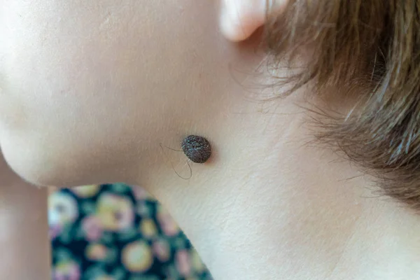
Why do i have a red birthmark
Birthmarks: Symptoms, Causes, Treatments
Overview
What are birthmarks?
There are two main categories of birthmarks — red birthmarks and pigmented birthmarks. Red birthmarks are a vascular (blood vessel) type of birthmark. Pigmented birthmarks are areas in which the color of the birthmark is different from the color of the rest of the skin.
What are red birthmarks?
Red birthmarks are colored, vascular (blood vessel) skin markings that develop before or shortly after birth. Red birthmarks are caused by an overgrowth of blood vessels.
What are the types of red birthmarks?
One common kind of vascular birthmark is the hemangioma. It usually is painless and harmless and its cause is unknown. Color from the birthmark comes from the extensive development of blood vessels at the site.
Strawberry hemangiomas (strawberry mark, nevus vascularis, capillary hemangioma, hemangioma simplex) might appear anywhere on the body, but are most common on the face, scalp, back, or chest. They consist of small, closely packed blood vessels. They might be absent at birth, and develop after several weeks. They usually grow rapidly, remain a fixed size, and then subside. In most cases, strawberry hemangiomas disappear by the time a child is 9 years old. Some slight discoloration or puckering of the skin might remain at the site of the hemangioma.
Cavernous hemangiomas (angioma cavernosum, cavernoma) are similar to strawberry hemangiomas but are more deeply situated. They might appear as a red-blue spongy mass of tissue filled with blood. Some of these lesions disappear on their own, usually as a child approaches school age.
Port-wine stains are flat, purple-to-red birthmarks made of dilated blood capillaries. These birthmarks occur most often on the face and might vary in size. Port-wine stains often are permanent (unless treated) and might thicken or darken over time, resulting in emotional distress.
Salmon patches (also called stork bites) appear on 30 percent to 50 percent of newborn babies. These marks are small blood vessels (capillaries) that are visible through the skin. They are most common on the forehead, eyelids, upper lip, between the eyebrows, and the back of the neck. Often, these marks fade as the infant grows.
What are pigmented birthmarks?
Pigmented birthmarks are skin markings that are present at birth. The marks might range from brown or black to bluish, or blue-gray in color.
What are the types of pigmented birthmarks?
Mongolian spots are usually bluish and look like bruises. They often appear on the buttocks and/or lower back, but they sometimes also appear on the trunk or arms. These spots are seen most often in people who have darker skin.
Pigmented nevi (moles) are growths on the skin that usually are flesh-colored, brown, or black. Moles can appear anywhere on the skin, alone or in groups.
Congenital nevi are moles that are present at birth. About 1 in 100 people are born with one or more moles. These birthmarks have a slightly increased risk of becoming skin cancer, depending on their size. Larger congenital nevi (>20 cm) have a greater risk of developing into skin cancer than do smaller congenital nevi. All congenital nevi should be examined by a healthcare provider, and any change in the birthmark should be reported.
Cafe-au-lait spots are light tan or light brown spots that are usually oval in shape. They usually appear at birth but might develop in the first few years of a child’s life. Cafe-au-lait spots might be a normal type of birthmark, but the presence of several cafe-au-lait spots larger than a quarter might occur in neurofibromatosis (a genetic disorder that causes abnormal cell growth of nerve tissues) and other conditions.
Symptoms and Causes
What causes pigmented birthmarks?
The cause of pigmented birthmarks is not known. However, the amount and location of melanin (a substance that determines skin color) determines the color of pigmented birthmarks. Cafe-au-lait spots might be a normal type of birthmark, but the presence of several cafe-au-lait spots larger than a quarter might occur in neurofibromatosis (a genetic disorder that causes abnormal cell growth of nerve tissues). Moles occur when cells in the skin grow in a cluster instead of being spread throughout the skin. These cells are called melanocytes, and they make the pigment that gives skin its natural color. Moles might darken after exposure to the sun, during the teen years, and during pregnancy.
What are the symptoms of pigmented birthmarks?
Pigmented birthmarks might increase in size as the child grows, change colors (especially after sun exposure and during the teen years as hormone levels change), become itchy, and might occasionally bleed.
What are the symptoms of red birthmarks?
Symptoms of red birthmarks include:
- Skin markings that develop before or shortly after birth
- Red skin rashes or lesions
- Skin markings that resemble blood vessels
- Possible bleeding
- Skin that might break open
Diagnosis and Tests
How are red birthmarks diagnosed?
In most cases, a healthcare professional can diagnose a red birthmark based on the appearance of the skin. Deeper birthmarks can be confirmed with imaging tests such as MRI, ultrasound, CT scans, or biopsies.
How are pigmented birthmarks diagnosed?
In most cases, healthcare professionals can diagnose birthmarks based on the appearance of the skin. If needed, a skin biopsy might be performed.
Management and Treatment
How are pigmented birthmarks treated?
In most cases, no treatment is needed for the birthmarks themselves. When birthmarks do require treatment, however, that treatment varies based on the kind of birthmark and its related conditions.
Large or prominent nevi that affect the appearance and self-esteem might be covered with special cosmetics.
Moles might be removed surgically if they affect the appearance or if they have an increased cancer risk.
What is the treatment for red birthmarks?
Many capillary birthmarks such as salmon patches and strawberry hemangiomas are temporary and require no treatment. For permanent lesions, concealing cosmetics might be helpful. Cortisone (oral or injected) can reduce the size of a hemangioma that is growing rapidly and obstructing vision or vital structures. Other oral medicines have been used experimentally with some success in these cases, as well.
Port-wine stains on the face can be treated at a young age with a yellow-pulsed dye laser for best results. Treatment of the birthmarks might help prevent psychosocial problems that can result in individuals who have port-wine stains.
Permanent red birthmarks might be treated with methods including:
- Cryotherapy (freezing)
- Laser surgery
- Surgical removal
In some cases, birthmarks are not treated until a child reaches school age. However, birthmarks are treated earlier if they result in unwanted symptoms or if they compromise vital functions such as vision or breathing.
Prevention
Can red birthmarks be prevented?
Currently, there is no known way to prevent red birthmarks.
Can pigmented birthmarks be prevented?
There is no known way to prevent birthmarks. People with birthmarks should use a good quality sunscreen when outdoors in order to prevent complications.
Living With
What are the complications of pigmented birthmarks?
Some complications of pigmented birthmarks can include psychological effects in cases in which the birthmark is prominent. Pigmented birthmarks also can pose an increased skin cancer risk.
A doctor should check any changes that occur in the color, size, or texture of a nevus or other skin lesion. See a doctor right away if there is any pain, bleeding, itching, inflammation, or ulceration of a congenital nevus or other skin lesion.
Birthmarks - red Information | Mount Sinai
Strawberry mark; Vascular skin changes; Angioma cavernosum; Capillary hemangioma; Hemangioma simplex
Red birthmarks are skin markings created by blood vessels close to the skin surface. They develop before or shortly after birth.
A stork bite is a vascular lesion quite common in newborns consisting of one or more pale red patches of skin. Most often stork bites appear on the forehead, eyelids, tip of the nose, upper lip or back of the neck. They are usually gone within 18 months of birth.
Hemangiomas are tumors made up of dilated blood vessels that usually appear shortly after birth, although they may be present at birth. Hemangiomas on the face can be disfiguring and may interfere with visual development or cause obstruction of the airway.
This child has a juvenile hemangioma (strawberry hemangioma) on the chin. These may begin as flat, red spots and later become larger and elevated. Juvenile hemangiomas often go away (involute) spontaneously.
Causes
There are two main categories of birthmarks:
- Red birthmarks are made up of blood vessels close to the skin surface. These are called vascular birthmarks.
- Pigmented birthmarks are areas in which the color of the birthmark is different from the color of the rest of the skin.

Hemangiomas are a common type of vascular birthmark. Their cause is unknown. Their color is caused by the growth of blood vessels at the site. Different kinds of hemangiomas include:
- Strawberry hemangiomas (strawberry mark, nevus vascularis, capillary hemangioma, hemangioma simplex) may develop several weeks after birth. They may appear anywhere on the body, but are most often found on the neck and face. These areas consist of small blood vessels that are very close together.
- Cavernous hemangiomas (angioma cavernosum, cavernoma) are similar to strawberry hemangiomas, but they are deeper and may appear as a red-blue spongy area of tissue filled with blood.
- Salmon patches (stork bites) are very common. Up to half of all newborns have them. They are small, pink, flat spots made up of small blood vessels that can be seen through the skin. They are most common on the forehead, eyelids, upper lip, between the eyebrows, and on the back of the neck. Salmon patches may be more noticeable when an infant cries or during temperature changes.

- Port-wine stains are flat hemangiomas made of expanded tiny blood vessels (capillaries). Port-wine stains on the face may be associated with Sturge-Weber syndrome. They are most often located on the face. Their size varies from very small to over half of the body's surface.
Symptoms
The main symptoms of birthmarks include:
- Marks on the skin that look like blood vessels
- Skin rash or lesion that is red
Exams and Tests
A health care provider should examine all birthmarks. Diagnosis is based on how the birthmark looks.
Tests to confirm deeper birthmarks include:
- Skin biopsy
- CT scan
- MRI of the area
Treatment
Many strawberry hemangiomas, cavernous hemangiomas, and salmon patches are temporary and do not need treatment.
Port-wine stains may not need treatment unless they:
- Affect your appearance
- Cause emotional distress
- Are painful
- Change in size, shape, or color
Most permanent birthmarks are not treated before a child reaches school age or the birthmark is causing symptoms. Port-wine stains on the face are an exception. They should be treated at a young age to prevent emotional and social problems. Laser surgery can be used to treat them.
Concealing cosmetics may hide permanent birthmarks.
Oral or injected cortisone may reduce the size of a hemangioma that is growing quickly and affecting vision or vital organs.
Other treatments for red birthmarks include:
- Beta-blocker medicines
- Freezing (cryotherapy)
- Laser surgery
- Surgical removal
Outlook (Prognosis)
Birthmarks rarely cause problems, other than changes in appearance. Many birthmarks go away on their own by the time a child reaches school age, but some are permanent. The following development patterns are typical for the different types of birthmarks:
- Strawberry hemangiomas usually grow quickly and stay the same size.
 Then they go away. Most strawberry hemangiomas are gone by the time a child is 9 years old. However, there may be a slight change in color or puckering of the skin where the birthmark was.
 - Some cavernous hemangiomas go away on their own, usually as a child is about school age.
- Salmon patches often fade as the infant grows. Patches on the back of the neck may not disappear. They usually are not visible as hair grows.
- Port-wine stains are often permanent.
Possible Complications
The following complications can occur from birthmarks:
- Emotional distress because of appearance
- Discomfort or bleeding from vascular birthmarks (occasional)
- Interference with vision or bodily functions
- Scarring or complications after surgery to remove them
When to Contact a Medical Professional
Have your provider look at all birthmarks.
Prevention
There is no known way to prevent birthmarks.
Dinulos JGH. Vascular tumors and malformations. In: Dinulos JGH, ed. Habif's Clinical Dermatology. 7th ed. Philadelphia, PA: Elsevier; 2021:chap 23.
Folpe AL, Kozakewich HP. Benign vascular tumors and malformations. In: Goldblum JR, Folpe AL, Weiss SW, eds. Enzinger and Weiss's Soft Tissue Tumors. 7th ed. Philadelphia, PA: Elsevier; 2020:chap 20.
Patterson JW. Vascular tumors. In: Patterson JW, ed. Weedon's Skin Pathology. 5th ed. Philadelphia, PA: Elsevier Limited; 2021:chap 39.
Last reviewed on: 11/10/2020
Reviewed by: Ramin Fathi, MD, FAAD, Director, Phoenix Surgical Dermatology Group, Phoenix, AZ. Also reviewed by David Zieve, MD, MHA, Medical Director, Brenda Conaway, Editorial Director, and the A.D.A.M. Editorial team.
Baby birthmarks, recommended medical supervision. Lakhta Junior in St. Petersburg
Let's talk about children's birthmarks, their possible and most common options.
“Like a baby,” we envy about the most healthy, satiny, velvety, uniform skin. But the idea that any spot on the skin of a baby is obviously a pathology is nothing more than a myth.
Birthmarks are called so because they are often visible already in the newborn, although sometimes they appear at later stages of development. The color, size, shape of a birthmark can be very different. Some of these marks disappear over time, others become brighter and/or remain permanently. nine0004 Much and completely justified attention is paid today to the dangers that moles carry in themselves in adulthood and old age. On the contrary, most children's moles do not pose any danger.
Vascular ("stork's mark", "angel's kiss", hemangiomas, venous malformations, etc.). The cause of such spots are problems with small blood vessels, usually transient. Vascular spots are distinguished by a red-violet color in one shade or another; may rise slightly above the surface of the skin or be flat. nine0004 Age spots (“café au lait”, melanocystosis, Mongolian spots, etc.). Formed by various skin cells; look, as a rule, brown-gray, usually do not rise above the skin.
Again, in most cases, both main varieties of birthmarks are quite safe and harmless. And yet, only a doctor can judge whether it is possible to confine oneself to observation or whether the stain is to be removed for medical reasons. This type of consultation is strictly required.
Let's take a closer look at common types of birthmarks.
Stork Mark (stork peck, stork mark, etc.).
This is the name in many countries for reddish-pink spots that a newborn may have on the back of the head or back of the neck. As a rule, such spots do not rise above the surface and have an uneven shape. Most often, the stork mark occurs in children with fair skin.
The explanation that this is a trace of the "transportation" of the baby by the stork, older brothers and sisters, as a rule, is accepted unconditionally, and even with delight. "Stork peck" usually disappears without a trace by 18 months of age. Otherwise, especially if the stain causes noticeable discomfort, it is removed (for example, using minimally invasive laser technology). nine0005
Port-wine stain
Port-wine stain or, in medical terminology, flaming nevus - flat capillary moles, outwardly and really resembling a puddle of spilled purple-red wine. They occur with a frequency of 3-5 cases per 1000 newborns. With age, they can increase and acquire a darker shade.
Treatment is not usually required, but the skin in this area tends to become dry and irritated. Sometimes they resort to the method of laser removal, which, however, does not always completely eliminate the cosmetic defect. nine0005
"Strawberry hemangioma"
Obsolete name for infantile hemangioma, a bright red congenital benign vascular tumor that rises above the surface of the skin and has a characteristic surface texture. It occurs in about 5% of newborns, more often in girls, twins and premature babies.
Usually localized on the face, head or chest; as a rule, decreases with age and finally disappears in the preschool period. It is possible to remove a hemangioma, especially if it is large and / or located near the eyes, nose, mouth. Both minimally invasive surgical and conservative methods of removal are practiced. nine0005
Venous malformations
These are blue-violet spots that can sometimes appear on the skin, especially during moments of intense crying. Venous malformations (lit. "malformations") are always congenital in nature, but sometimes remain almost invisible until adolescence. They can be located on any part of the body. Compared to other types of birthmarks, they are rare - in one case in about five thousand.
Coffee with milk
Localized abnormal pigmentation that occurs in 30% of people. The color of such a spot is usually light brown or beige, which is reflected in the name. The edges are usually even, the perimeter is sometimes darker than the inner area. In most cases, a café au lait pigment spot is quite harmless, but it can enlarge and/or darken with age. Such spots are subject to observation in dynamics, in some cases they can be removed.
Moles proper
Well-known round or oval freckle-like formations. The color varies from light brown or pink-red to almost black. Sizes in individual cases also vary widely. They can be both flat and protruding above the surface. nine0004 Moles should be inspected regularly to prevent any damage (mechanical, chemical, etc.). If a mole begins to bleed, change color, shape, appearance and any other characteristics, you should see a specialist as soon as possible - a dermatologist or oncologist at the Lakhta Junior Children's Clinic in St. Petersburg.
Birthmarks are beautiful and dangerous
12/29/2017
184955
Since ancient times, moles and birthmarks have received special attention. The most famous folk sign on this subject says that if a person without a mirror can see a lot of moles on himself, he is considered lucky. nine0005
Surely even today there will be those who believe in the karmic influence of birthmarks on a person's destiny. Skeptics will decide that it is not worth paying attention to. Deputy chief physician of the GBU RO "OKKVD" for organizational and methodological work, dermatovenereologist Mikhail Zhuchkov is in a hurry to convince: it's worth it, and how! “Each pigmented neoplasm on the surface of the skin,” he says, “is not just an ornament, but a full-fledged tumor. ” nine0005
Take a picture of your mole!
We must immediately make a reservation: any congenital mole during its life is doomed to turn into the most terrible malignant tumor of the skin - melanoma, which grows from cells that produce pigment (melanocytes). This transformation occurs during the life of the pigment formation, which can reach 110-150 years, or maybe more. A person, due to objective reasons, often does not live up to this transformation. But it may also happen that some age spots are in a hurry to "live" and acquire "malignancy" much earlier, that is, during a person's life. nine0005
Moles are divided into two main types: congenital and acquired. From the names it follows that congenital birthmarks are those with which a person is born, acquired ones occur during life. But their character is different. To begin with, it is worth learning to distinguish the first from the second. This is as easy as shelling pears: if the mole is less than a centimeter, then it is acquired. Exceptions certainly exist, but they are quite rare.
Acquired moles first appear as a brown spot that does not rise above the surface of the skin. Then the pigment cells go deep into the skin. There, in the process of their destruction, the so-called "epidermal growth factors" are released - substances that make the mole grow. Over time, an intangible speck of dark brown color begins to rise, without changing in total size, to lose shades of brown. Over the course of life, this mole can become benign if every one of the pigment cells leaves it. Then it can be removed in any cosmetic way. But to determine the presence of pigment in a mole is possible only with the help of a special procedure - confocal epiluminescent dermatoscopy. The combination of words for the layman is frightening. In fact, it is a simple procedure. nine0005
Look inside the mole
Using a special microscope that is placed directly on the mole, the dermatoscope, you can look inside the formation. A research method that allows you to look deep into the skin without making incisions with a scalpel and without using any "irradiation" is called dermatoscopy. This technique is already about a century old, but it has become relatively recent to be used to determine the malignancy of birthmarks. It was first used in clinical practice for the diagnosis of one of the most severe diseases of the connective tissue - systemic scleroderma. And only a few years later it became obvious that it was in this way that many questions regarding moles could be answered: is there a threat in this or that pigmented formation? nine0005
Before the widespread introduction of dermatoscopy into practice, the only way to know the truth was to remove the birthmark with a scalpel and conduct a subsequent histological examination. Moreover, today it has become possible with a certain degree of probability to predict the risk of developing malignant neoplasms that develop against the background of birthmarks.
Tricolor and ugly ducklings
Any changes in congenital moles: the appearance of shades of black, peripheral growth, discoloration, is a reason for an urgent appeal for dermato-oncological care. Abroad, in almost all EU countries, congenital moles are removed in a planned manner before the child reaches the age of 14. The procedure is performed by dermato-oncologists. Strange as it may seem, there is no such specialty in Russia yet. nine0005
But foreign experience in self-control of moles could well be useful. Abroad, people come to an appointment with a dermato-oncologist with photographs of their pigmentation. The benefits of this are enormous. Studies show that self-monitoring can detect melanoma 2-2.5 years earlier. Therefore, you should remember rule number one: to independently compare changes on your own skin is a simple matter, but effective.
Here is a short set of rules for observing birthmarks from Mikhail Zhuchkov :
- Each mole should rise and fade, but not grow in width.

- The appearance of shades of black is a cause for concern. This indicates that the pigment cells do not go deep, but approach the surface.
- The "colors of the Russian flag" should not appear: shades of white, blue, red. It doesn't have to be a tricolor. But, white-pink, blue-violet and so on are the characteristic colors of melanoma. As well as two or more shades of brown. nine0099
- Spontaneous bleeding is also a reason for an extraordinary examination.
There is also a generally accepted rule that is available to an ordinary person who does not have special knowledge in dermatology and dermato-oncology. It tells you which acquired mole should be observed first. It turns out that all moles satisfy the ABCD rule, which includes the assessment of a pigmented skin neoplasm according to 4 parameters:
A - pigment spot asymmetry;
B - border roughness; nine0004 C - color unevenness;
D - diameter more than 6 mm.
Mikhail Valeryevich recommends paying attention to one more nuance. A mandatory examination of moles that demonstrate the "ugly duckling syndrome", that is, they differ in their development dynamics from their "brothers", is necessary. For example, if the birthmark did not begin to rise above the surface of the skin at a time when the rest rushed up.
Attention, deletion! nine0011
How to get rid of moles? It is necessary to contact the oncology clinic, where the objectionable mole will be removed with a scalpel within healthy tissue and a few barely noticeable stitches will be applied. Dermatologists remove only those moles from which the pigment is completely removed, that is, those that no longer pose any oncological danger. Other birthmarks are the prerogative of oncologists. In no case should you do this in beauty salons and other non-specialized places! Finding out everything about your mole without an incision is possible only with the help of a dermatoscopy procedure. In any case, consult your doctor. nine0005
Persons and organizations of the article
Deputy chief physician for organizational and methodological work
Zhuchkov Mikhail Valerievich The highest qualification category,
The highest qualification category, Dermatovenereology, Health Organization and Public Health
nine0003 Author: Mikhail Zhuchkov Source: Portal UZRF.


