When is chorionic villus sampling done
Chorionic villus sampling - NHS
Chorionic villus sampling (CVS) is a test you may be offered during pregnancy to check if your baby has a genetic or chromosomal condition, such as Down's syndrome, Edwards' syndrome or Patau's syndrome.
It involves removing and testing a small sample of cells from the placenta, the organ linking the mother's blood supply with the unborn baby's.
When CVS is offered
CVS is not routinely offered in pregnancy.
It's only offered if there's a high chance your baby could have a genetic or chromosomal condition.
This could be because:
- an antenatal screening test has suggested your baby may be born with a condition, such as Down's syndrome, Edwards' syndrome or Patau's syndrome
- you had a previous pregnancy affected by a genetic condition
- you have a family history of a genetic condition, such as sickle cell disease, thalassaemia, cystic fibrosis or muscular dystrophy
It's important to remember that you do not have to have CVS if it's offered. It's up to you to decide whether you want it.
A midwife or doctor will speak to you about what the test involves, and let you know what the possible benefits are, to help you make a decision.
Find out more about why CVS is offered and deciding whether to have it
How CVS is performed
CVS is usually carried out between the 11th and 14th weeks of pregnancy, although it's sometimes performed later than this if necessary.
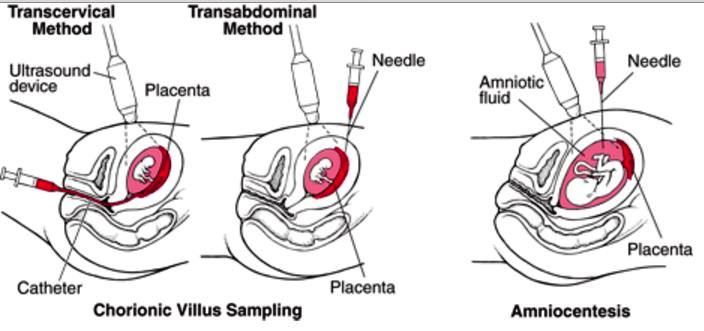
During the test, a small sample of cells is removed from the placenta using 1 of 2 methods:
- transabdominal CVS – a needle is inserted through your tummy (this is the most common method used)
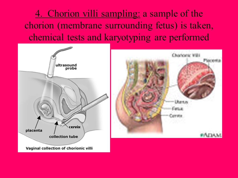
- transcervical CVS – a tube or small forceps (smooth metal instruments that look like tongs) are inserted through the cervix (the neck of the womb)
The test itself takes about 10 minutes, although the whole consultation may take about 30 minutes.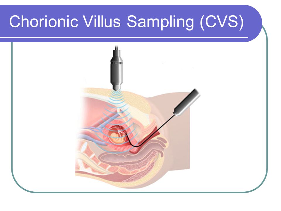
The CVS procedure is usually described as being uncomfortable rather than painful, although you may experience some cramps that are similar to period pains for a few hours afterwards.
Find out more about what happens during CVS
Getting your results
The first results of the test should be available in about 3 days. This is known as the rapid CVS result.
A more detailed set of CVS results will be available after 2 weeks.
If the rapid CVS result and a previous ultrasound scan both indicate your baby has a condition, your doctor will discuss your options with you straightaway.
If your previous ultrasound did not find anything unexpected, it’s recommended you wait until the more detailed set of CVS results before making a decision about ending your pregnancy.
If the results of these tests suggest it is highly likely your baby has a genetic condition, a specialist doctor (obstetrician) or midwife will explain what the screening results mean and talk to you about your options.
There's no cure for most of the conditions found by CVS, so you'll need to consider your options carefully.
You may decide to continue with your pregnancy while gathering information about the condition so you're fully prepared.
Find out more about having a baby that might be born with a genetic condition
Or you may consider ending your pregnancy (having a termination).
Find out more about the results of CVS
Miscarriage and infections.
Before you decide to have CVS, the possible complications will be discussed with you.
CVS can cause miscarriage, the loss of the pregnancy in the first 23 weeks. The chance of miscarrying after CVS is thought to be less than 1 in 200 for most pregnancies, and at around 1 in 100 for multiple pregnancies (such as twins).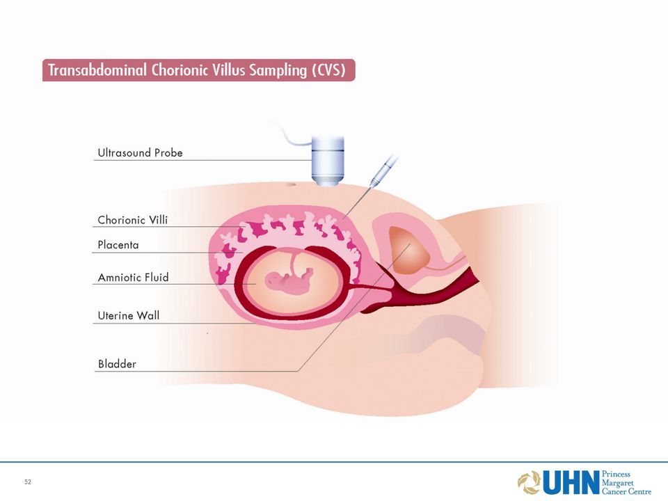
You might also get an infection, or need to have CVS again because it was not successful the first time.
Read more about the complications of CVS
What are the alternatives?
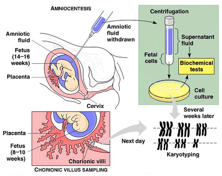
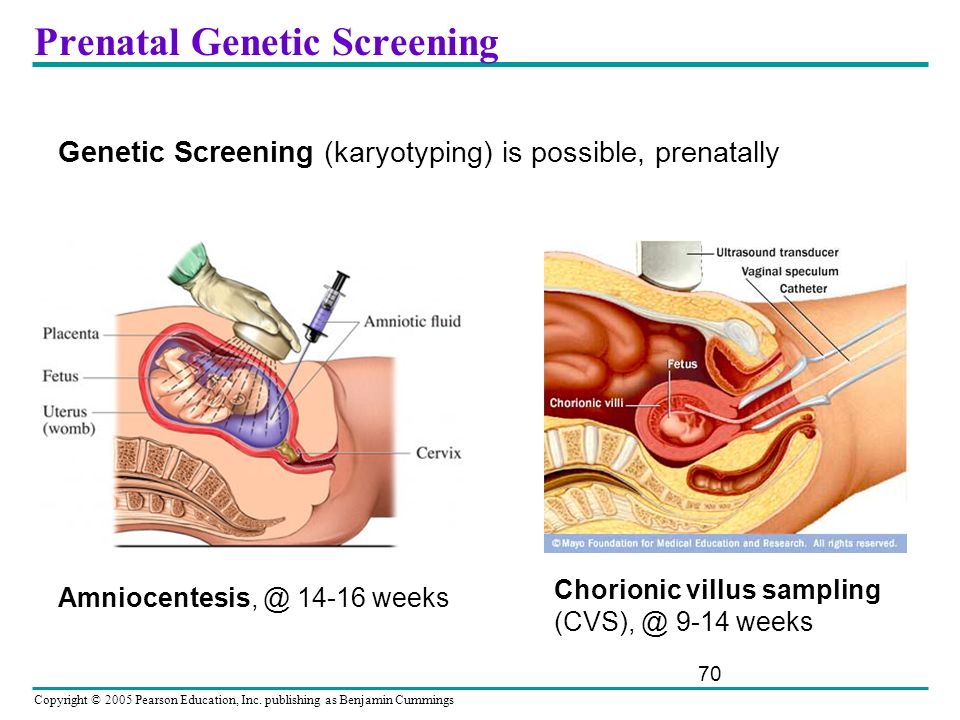
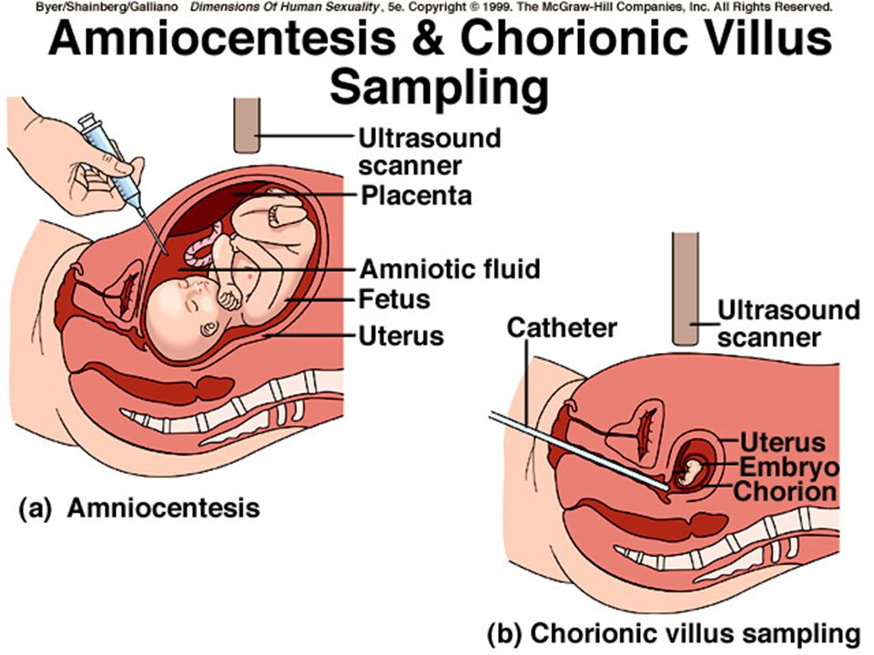
An alternative to CVS is a test called amniocentesis.
This is where a small sample of amniotic fluid, the fluid that surrounds the baby in the womb, is removed for testing.
It's usually carried out between the 15th and 18th week of pregnancy, although it can be performed later than this if necessary.
This test can also cause a miscarriage, but your pregnancy will be at a more advanced stage before you can get the results, so you'll have less time to consider your options.
If you're offered tests to look for a genetic or chromosomal condition in your baby, a specialist involved in carrying out the test will be able to discuss the different options with you and help you make a decision.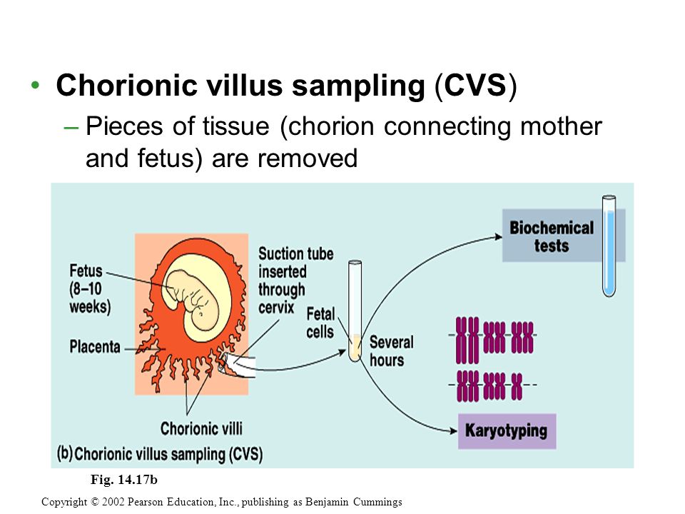
Page last reviewed: 03 January 2023
Next review due: 03 January 2026
Chorionic Villus Sampling (CVS) | Johns Hopkins Medicine
What is chorionic villus sampling?
Chorionic villus sampling (CVS), or chorionic villus biopsy, is a prenatal test that involves taking a sample of tissue from the placenta to test for chromosomal abnormalities and certain other genetic problems. The placenta is a structure in the uterus that provides blood and nutrients from the mother to the fetus,
The chorionic villi are tiny projections of placental tissue that look like fingers and contain the same genetic material as the fetus. Testing may be available for other genetic defects and disorders depending on the family history and availability of lab testing at the time of the procedure.
CVS is usually done between the 10th and 12th weeks of pregnancy. Unlike amniocentesis (another type of prenatal test), CVS does not provide information on neural tube defects, such as spina bifida.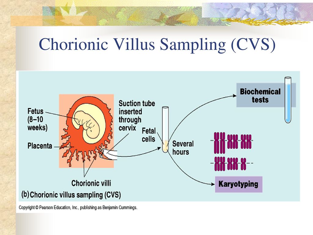 For this reason, women who undergo CVS also need a follow-up blood test between 16 to 18 weeks of their pregnancy to screen for neural tube defects.
There are two types of CVS procedures:
-
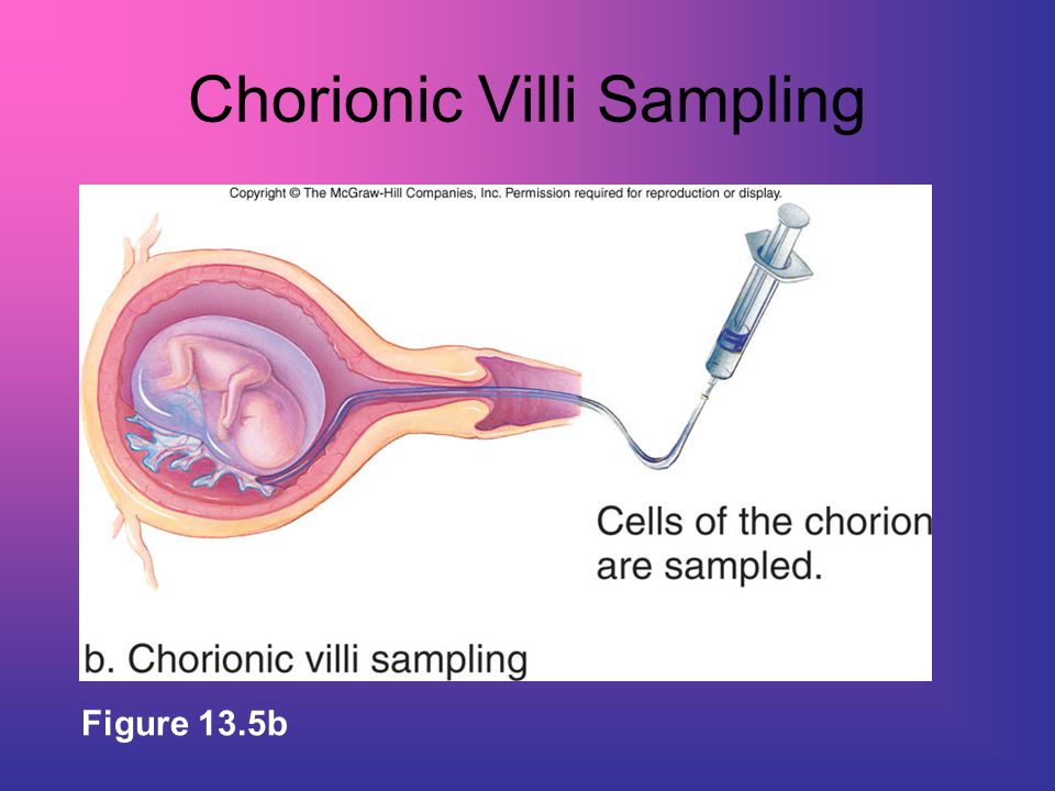
Transcervical. In this procedure, a catheter is inserted through the cervix into the placenta to obtain the tissue sample
-
Transabdominal. In this procedure, a needle is inserted through the abdomen and uterus into the placenta to obtain the tissue sample
Another related procedure that may be used to diagnose genetic and chromosomal defects is amniocentesis.
Anatomy of the fetus in utero
Click image to enlarge.
-
Amniotic sac. This is a thin-walled sac that surrounds the fetus during pregnancy. The sac is filled with amniotic fluid (liquid made by the fetus) and the amnion (the membrane that covers the fetal side of the placenta), which protects the fetus from injury and helps to regulate the temperature of the fetus.
-
Anus. This is the opening at the end of the anal canal.
-
Cervix. This is the lower part of the uterus that projects into the vagina. Made up of mostly fibrous tissue and muscle, the cervix is circular in shape.
-
Fetus. An unborn baby from the eighth week after fertilization until birth is called a fetus.
-
Placenta. This is an organ, shaped like a flat cake that only grows during pregnancy and provides a metabolic interchange between the fetus and mother. (The fetus takes in oxygen, food, and other substances and eliminates carbon dioxide and other wastes.)
-
Umbilical cord. This is a rope-like cord connecting the fetus to the placenta. The umbilical cord contains 2 arteries and a vein, which carry oxygen and nutrients to the fetus and waste products away from the fetus.
-
Uterine wall. This is the wall of the uterus.
-
Uterus (also called the womb). The uterus is a hollow, pear-shaped organ located in a woman's lower abdomen, between the bladder and the rectum. It sheds its lining each month during menstruation and in which a fertilized egg (ovum) becomes implanted and the fetus develops.
-
Vagina. This is part of the female genitals sits behind the bladder and in front of the rectum. It forms a canal extending from the uterus to the vulva.
Reasons for the procedure
Chorionic villus sampling may be used for genetic and chromosome testing in the first trimester of pregnancy . Here are some reasons that a woman might elect to undergo CVS:
-
Previously affected child or a family history of a genetic disease, chromosomal abnormalities, or metabolic disorder
-
Maternal age over 35 years by the pregnancy due date
-
Risk of a sex-linked genetic disease
-
Previous ultrasound with questionable or abnormal findings
-
Abnormal cell-free DNA test
There may be other reasons for your doctor to recommend a chorionic villus sampling.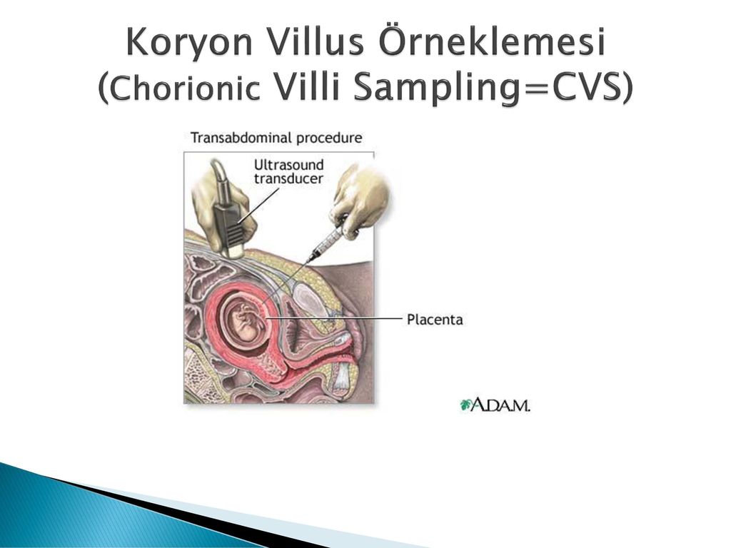
Risks of the procedure
As with any invasive procedure, complications may occur. Some possible complications may include, but are not limited to, the following:
-
Cramping, bleeding, or leaking of amniotic fluid (water breaking)
-
Infection
-
Miscarriage
-
Preterm labor
-
Limb defects in infants, especially in CVS procedures done before 9 weeks (rare)
People who are allergic to or sensitive to medications or latex should notify their doctor.
Women with twins or other multiples will need sampling from each placenta in order to study each baby.
There may be other risks depending upon your specific medical condition. Be sure to discuss any concerns with your doctor prior to the procedure.
Certain factors or conditions may interfere with CVS. These factors include, but are not limited to, the following:
-
Pregnancy earlier than seven weeks or later than 13 weeks
-
Position of the baby, placenta, amount of amniotic fluid, or mother's anatomy
-
Vaginal or cervical infection
-
Samples that are inadequate for testing, or that may contain maternal tissue
Before the procedure
-
The doctor will explain the procedure to you and offer you the opportunity to ask any questions that you might have about the procedure.
-
You will be asked to sign a consent form that gives your permission to do the procedure. Read the form carefully and ask questions if something is not clear.
-
Generally, there is no special restriction on diet or activity prior to chorionic villus sampling.
-
Tell your doctor if you are sensitive to or are allergic to any medications, latex, iodine, tape, and anesthetic agents (local and general).
-
Tell your doctor of all medications (prescribed and over-the-counter) and herbal supplements that you are taking.
-
Tell your doctor if you have a history of bleeding disorders or if you are taking any anticoagulant (blood-thinning) medications, aspirin, or any other medications that may affect blood clotting. It may be necessary for you to stop these medications prior to the procedure.
-
Tell your doctor if you are Rh negative.
 During the CVS procedure, blood cells from the mother and fetus can mix. This may lead to Rh sensitization and breaking down of fetal red blood cells. In most cases, prenatal blood tests will have determined whether you are Rh negative. You may be asked to provide these lab results before the procedure.
 -
You may or may not be asked to have a full bladder right before the procedure. Depending on the position of the uterus and placenta, a full or empty bladder may help move the uterus into a better position for the procedure.
-
Based upon your medical condition, your doctor may request other specific preparation.
During the procedure
Click image to enlarge.
A CVS procedure may be done on an outpatient basis, or as part of your stay in a hospital. Procedures may vary depending on your condition and your doctor’s practices.
Generally, a CVS procedure follows this process:
-
You will be asked to undress completely, or from the waist down, and put on a hospital gown.
-
You will be asked to lie down on an exam table and place your hands behind your head.
-
Your vital signs (blood pressure, heart rate, and breathing rate) will be checked.
-
An ultrasound will be performed to check the fetal heart rate, and the position of the placenta, fetus, and umbilical cord.
-
Based on the location of the placenta, the CVS procedure will be performed through your cervix (transcervical) or through your abdominal wall (transabdominal).
For a transcervical CVS procedure:
-
The doctor will insert an instrument called a speculum into your vagina so that he or she can see your cervix.
-
Your vagina and cervix will be cleansed with an antiseptic solution.
-
Using ultrasound guidance, a thin tube will be guided through the cervix to the chorionic villi.
-
Cells will be gently suctioned through the tube into a syringe.
 You may feel a twinge or slight cramping. More than one sample may be needed to obtain enough tissue for testing.
 -
The tube will then be removed.
For a transabdominal CVS procedure:
-
For an abdominal CVS, your abdomen will be cleansed with an antiseptic. You will be instructed not to touch the sterile area on your abdomen during the procedure.
-
The doctor may inject a local anesthetic to numb the skin. If a local anesthetic is used, you will feel a needle stick when the anesthetic is injected. This may cause a brief stinging sensation.
-
Ultrasound will be used to help guide a long, thin, hollow needle through your abdomen and into the uterus and placenta. This may be slightly painful, and you may feel a cramp as the needle enters the uterus.
-
Cells will be gently suctioned into a syringe. More than one sample may be needed to obtain enough tissue for testing.

-
The needle will then be removed. An adhesive bandage will be placed over the abdominal needle insertion site.
Procedure completion, both methods:
-
The fetus’ heart rate and your vital signs will be reassessed.
-
If you are Rh negative, you may be given Rho(D) immune globulin. This is a specially developed blood product that can prevent an Rh negative mother's antibodies from reacting to Rh positive fetal cells.
-
The chorionic villus tissue will be sent to the lab.
After the procedure
You and your fetus will be monitored for a while after the procedure. Your vital signs and the fetal heart rate will be checked periodically for an hour or longer.
The CVS tissue will be sent to a specialty genetics lab for analysis. Counseling with a genetics specialist may be recommended depending on the test results.
You may experience some slight cramping and light spotting for a few hours after CVS.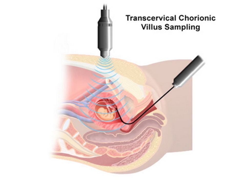
You should rest at home and avoid strenuous activities for at least 24 hours. You should not douche or have sexual intercourse for 2 weeks, or until directed by your doctor.
Tell your doctor to report any of the following:
If a transabdominal procedure was performed, check the bandaged needle site on your abdomen for any bleeding or drainage of fluid.
Your doctor may give you additional or alternate instructions after the procedure, depending on your particular situation.
Chorion biopsy procedure: essence and preparation
Pregnancy is always an unforeseen and sometimes difficult process. It happens that in order for it to pass calmly and comfortably for future parents, it is necessary to do some research. They will be able to show if the baby has any abnormalities. And depending on the results, parents will be able to decide whether to continue the pregnancy. One of the most important studies is the chorionic biopsy procedure.
What is the purpose of the procedure?
Chorionic biopsy is one of the most modern methods of prenatal diagnosis, which allows to identify genetic disorders in the fetus. This is an invasive diagnosis. With it, a special needle is inserted through the wall of the abdominal cavity or through the cervix, with which biological material is taken from the chorion (future placenta). This organ has the same chromosomal material as the embryo. That is why a biopsy helps to find out accurate information about the unborn child. At the same time, without affecting it and conducting research only with the help of tissues taken from the future placenta.
There are two types of chorionic villus biopsy:
- Transcervical biopsy – a needle is inserted through the cervix.
- Abdominal biopsy - the needle is inserted through the abdomen.
Both one and the second method are very informative and able to indicate the presence or absence of genetic problems with an accuracy of 99%.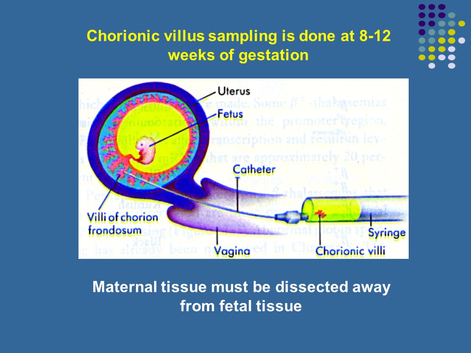 But which research method to choose depends on the location of the chorion.
What is the purpose of the procedure?
Chorionic biopsy is a voluntary test. It is not mandatory for everyone. It is carried out only at the request of future parents. But this method of prenatal diagnosis sets itself very specific goals. They consist in identifying:
- Down syndrome, Turner, Edwards.
- Klinefelter syndrome.
- Hemophilia.
- Amavrotic idiocy.
- Sickle cell anemia.
- Other rarer genetic diseases.
What are the indications for testing?
This procedure is not for everyone. It is carried out exclusively under certain circumstances, but they cannot serve as a mandatory reason to carry out the study. It should only be done with parental consent. So, the indications for a chorionic biopsy can be as follows:
- Mother of a child over 35 years of age. It has been proven that after this age, the risk of having a child with Down syndrome increases every year.
 It is especially important to undergo this study if a pregnant woman is older than 40-45 years.
 - If the couple who is expecting a baby is an older one. For example, the mother is over 35 years old, the father is 40. The older both parents are, the greater the risk of having a baby with various genetic abnormalities.
- If prenatal screening has identified a high risk of having a child with various genetic diseases. This can usually be suspected if a blood test for certain hormones shows poor results. Then additional studies may be prescribed, one of which is a chorion biopsy. Sometimes some syndromes can be suspected during an ultrasound examination. For example, inconsistencies of the nasal bone with the gestational age or other abnormal inconsistencies that can be noticed by an uzist.
- If there is an urgent need to know the exact gender of the baby. This is the case if a genetic disorder that is only found in girls or only in boys is suspected (eg, hemophilia).
- In the case when the family already has one child with genetic "breakdowns".
- When one of the relatives suffered from genetic diseases.
- If a doctor suspects a genetic disorder during routine ultrasound.
It should be noted that even in the presence of such problems, there is no guarantee that the child will be sick or born with some genetic problems. Carrying out a biopsy of the chorion is currently only advisory. No doctor can force a future mother to undergo such a study. In addition, this is a rather expensive procedure and not every clinic can perform it.
Are there any contraindications?
Chorionic villus sampling, like any procedure, has its contraindications. In some cases, it cannot be done, otherwise it can harm the mother or child. These are situations when:
- There is an exacerbation of chronic diseases in the mother.
- The threat of miscarriage persists.
- The uterus is in constant tone.

- There is a curved cervix
- A woman has bleeding from the vagina.
- There are uterine fibroids.
- Diagnosed adhesions in the pelvic organs.
- If a woman is expecting twins.
- In case there is no possibility of normal access to the chorion.
In some of the situations listed above, a biopsy may be performed after symptoms are relieved or there is no threat of miscarriage. And in some, like fibroids or multiple pregnancies, such a procedure cannot be done at all.
When to do the procedure?
The timing of the chorion biopsy is quite specific. The most optimal are the periods from 11 to 13 weeks. According to research, it is precisely at these times that the presence of genetic abnormalities can be accurately established. But sometimes such a procedure can be done from the 9th to the 12th week.
Sometimes it happens that the period for applying this method can be "stretched" from the 10th to the 19th week.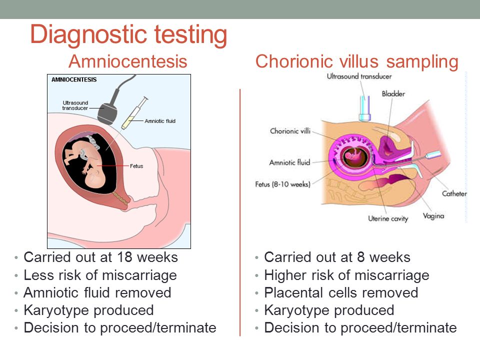 This happens in purely individual cases, and only a doctor can choose the most optimal time to do this study.
From a psychological point of view, it is best to take a biopsy before the 12th week. After all, if a negative diagnosis is confirmed, then it will be much easier and less painful to terminate such a pregnancy at an earlier date than to do a similar procedure at a later period.
What are the steps for performing a biopsy?
The procedure must be carried out in specialized clinics. It does not require a woman to stay in a hospital, but at the same time, it should be done only according to strict indications and after all the necessary tests. Preparation for a chorion biopsy includes an ultrasound of the pelvic organs (to accurately find out the number of fetuses), to understand exactly where the chorion is located. Also taking blood, urine and vaginal smear tests. In addition, the Rh factor and blood type must be accurately established.
The procedure can be performed through the abdominal wall or through the cervix. This is decided by the doctor.
If the examination is done through the abdominal wall, the puncture site must be lubricated with an antiseptic. Then the abdominal cavity is pierced with a special needle and biological material is taken from the chorion. It needs quite a bit, but at the same time the information content is very high.
If the examination is performed transcervically, the cervix is also lubricated with an antiseptic. Then the doctor takes a piece of tissue for examination with a special probe.
For all biopsy procedures, an ultrasound doctor must be present, who can control the process with the help of an ultrasound machine.
After the examination, the woman can lead a normal life, although on the first day after the biopsy she should limit physical activity, intimate life and be especially attentive to herself.
Are the results accurate?
Biopsy is a relatively new method of prenatal diagnosis. But she has established herself as one of the most accurate and reliable ways.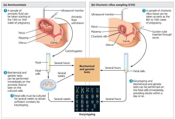 Chorionic biopsy results almost always show an accuracy of 99%.
All biological materials after they have been taken are placed in a special environment. Then they go to the laboratory. There, experienced specialists conduct research using the most modern methods and methods that allow you to make a qualitative analysis for genetic abnormalities.
Preliminary results can be known after 3 days, and the final and exact results become known after about 10 days. Sometimes it happens that due to the complexity of the analysis, the result may be uncertain. Then you need to undergo another biopsy at 16 weeks.
A biopsy is only accurate for the presence or absence of certain genetic diseases. But this method cannot give an absolute guarantee that the child will be born absolutely healthy without any other deviations.
Can there be complications?
This procedure is not completely safe. This is an invasive method that can have its negative consequences. But they are found only in rare cases.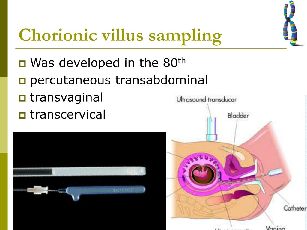 Only 2-3% of women can have serious troubles. But at the same time, complications after a chorionic biopsy are as follows:
- Abdominal pain in a woman during the first few days after the procedure. If these are small cramping discomforts, this should not be feared. It'll all be over in a couple of days.
- Amniotic fluid may leak in the early days.
- Membrane integrity may be compromised.
- Intrauterine infection of the fetus sometimes occurs.
- Severe bleeding may occur.
- Spontaneous miscarriage can sometimes occur.
According to statistics, only 0.4% of women can miscarry after a biopsy. And often this happens when the diagnosis for the presence of genetic defects is confirmed. Thus, there is a kind of "natural selection". In other cases, the risk of complications is minimal. But in any case, if after the procedure a woman has severe pain, bleeding or abundant watery discharge, it is necessary to urgently consult a doctor.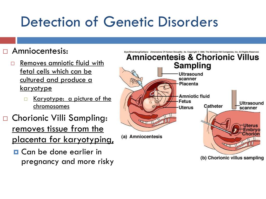
How much does it cost?
The biopsy procedure is not cheap, but it should be done only in trusted clinics that have all the necessary equipment and specialists. Chorionic biopsy price in Kyiv is quite affordable at the IPF clinic. Here you will receive high-quality services and at the same time you can be sure that the procedure will be carried out by highly qualified specialists. In addition, the constant support of a doctor, friendly staff and the most modern equipment will allow you to be confident and calm, focused on getting a good result.
If you are interested in chorion biopsy in Kyiv, always contact only trusted clinics, because this procedure requires special knowledge, equipment and skill.
Chorionic biopsy is an opportunity to understand whether the unborn baby is healthy. It has certain risks, but at the same time it allows you to determine the presence of incurable genetic diseases. This method gives parents a chance to choose whether to terminate or continue the pregnancy, prepare for the birth of a special child, or simply enjoy the pregnancy happily, knowing that the baby will be born healthy and without pathologies.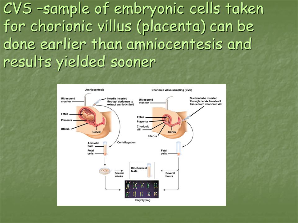
Chorionic biopsy | Ida-Tallinna Keskaigla
The purpose of this leaflet is to provide the patient with information about the purpose, nature and risks of chorionic biopsy.
Chorionic biopsy is performed on pregnant women whose primary tests (OSCAR test, NIPTIFY, Panorama or twin test) indicate a possible increased risk of a chromosomal disorder or whose history indicates the possibility of a hereditary disorder.
Chorionic biopsy is a voluntary procedure and you can decide for yourself whether you want to have this procedure or not. Before the study, it is necessary to sign an informed voluntary consent to the procedure.
Please note that the procedure takes two to three hours.
How is a chorion biopsy performed?
In the case of a chorionic biopsy, the cells of the chorionic villi, that is, the developing placenta, are examined.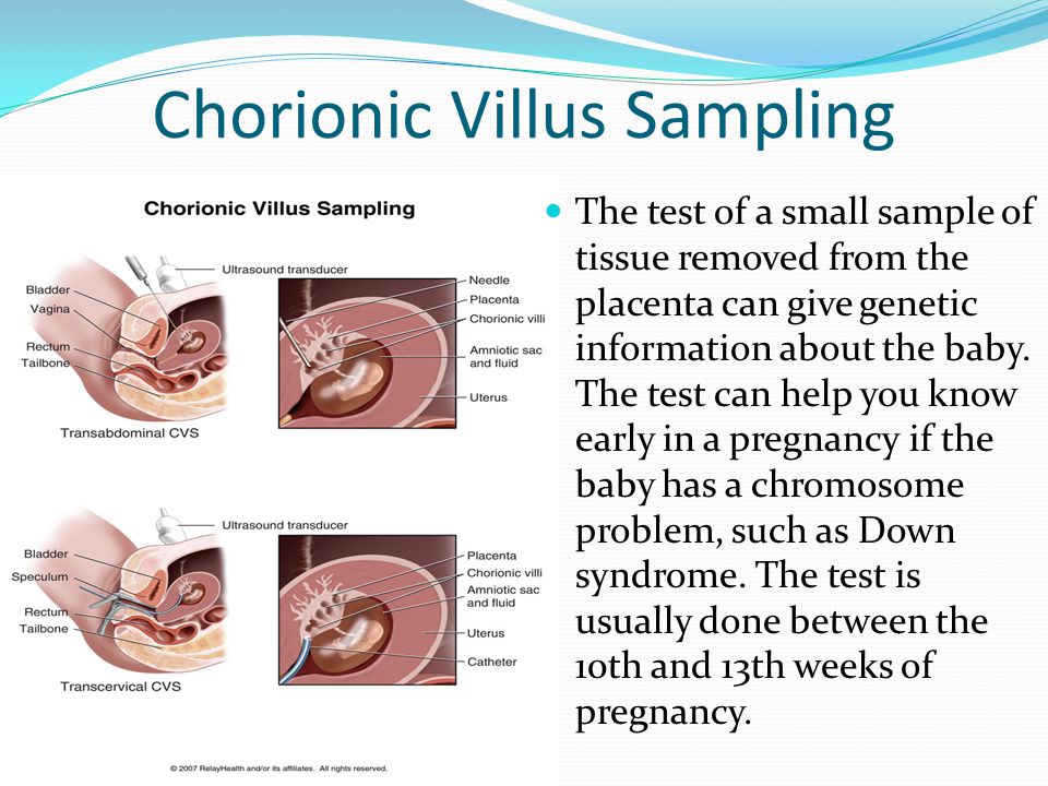 The chromosomes contained in the chorion cells coincide with the cells of the fetus. In order to examine these cells, a small piece of placental tissue is taken through the abdominal wall under ultrasound guidance with a thin needle. The collected material is sent to the laboratory for analysis.
When is the best time to do a chorionic biopsy?
Chorionic biopsy is usually performed from the 12th week of pregnancy, but may be performed later. The safest are the 12-13th weeks of pregnancy.
Is the examination painful?
The test can be uncomfortable, but most women find it no more painful than drawing blood from a vein. After the test, you may experience some spotting and you may feel some tension in your abdomen, which is normal. If the bleeding increases, you should immediately consult a doctor.
What are the risks associated with a chorionic biopsy?
Chorionic biopsy is a common test and complications are rare.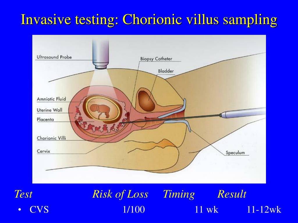 For most women, the importance of the information obtained from the study far outweighs the risks of the study.
-
Chorion biopsy increases the risk of miscarriage by 1–2%. The vast majority of pregnancies continue without problems.
-
Chorionic biopsy is performed under sterile conditions to prevent inflammation, but in spite of this, inflammation can occasionally develop, which is manifested by fever, uterine contractions, and abdominal pain.
-
In Rh-negative women, chorionic biopsy carries the risk that fetal blood cells enter the pregnant woman's bloodstream and develop antibodies to them. In order to prevent this, after the study, a pregnant woman is given an injection of antibodies, which reduces the risk of an Rhesus conflict.
-
Occasionally, chorionic biopsy (more often than amniocentesis) produces cells unsuitable for examination.