What is ultrasound scanner
Ultrasound scans: How do they work?
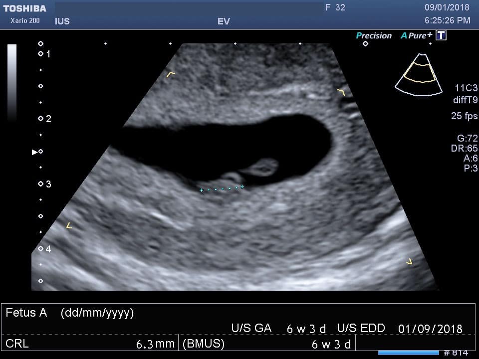
An ultrasound scan uses high-frequency sound waves to create images of the inside of the body. It is suitable for use during pregnancy.
Ultrasound scans, or sonography, are safe because they use sound waves or echoes to make an image, instead of radiation.
Ultrasound scans are used to evaluate fetal development, and they can detect problems in the liver, heart, kidney, or abdomen. They may also assist in performing certain types of biopsy.
The image produced is called a sonogram.
Fast facts on ultrasound scans
- Ultrasound scans are safe and widely used.
- They are often used to check the progress of a pregnancy.
- They are used for diagnosis or treatment.
- No special preparation is normally necessary before an ultrasound scan.
The person who performs an ultrasound scan is called a sonographer, but the images are interpreted by radiologists, cardiologists, or other specialists.
The sonographer usually holds a transducer, a hand-held device, like a wand, which is placed on the patient’s skin.
Ultrasound is sound that travels through soft tissue and fluids, but it bounces back, or echoes, off denser surfaces. This is how it creates an image.
The term “ultrasound” refers to sound with a frequency that humans cannot hear.
For diagnostic uses, the ultrasound is usually between 2 and 18 megahertz (MHz).
Higher frequencies provide better quality images but are more readily absorbed by the skin and other tissue, so they cannot penetrate as deeply as lower frequencies.
Lower frequencies penetrate deeper, but the image quality is inferior.
How does it capture an image?
Ultrasound will travel through blood in the heart chamber, for example, but if it hits a heart valve, it will echo, or bounce back.
It will travel straight through the gallbladder if there are no gallstones, but if there are stones, it will bounce back from them.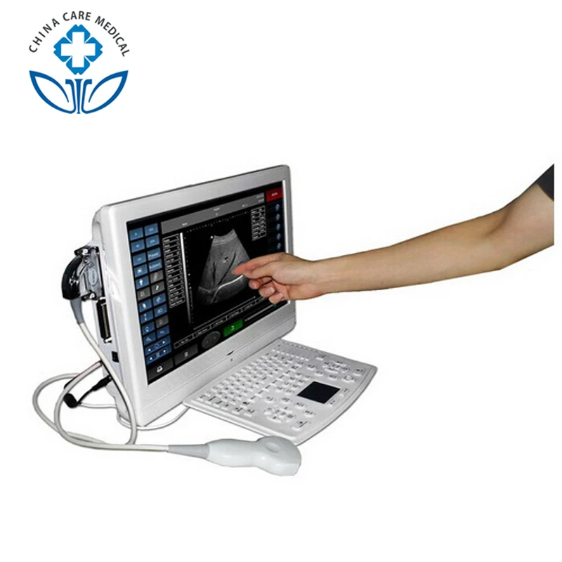
The denser the object the ultrasound hits, the more of the ultrasound bounces back.
This bouncing back, or echo, gives the ultrasound image its features. Varying shades of gray reflect different densities.
Ultrasound transducers
The transducer, or wand, is normally placed on the surface of the patient’s body, but some kinds are placed internally.
These can provide clearer, more informative images.
Examples are:
- an endovaginal transducer, for use in the vagina
- an endorectal transducer, for use in the rectum
- a transesophageal transducer, passed down the patient’s throat for use in the esophagus
Some very small transducers can be placed onto the end of a catheter and inserted into blood vessels to examine the walls of blood vessels.
Share on PinterestUltrasound images are made from reflected sound, and a diagnosis can then be made.
Ultrasound is commonly used for diagnosis, for treatment, and for guidance during procedures such as biopsies.
It can be used to examine internal organs such as the liver and kidneys, the pancreas, the thyroid gland, the testes and the ovaries, and others.
An ultrasound scan can reveal whether a lump is a tumor. This could be cancerous, or a fluid-filled cyst.
It can help diagnose problems with soft tissues, muscles, blood vessels, tendons, and joints. It is used to investigate a frozen shoulder, tennis elbow, carpal tunnel syndrome, and others.
Circulatory problems
Doppler ultrasound can assess the flow of blood in a vessel or blood pressure. It can determine the speed of the blood flow and any obstructions.
An echocardiogram (ECG) is an example of Doppler ultrasound. It can be used to create images of the cardiovascular system and to measure blood flow and cardiac tissue movement at specific points.
A Doppler ultrasound can assess the function and state of cardiac valve areas, any abnormalities in the heart, valvular regurgitation, or blood leaking from valves, and it can show how well the heart pumps out blood.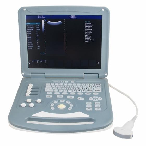
It can also be used to:
- examine the walls of blood vessels
- check for DVT or an aneurysm
- check fetal heart and heartbeat
- evaluate for plaque buildup and clots
- assess for blockages or narrowing of arteries
A carotid duplex is a form of carotid ultrasonography that may include a Doppler ultrasound. This would reveal how blood cells move through the carotid arteries.
Ultrasound in anesthesiology
Ultrasound is often used by anesthetists to guide a needle with anesthetic solutions near nerves.
An ultrasound can be done at a doctor’s office, at an outpatient clinic, or in the hospital.
Most scans take between 20 and 60 minutes. It is not normally painful, and there is no noise.
In most cases, no special preparation is needed, but patients may wish to wear loose-fitting and comfortable clothing.
If the liver or gallbladder is affected, the patient may have to fast, or eat nothing, for several hours before the procedure.
For a scan during pregnancy, and especially early pregnancy, the patient should drink plenty of water and try to avoid urinating for some time before the test.
When the bladder is full, the scan produces a better image of the uterus.
The scan usually takes place in the radiology department of a hospital. A doctor or a specially-trained sonographer will carry out the test.
External ultrasound
The sonographer puts a lubricating gel onto the patient’s skin and places a transducer over the lubricated skin.
The transducer is moved over the part of the body that needs to be examined. Examples include ultrasound examinations of a patient’s heart or a fetus in the uterus.
The patient should not feel discomfort or pain. They will just feel the transducer over the skin.
During pregnancy, there may be slight discomfort because of the full bladder.
Internal ultrasound
If the internal reproductive organs or urinary system need to be evaluated, the transducer may be placed in the rectum for a man or in the vagina for a woman.
To evaluate some part of the digestive system, for example, the esophagus, the chest lymph nodes, or the stomach, an endoscope may be used.
A light and an ultrasound device are attached to the end of the endoscope, which inserted into the patient’s body, usually through the mouth.
Before the procedure, patients are given medications to reduce any pain.
Internal ultrasound scans are less comfortable than external ones, and there is a slight risk of internal bleeding.
Most types of ultrasound are noninvasive, and they involve no ionizing radiation exposure. The procedure is believed to be very safe.
However, since the long-term risks are not established, unnecessary “keepsake” scans during pregnancy are not encouraged. Ultrasound during pregnancy is recommended only when medically needed.
Anyone who is allergic to latex should inform their doctor so that they will not use a latex-covered probe.
Ultrasound scans: How do they work?
An ultrasound scan uses high-frequency sound waves to create images of the inside of the body. It is suitable for use during pregnancy.
Ultrasound scans, or sonography, are safe because they use sound waves or echoes to make an image, instead of radiation.
Ultrasound scans are used to evaluate fetal development, and they can detect problems in the liver, heart, kidney, or abdomen. They may also assist in performing certain types of biopsy.
The image produced is called a sonogram.
Fast facts on ultrasound scans
- Ultrasound scans are safe and widely used.
- They are often used to check the progress of a pregnancy.
- They are used for diagnosis or treatment.
- No special preparation is normally necessary before an ultrasound scan.
The person who performs an ultrasound scan is called a sonographer, but the images are interpreted by radiologists, cardiologists, or other specialists.
The sonographer usually holds a transducer, a hand-held device, like a wand, which is placed on the patient’s skin.
Ultrasound is sound that travels through soft tissue and fluids, but it bounces back, or echoes, off denser surfaces. This is how it creates an image.
The term “ultrasound” refers to sound with a frequency that humans cannot hear.
For diagnostic uses, the ultrasound is usually between 2 and 18 megahertz (MHz).
Higher frequencies provide better quality images but are more readily absorbed by the skin and other tissue, so they cannot penetrate as deeply as lower frequencies.
Lower frequencies penetrate deeper, but the image quality is inferior.
How does it capture an image?
Ultrasound will travel through blood in the heart chamber, for example, but if it hits a heart valve, it will echo, or bounce back.
It will travel straight through the gallbladder if there are no gallstones, but if there are stones, it will bounce back from them.
The denser the object the ultrasound hits, the more of the ultrasound bounces back.
This bouncing back, or echo, gives the ultrasound image its features. Varying shades of gray reflect different densities.
Ultrasound transducers
The transducer, or wand, is normally placed on the surface of the patient’s body, but some kinds are placed internally.
These can provide clearer, more informative images.
Examples are:
- an endovaginal transducer, for use in the vagina
- an endorectal transducer, for use in the rectum
- a transesophageal transducer, passed down the patient’s throat for use in the esophagus
Some very small transducers can be placed onto the end of a catheter and inserted into blood vessels to examine the walls of blood vessels.
Share on PinterestUltrasound images are made from reflected sound, and a diagnosis can then be made.
Ultrasound is commonly used for diagnosis, for treatment, and for guidance during procedures such as biopsies.
It can be used to examine internal organs such as the liver and kidneys, the pancreas, the thyroid gland, the testes and the ovaries, and others.
An ultrasound scan can reveal whether a lump is a tumor. This could be cancerous, or a fluid-filled cyst.
It can help diagnose problems with soft tissues, muscles, blood vessels, tendons, and joints. It is used to investigate a frozen shoulder, tennis elbow, carpal tunnel syndrome, and others.
Circulatory problems
Doppler ultrasound can assess the flow of blood in a vessel or blood pressure. It can determine the speed of the blood flow and any obstructions.
An echocardiogram (ECG) is an example of Doppler ultrasound. It can be used to create images of the cardiovascular system and to measure blood flow and cardiac tissue movement at specific points.
A Doppler ultrasound can assess the function and state of cardiac valve areas, any abnormalities in the heart, valvular regurgitation, or blood leaking from valves, and it can show how well the heart pumps out blood.
It can also be used to:
- examine the walls of blood vessels
- check for DVT or an aneurysm
- check fetal heart and heartbeat
- evaluate for plaque buildup and clots
- assess for blockages or narrowing of arteries
A carotid duplex is a form of carotid ultrasonography that may include a Doppler ultrasound.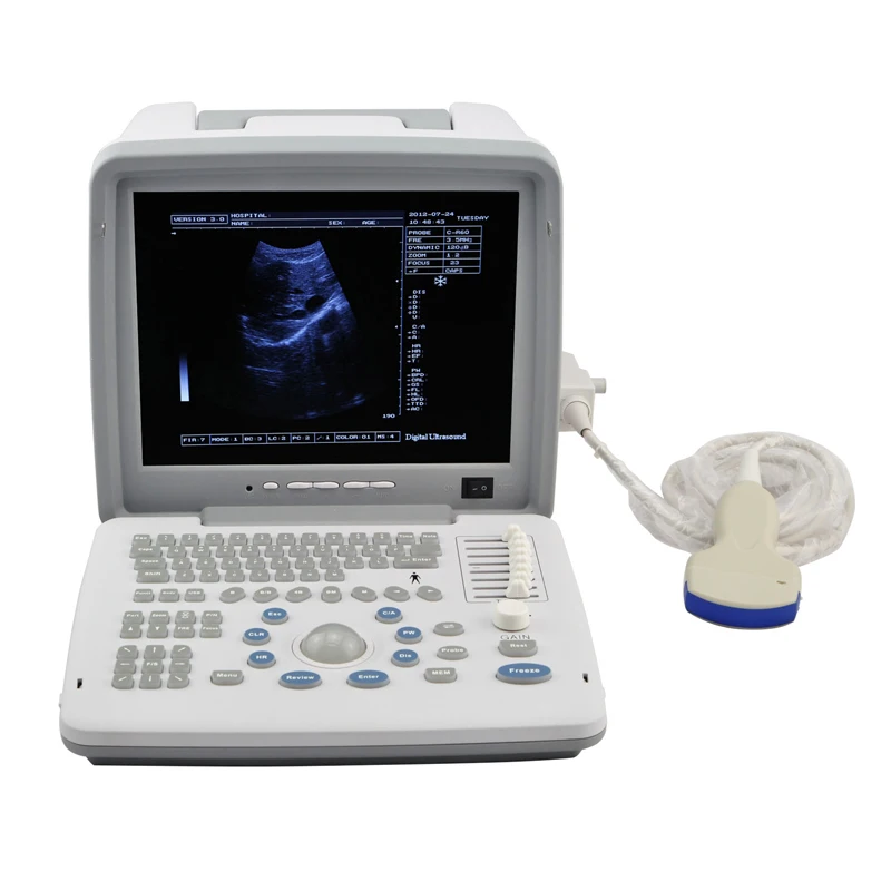 This would reveal how blood cells move through the carotid arteries.
Ultrasound in anesthesiology
Ultrasound is often used by anesthetists to guide a needle with anesthetic solutions near nerves.
An ultrasound can be done at a doctor’s office, at an outpatient clinic, or in the hospital.
Most scans take between 20 and 60 minutes. It is not normally painful, and there is no noise.
In most cases, no special preparation is needed, but patients may wish to wear loose-fitting and comfortable clothing.
If the liver or gallbladder is affected, the patient may have to fast, or eat nothing, for several hours before the procedure.
For a scan during pregnancy, and especially early pregnancy, the patient should drink plenty of water and try to avoid urinating for some time before the test.
When the bladder is full, the scan produces a better image of the uterus.
The scan usually takes place in the radiology department of a hospital. A doctor or a specially-trained sonographer will carry out the test.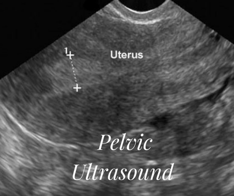
External ultrasound
The sonographer puts a lubricating gel onto the patient’s skin and places a transducer over the lubricated skin.
The transducer is moved over the part of the body that needs to be examined. Examples include ultrasound examinations of a patient’s heart or a fetus in the uterus.
The patient should not feel discomfort or pain. They will just feel the transducer over the skin.
During pregnancy, there may be slight discomfort because of the full bladder.
Internal ultrasound
If the internal reproductive organs or urinary system need to be evaluated, the transducer may be placed in the rectum for a man or in the vagina for a woman.
To evaluate some part of the digestive system, for example, the esophagus, the chest lymph nodes, or the stomach, an endoscope may be used.
A light and an ultrasound device are attached to the end of the endoscope, which inserted into the patient’s body, usually through the mouth.
Before the procedure, patients are given medications to reduce any pain.
Internal ultrasound scans are less comfortable than external ones, and there is a slight risk of internal bleeding.
Most types of ultrasound are noninvasive, and they involve no ionizing radiation exposure. The procedure is believed to be very safe.
However, since the long-term risks are not established, unnecessary “keepsake” scans during pregnancy are not encouraged. Ultrasound during pregnancy is recommended only when medically needed.
Anyone who is allergic to latex should inform their doctor so that they will not use a latex-covered probe.
How it works. Ultrasound scanner
Today, ultrasound examinations are one of the most popular methods of medical diagnostics. Ultrasound scanners have firmly established themselves in every modern clinic, diagnostic room, and even reanimobile. Ultrasound diagnostics continues to develop rapidly - new technologies are coming to replace the usual two-dimensional picture. Many of them are presented in expert-class ultrasound scanners manufactured by Rostec. But whatever the ultrasound machines, they all work on the same principle, which we will consider in this material.
silent sound
Ultrasound is such sound vibrations that, due to their high frequency, are not perceived by the human ear. Both natural and man-made sources can emit ultrasound. Some animals use high-frequency sounds for communication and orientation in space. For example, ultrasound helps bats avoid obstacles in flight and catch insects. And scientists, engineers and physicians use high-frequency sounds to study various physical environments.
Like many inventions, the medical application of ultrasound does not have a single parent. Its appearance was preceded by a number of discoveries. So, the very phenomenon of ultrasound was first studied at the end of the 18th century by the Italian scientist Lazzaro Spallanzani. While experimenting with bats, he noticed that mice with their ears closed lose their ability to orient themselves. The Italian suggested that these animals make and perceive special sounds.
Echolocation in bats. Image: wikimedia.org
The next important step towards the emergence of ultrasound machines was the research of Pierre Curie, a French physicist, the future husband of the famous Marie Sklodowska-Curie. In 1880, together with his older brother, they discover the piezoelectric effect, due to which electricity arises in deformable crystals. Based on this effect, modern ultrasound transducers work.
The medical use of ultrasound began in the 20th century. At first, it was considered as a method of therapy and was used to treat arthritis, stomach ulcers, and asthma. Then at 19In the 1940s, Austrian neurologist Carl Frederick Dussick pioneered the use of ultrasound to study the brain. Observing the passage of a sound wave through the patient's skull, he concluded that there was a brain tumor, which, however, was not confirmed. Around the same time, the Englishman John Wild, using ultrasound, determines the thickness of the intestine.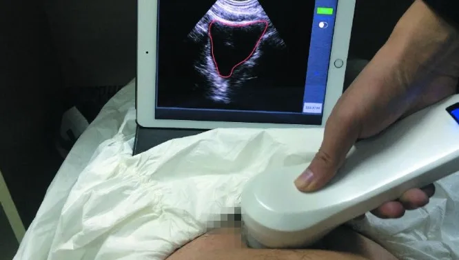
It must be said that the first ultrasound devices were very different from those that we are used to seeing in clinics today. In some cases, patients were placed in water baths, in others they were forced to sit in one place for a long time while sensors and photo devices rotated around them. Ultrasound technology, which works like modern real-time scanners, appeared only in 1960s, but it was also very bulky.
In 1967, the US began licensing the activities of ultrasound diagnosticians, and the first standards appeared. Ultrasound is spreading most rapidly in gynecology: already in the first years of its use, the new technology made it possible to almost completely abandon radiography, which is unsafe for pregnant women.
The principle of operation of the ultrasound scanner
Let's see how modern medical ultrasound scanners work. The main element of any such device is an ultrasonic sensor. It generates and perceives ultrasound, working on the same principle of the piezoelectric effect discovered by the Curie brothers.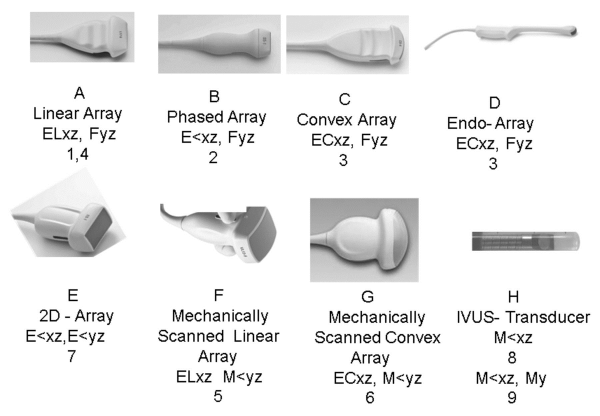
Most often, the sensor uses crystals of piezoelectric elements, the nature of which allows them to generate ultrasound under the influence of electricity. The crystals also “work” in the opposite direction - having received the reflected ultrasound at the input, they convert it into an electrical signal that forms a picture on the display of the ultrasound machine.
Scheme of operation of the ultrasonic sensor. Image: wikimedia.org
It is worth noting here that if the result of modern ultrasound is a two-dimensional or even three-dimensional image, then the first devices worked, one might say, in a one-dimensional coordinate system. Instead of a picture of one or another organ, doctors saw a graph of the passage of sound through tissues to a certain depth, similar to an oscilloscope graph. By measuring the sound intensity, it was possible to assess the condition of the patient's tissues at different depths. Today, this method, which is called A-mode (from amplitude), is used in ophthalmology.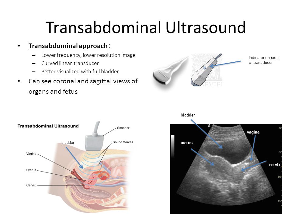
Over time, sensors have improved. In the 1970s, an electric motor began to be added to ultrasound sensors, which made it possible to rotate the device and obtain a two-dimensional image. Then the so-called phased sensors appeared, containing many elements, with the help of which a better image was obtained. And at the turn of the 1990-2000s, the results of ultrasound became three-dimensional. Today, the speed and quality of ultrasound scanners make it possible to obtain even a moving image.
4D echocardiogram. Author: Kjetil Lenes, wikimedia.org
How exactly does ultrasound help us see what is hidden from the human eye? The fact is that gases, liquids and solids react differently to a sound wave. The bones, tissues, and fluids of our body can transmit, reflect, absorb, or scatter ultrasound. The sensor alternately sends sound impulses to the body and receives reflected ones. The received data is processed by the processor, which, knowing the speed of ultrasound and the time of the wave, determines the distance to the object and forms its model.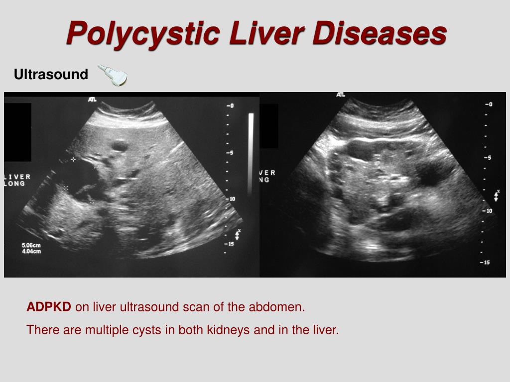 All this happens in millionths of a second.
Most often, the ultrasound result on the screen is displayed in black and white, since our eye distinguishes these colors more clearly. Bone tissues reflect sound better than others and are displayed in white. Fluids and voids are rendered in black, while soft tissues are rendered in grayscale. Some studies use color modes.
In addition to the sensors themselves, the ultrasound scanner includes special lenses, a voltage source, a computer with a display, a keyboard and a printer. To improve sound transmission and more comfortable contact with the skin, a gel is used. The transducer controls and wide customization options of modern ultrasound machines allow physicians to use a variety of modes suitable for a particular study, and a high level of automation facilitates their work.
Ultrasound in Russian
The first domestic experiments on the medical application of ultrasound were carried out as early as the 1950s. In the 1960s, original devices were developed, but, unfortunately, they did not become widespread and went on the shelf. The gradual introduction of ultrasound diagnostics began only in the 1980s, mainly Japanese-made devices were used. Until recently, almost all Russian clinics used foreign ultrasound equipment, the leader in the creation of which today is China and South Korea. By 2018, Russian healthcare annually purchased about 3,000 scanners from abroad.
In the past few years, the Russian government has taken a number of measures to stimulate the domestic production of ultrasound equipment. Since 2018, Rostec, as part of the import substitution program, has been mass-producing Russian ultrasound scanners RuScan at the facilities of the Kalugapribor plant, which is part of the Avtomatika concern. The work is carried out jointly with the technological partner NPO Scaner.
The enterprise of the State Corporation produces ultrasound scanners of medium (RuScan 50), high (RuScan 60), and expert (RuScan 65/65M) classes, as well as a portable model, RuScan70P, which can be used in medical transport and sports facilities. .
.
Rostec ultrasound scanners have proven themselves in Russian medical institutions. When talking about RuScan devices, doctors note high-quality visualization, ease of use and ergonomic design. Among the advantages of Rostec's development is a professional Russian-language interface and integration into the software shell of scanners of the results of domestic population-statistical studies. This data will help Russian doctors make decisions based not only on their experience, but also on the knowledge of the entire industry. It is noteworthy that RuScan devices became the first domestic expert-class scanners with Russian software.
In 2021, the State Corporation plans to supply more than 1,000 scanners to the country's medical institutions. In total, the capacities of Rostec allow to produce up to 2000 products per year.
Veterinary Ultrasound Scanner KAIXIN KX-5200V
Portable Veterinary Ultrasound Scanner KAIXIN KX -5200V for diagnosing pregnancy in the early stages, identifying pathologies of the reproductive system of animals.
Modern ultrasound scanner for all types of animals.
With a replaceable sensor, it allows you to determine the pregnancy of animals, as well as the pathology of the ovaries and other organs in high quality with the ability to save and print images.
6.5 MHz rectal linear probe included as standard. It is possible to complete with a 4.0 MHz convex probe , the difference is in the display of the scan field. (when using a convex probe, a cone-shaped display field).
Color rendering on par with more expensive competitors! Features edge refinement to improve image quality.
Pseudo-color image makes it much easier to work in bright sunlight. The body of the scanner is hermetically sealed and waterproof, suitable for use in aggressive environments.
Using this model, it is possible to determine pregnancy from the 30th day of pregnancy, the sex of the calf from the 60th day of pregnancy, the pathology of the reproductive system.
- Lightweight, handheld, easy to diagnose
- Waterproof housing
- Automatic detection of the connected sensor
- USB port with the ability to connect a laser printer and save images to a USB stick
- Video output
Standard:
- Main unit
- Rectal linear transducer 6.5 MHz (or other transducer of your choice)
- 2 lithium batteries (2400 MAh), operating time without recharging 5 hours
- Arm cuff
- Neck straps
- Charger
- Stand
- Impact resistant carrying case
Optional :
Sensors:
6.5 MHz Linear Rectal Probe
4.0 MHz Convex Rectal Probe
Convex probe 3.5 MHz
Micro Convex Probe 5.0 MHz - 6.5 MHz
Linear transducer 7.5 MHz
Transvaginal transducer 6.