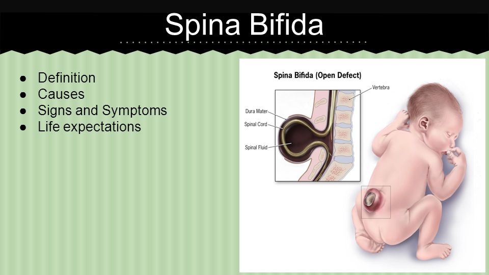
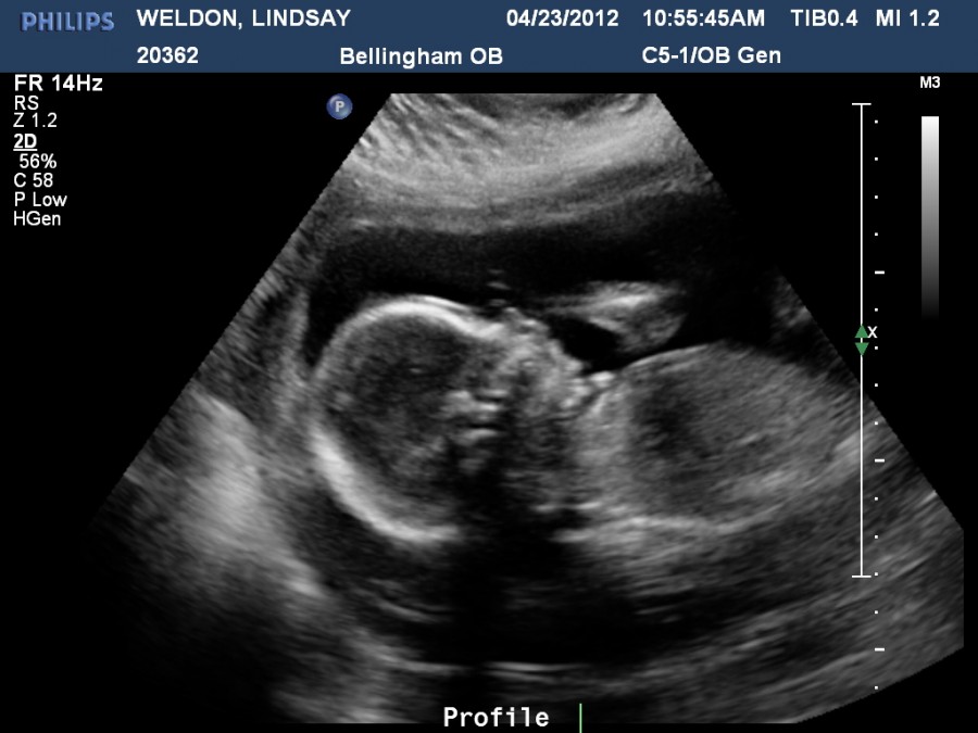
Spina bifida ultrasound 12 weeks
Prenatal diagnosis of neural tube defect before 12 weeks' gestation: direct and indirect ultrasonographic semeiology
Case Reports
. 1997 Dec;10(6):406-9.
doi: 10.1046/j.1469-0705.1997.10060406.x.
J P Bernard 1 , B Suarez, C Rambaud, F Muller, Y Ville
Affiliations
Affiliation
- 1 Fetal Medicine Unit, Hopital Antoine Beclere, Clamart, France.
- PMID: 9476326
- DOI: 10.1046/j.1469-0705.1997.10060406.x
Free article
Case Reports
J P Bernard et al. Ultrasound Obstet Gynecol. 1997 Dec.
Free article
. 1997 Dec;10(6):406-9.
doi: 10.1046/j.1469-0705.1997.10060406.x.
Authors
J P Bernard 1 , B Suarez, C Rambaud, F Muller, Y Ville
Affiliation
- 1 Fetal Medicine Unit, Hopital Antoine Beclere, Clamart, France.
- PMID: 9476326
- DOI: 10.1046/j.1469-0705.1997.10060406.x
Abstract
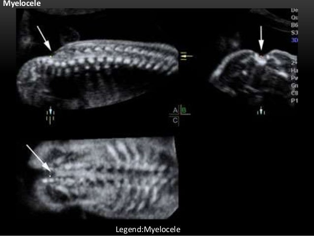
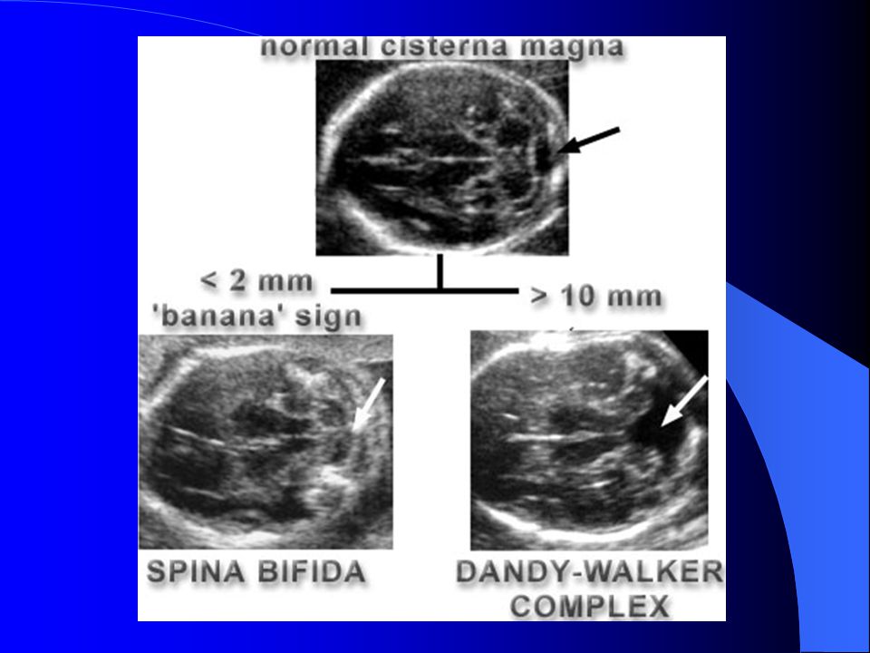
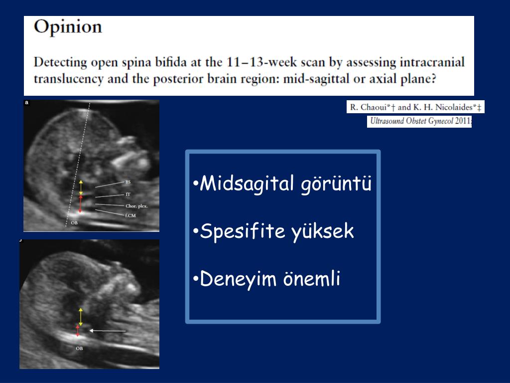
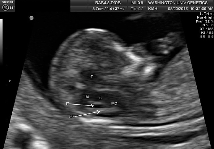
We describe the direct and indirect ultrasonographic features of a case of lumbar open spina bifida. The spinal defect was the prominent feature at 10 weeks + 5 days' gestation; however, cranial signs including narrowing of the frontal bones and flattening of the occiput were helpful at 12 weeks. This 'acorn' sign is likely to precede the 'lemon' sign, describing scalloping of the frontal bones at a later gestation. The diagnosis of spina bifida was confirmed by electrophoresis of the amniotic fluid, which showed an abnormal migration of acetylcholinesterase. Postmortem ultrasound examination of the same fetus proved useful in refining the diagnosis and also revealed the presence of the Arnold-Chiari malformation. Development of ultrasound screening in the first trimester of pregnancy should allow further evaluation of these findings. It seems reasonable to confirm such an early diagnosis by electrophoresis of the amniotic fluid as an alternative to ultrasonographic confirmation at 13-14 weeks.
Similar articles
-
Prenatal diagnosis of spina bifida: from intracranial translucency to intrauterine surgery.
Sepulveda W, Wong AE, Sepulveda F, Alcalde JL, Devoto JC, Otayza F. Sepulveda W, et al. Childs Nerv Syst. 2017 Jul;33(7):1083-1099. doi: 10.1007/s00381-017-3445-7. Epub 2017 Jun 7. Childs Nerv Syst. 2017. PMID: 28593553 Review.
-
Presence of the 'lemon' sign in fetuses with spina bifida at the 10-14-week scan.
Sebire NJ, Noble PL, Thorpe-Beeston JG, Snijders RJ, Nicolaides KH. Sebire NJ, et al. Ultrasound Obstet Gynecol. 1997 Dec;10(6):403-5. doi: 10.1046/j.1469-0705.1997.10060403.x. Ultrasound Obstet Gynecol. 1997. PMID: 9476325
-
New ultrasonographic criteria for the prenatal diagnosis of Chiari type 2 malformation.
Fujisawa H, Kitawaki J, Iwasa K, Honjo H. Fujisawa H, et al.
 Acta Obstet Gynecol Scand. 2006;85(12):1426-9. doi: 10.1080/00016340600645602. Acta Obstet Gynecol Scand. 2006. PMID: 17260216
 -
The detection of spina bifida before 10 gestational weeks using two- and three-dimensional ultrasound.
Blaas HG, Eik-Nes SH, Isaksen CV. Blaas HG, et al. Ultrasound Obstet Gynecol. 2000 Jul;16(1):25-9. doi: 10.1046/j.1469-0705.2000.00149.x. Ultrasound Obstet Gynecol. 2000. PMID: 11084961
-
Sonographic detection of open spina bifida in the first trimester: review of the literature.
Meller C, Aiello H, Otaño L. Meller C, et al. Childs Nerv Syst. 2017 Jul;33(7):1101-1106. doi: 10.1007/s00381-017-3443-9. Epub 2017 May 16. Childs Nerv Syst. 2017. PMID: 28510070 Review.
See all similar articles
Cited by
-
Prenatal diagnosis of spina bifida: from intracranial translucency to intrauterine surgery.
Sepulveda W, Wong AE, Sepulveda F, Alcalde JL, Devoto JC, Otayza F. Sepulveda W, et al. Childs Nerv Syst. 2017 Jul;33(7):1083-1099. doi: 10.1007/s00381-017-3445-7. Epub 2017 Jun 7. Childs Nerv Syst. 2017. PMID: 28593553 Review.
-
Imaging the fetal central nervous system.
De Keersmaecker B, Claus F, De Catte L. De Keersmaecker B, et al. Facts Views Vis Obgyn. 2011;3(3):135-49. Facts Views Vis Obgyn. 2011. PMID: 24753859 Free PMC article. Review.
-
Association of Chiari malformation and vitamin B12 deficit in a family.

Welsch M, Antes S, Kiefer M, Meyer S, Eymann R. Welsch M, et al. Childs Nerv Syst. 2013 Jul;29(7):1193-8. doi: 10.1007/s00381-013-2056-1. Epub 2013 Mar 7. Childs Nerv Syst. 2013. PMID: 23468202
-
Prenatal neurologic anomalies: sonographic diagnosis and treatment.
De Catte L, De Keersmaeker B, Claus F. De Catte L, et al. Paediatr Drugs. 2012 Jun 1;14(3):143-55. doi: 10.2165/11597030-000000000-00000. Paediatr Drugs. 2012. PMID: 22242843 Review.
Publication types
MeSH terms
Substances
Neural tube defects | Pregnancy Birth and Baby
Neural tube defects | Pregnancy Birth and Baby beginning of content4-minute read
Listen
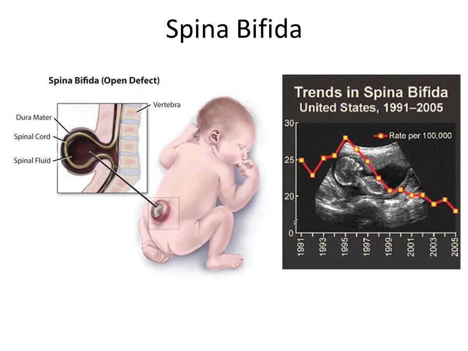
Neural tube defects are abnormalities that occur in the development of the spinal cord and brain of some babies. The most common defects are spina bifida (abnormal development of part of the spine and spinal cord) and anencephaly (severely abnormal development of the brain).
What are neural tube defects?
During the first month of life, an embryo (developing baby) grows a primitive tissue structure called the ‘neural tube’. As the embryo develops, the neural tube begins to change into a more complicated structure of bones, tissue and nerves that will eventually form the spine and nervous system.
However, in cases of spina bifida, something goes wrong with the development of the neural tube and the spinal column (the ridge of bone that surrounds and protects the nerves) does not fully close. Spina bifida is a Latin term that means ‘split spine’.
The chance that a pregnancy will be affected by a neural tube defect is less than one in 1,000.
What causes neural tube defects?
The cause of neural tube defects is not certain, but it appears to be due to a combination of genetic and environmental factors.
Women are at increased risk of having a baby with a neural tube defect if:
- they have already had a baby with a neural tube defect
- they or their partner have a close relative born with a neural tube defect
- they have type 1 (insulin dependent) diabetes (not gestational diabetes)
- they are obese, or take certain anti-epileptic medications, especially those containing sodium valproate or valproic acid
Prevention
About 2 in 3 neural tube defects can be prevented through increasing folate (folic acid) intake at least a month before pregnancy and during the first 3 months of pregnancy. Adequate folate levels are critical during the early days of the developing embryo, particularly the 3rd and 4th week, the period in which neural tube defects occur and when many women won't know they are pregnant.
You can increase your folate intake by eating folate-rich foods, including folate-fortified foods in your daily diet, or by taking a folic acid supplement.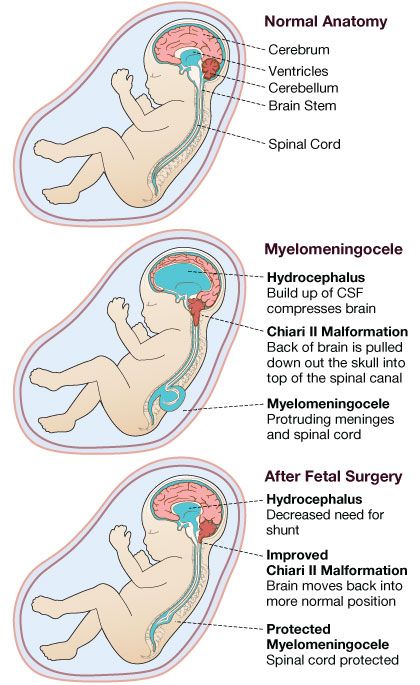 Good sources of folate include green leafy vegetables, fruit (citrus, berries and bananas), legumes and some cereals (bread and many breakfast cereals now have added folate).
Women who take medicines to control epilepsy, seizures or psychiatric disorders should talk to their doctor before taking folate because it can interfere with how their medications work.
For more information see folate and pregnancy.
Diagnosis
Neural tube defects may be diagnosed during the ultrasound scan that is carried out around week 12 of the pregnancy or, more likely, during the anomaly scan that is carried out at around weeks 18 to 20.
Ultrasound scans
An ultrasound scan is a safe procedure that uses sound waves to create an image of the inside of your body. Most hospitals will offer women at least 2 ultrasound scans during their pregnancy. The first is usually at around 8 to 14 weeks and is sometimes called the ‘dating scan’ because it can help to determine when the baby is due.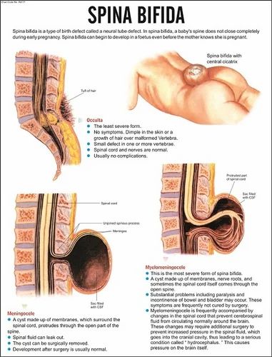 This first scan may be able to detect problems with your baby’s spine that could indicate spina bifida if the condition is severe. If a dating scan is done earlier than 12 weeks, another ultrasound called the nuchal translucency scan is done at 12 weeks to check amongst other things for signs of Down Syndrome, and if a dating scan is not done prior to this then this scan can be used as the dating scan.
Morphology scan
The morphology or anomaly scan is an ultrasound scan that is carried out around weeks 18 to 20 of your pregnancy. This scan aims to identify any physical problems with your baby. It is usually during this scan that spina bifida is diagnosed.
Coping with the results
If tests confirm that your baby has spina bifida, the implications will be fully discussed with you. You will need to consider your options carefully. Your options are to:
- continue with your pregnancy while getting information and advice so that you are prepared for caring for your baby
- end your pregnancy
If you are considering ending your pregnancy, you should talk to your doctor or midwife.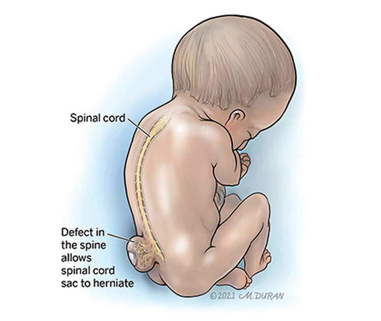 They will be able to provide you with important information and advice.
Your options for ending your pregnancy will depend on how many weeks pregnant you are when you make the decision. If you decide to end your pregnancy, you may wish to talk to a counsellor afterwards. Your doctor or midwife will be able to arrange this for you.
Call Pregnancy, Birth and Baby on 1800 882 436 to discuss your options regarding your pregnancy with a maternal child health nurse.
Sources:
Australian Institute of Health and Welfare (Neural tube defects in Australia), The Centre for Genetics Education (Neural tube defects – spina bifida and anencephaly), RANZCOG (Planning for pregnancy), Women's and Children's Health Network (Screening tests for neural tube defects), Sydney Children's Hospital Network (Factsheet: What is spina bifida?)Learn more here about the development and quality assurance of healthdirect content.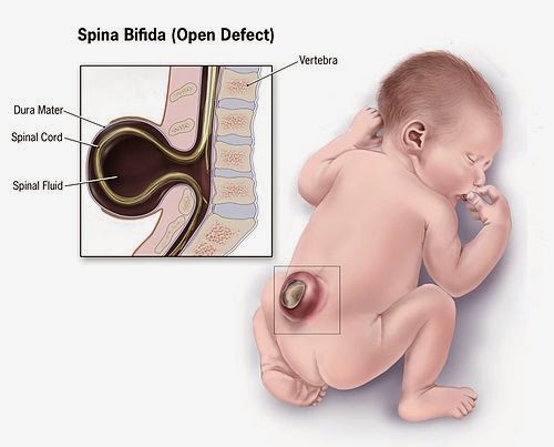
Last reviewed: March 2020
Back To Top
Need more information?
Pregnancy checkups, screenings and scans
Knowing what check-ups, screenings and scans to have and when to have them during your pregnancy is important information for every pregnant woman.
Read more on Pregnancy, Birth & Baby website
Neural tube defects: children & teens | Raising Children Network
Neural tube defects are brain and spinal cord abnormalities, including spina bifida, encephalocele and anencephaly. Read how they affect children.
Read more on raisingchildren.net.au website
Folate and pregnancy
Folate and folic acid are important for pregnancy since they can help prevent birth defects known as neural tube defects, such as spina bifida.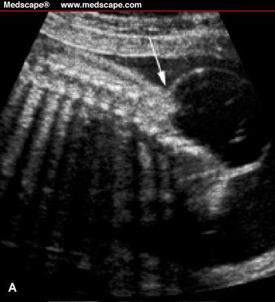
Read more on Pregnancy, Birth & Baby website
Spina bifida
Spina bifida is a condition that affects the normal development of a baby’s spine early in pregnancy.
Read more on Pregnancy, Birth & Baby website
Spina Bifida | Sydney Children's Hospitals Network
Spina Bifida comes from a latin term which means “split spine”
Read more on Sydney Children's Hospitals Network website
Maternal screening - Pathology Tests Explained
Why and when to get tested for maternal screening
Read more on Pathology Tests Explained website
Folic acid & iodine fortification, Summary - Australian Institute of Health and Welfare
Mandatory folic acid and iodine fortification of bread resulted in increased levels of folic acid and iodine in the food supply, increased folic acid and iodine intakes, a decreased rate of neural.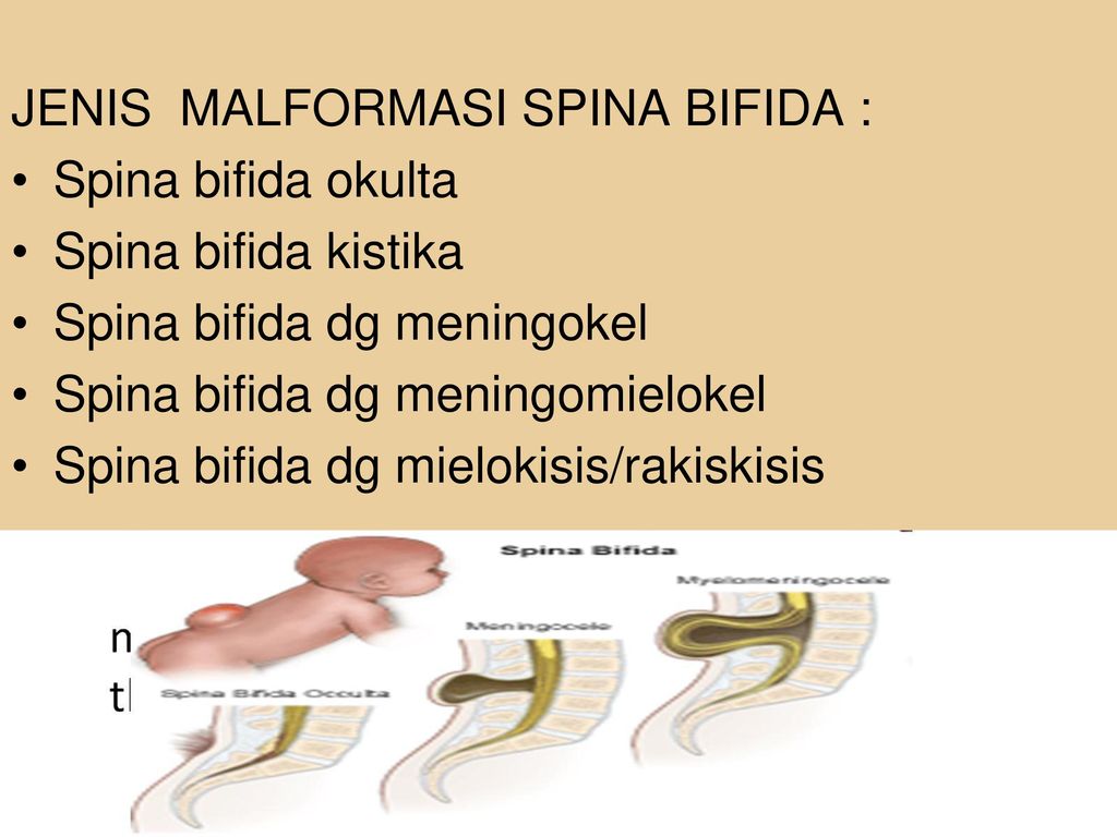 ..
..
Read more on AIHW – Australian Institute of Health and Welfare website
Folate | Jean Hailes
Folate is a B vitamin needed for healthy growing, in particular for the nervous system.
Read more on Jean Hailes for Women's Health website
What supplements should I take during pregnancy? | Queensland Health
Find out what supplements and vitamins you need to take when trying to get pregnant, during pregnancy, after pregnancy and while breastfeeding.
Read more on Queensland Health website
Pregnancy at weeks 1 to 4
When you conceive, your body’s hormone levels change, but you may not notice any signs that you’re pregnant yet.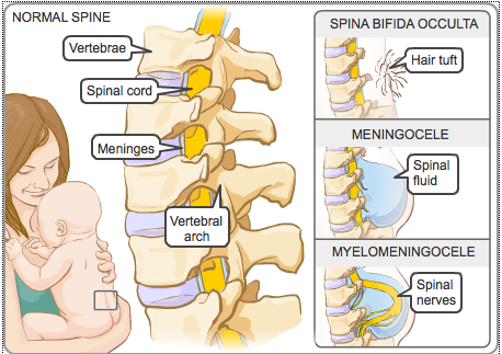
Read more on Pregnancy, Birth & Baby website
Disclaimer
Pregnancy, Birth and Baby is not responsible for the content and advertising on the external website you are now entering.
OKNeed further advice or guidance from our maternal child health nurses?
1800 882 436
Video call
- Contact us
- About us
- A-Z topics
- Symptom Checker
- Service Finder
- Linking to us
- Information partners
- Terms of use
- Privacy
Pregnancy, Birth and Baby is funded by the Australian Government and operated by Healthdirect Australia.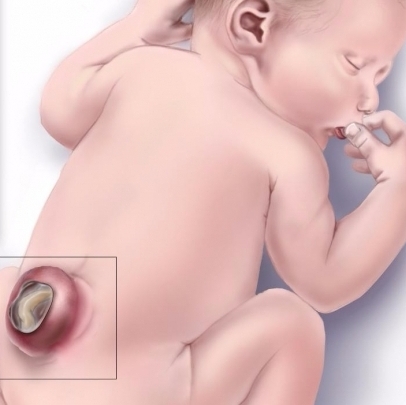
Pregnancy, Birth and Baby is provided on behalf of the Department of Health
Pregnancy, Birth and Baby’s information and advice are developed and managed within a rigorous clinical governance framework. This website is certified by the Health On The Net (HON) foundation, the standard for trustworthy health information.
This site is protected by reCAPTCHA and the Google Privacy Policy and Terms of Service apply.
This information is for your general information and use only and is not intended to be used as medical advice and should not be used to diagnose, treat, cure or prevent any medical condition, nor should it be used for therapeutic purposes.
The information is not a substitute for independent professional advice and should not be used as an alternative to professional health care. If you have a particular medical problem, please consult a healthcare professional.
Except as permitted under the Copyright Act 1968, this publication or any part of it may not be reproduced, altered, adapted, stored and/or distributed in any form or by any means without the prior written permission of Healthdirect Australia.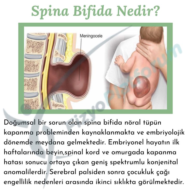
Support this browser is being discontinued for Pregnancy, Birth and Baby
Support for this browser is being discontinued for this site
- Internet Explorer 11 and lower
We currently support Microsoft Edge, Chrome, Firefox and Safari. For more information, please visit the links below:
- Chrome by Google
- Firefox by Mozilla
- Microsoft Edge
- Safari by Apple
You are welcome to continue browsing this site with this browser. Some features, tools or interaction may not work correctly.
symptoms, causes, treatment and surgery, complications, fund for helping children with spina bifida
Inna Inyushkina
founder of the Spina Bifida Charitable Foundation
Author profile
Editorial staff "About important"
spoke with Inna
According to statistics, about 1,500 children with spina bifida are born every year in Russia.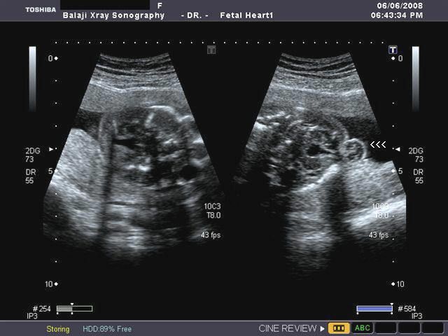 And many doctors still do not know that one can live with such a disease.
In 2012, at one of the planned ultrasounds during pregnancy, I learned that my child would have a severe form of disability. At that time, practically nothing was known about the diagnosis of spina bifida in our country, so I had to figure it out on my own. nine0003
In this article I will tell you what spina bifida is, what problems parents of children with this disease face, what kind of support they receive in Russia now, and how the Spina bifida foundation, which I founded in 2016, helps with this.
About important things
This article is part of the T—Zh “On Important” charity support program. As part of the program, we choose topics in the field of charity and publish stories about the work of foundations, the lives of their wards, and significant social projects. You can read all the materials about those who need help and those who help in the “About the Important” thread. nine0003
What is spina bifida
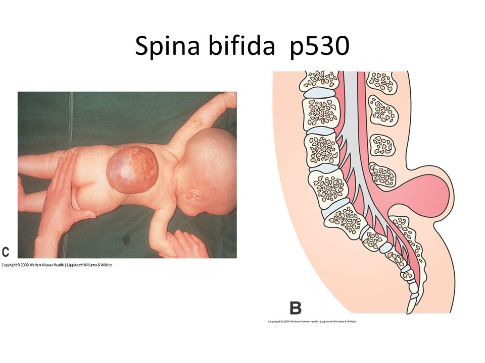
Spina bifida is Latin for "split spine".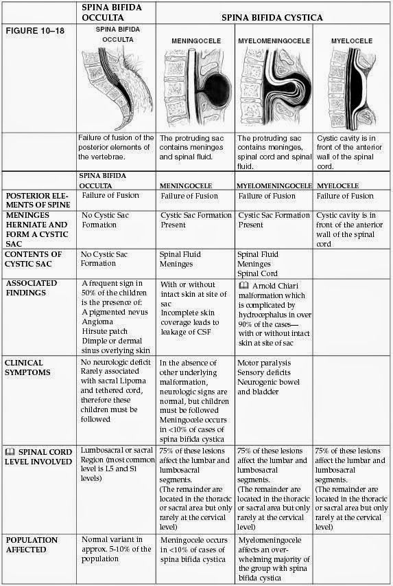 This is a developmental defect in which the vertebrae do not heal, and as a result, incomplete closure of the spinal canal occurs. Because of this, a hernial sac with cerebrospinal fluid - cerebrospinal fluid - can form on the back. Spina bifida leads to orthopedic, urological and neurosurgical problems.
Some patients with spina bifida move in a wheelchair, some have problems with fecal and urinary incontinence, others have hydrocephalus - accumulation of CSF inside the brain. At the same time, if all medical examinations are carried out on time, the intelligence of such patients remains intact. nine0027 The disease does not affect a person's life expectancy in any way, especially if you do not skip examinations and prevent secondary disorders.
/tsr/
What are technical means of rehabilitation
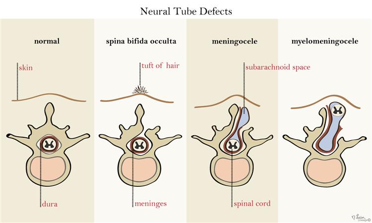
There are three most common forms of spina bifida: occult, meningocele and myelomeningocele. Occult, or hidden spina bifida, is a mild form of the disease without a spinal hernia: the spinal cord and surrounding tissues remain inside, but the bones in the lower back cannot form normally. At the site of splitting, there may be a hairy area, a dimple, or a birthmark. You can live with this form of the disease and not even know about it. nine0003
In case of meningocele, a sac filled with CSF appears on the outer surface of the back, but there is no spinal cord and nerve tissue in it. In myelomeningocele, the spinal cord and nerves develop outside the body - in a sac on the back. My son has just such a form of the disease.
These are the three most common forms of spina bifida. It can be seen how, depending on the degree of spina bifida, the spinal cord and nerves develop inside or outside the body - on the outer part of the back. This is how the three most common forms of spina bifida look like. It can be seen how, depending on the degree of spina bifida, the spinal cord and nerves develop inside or outside the body - on the outer part of the back Spina bifida is a congenital pathology: a spinal defect is formed in a child in the womb, as a rule, when the fetus is not yet 40 days old.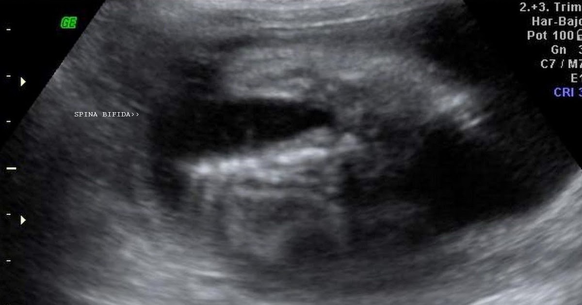
According to Russian clinical guidelines, the disease occurs with a frequency of 1-3 people per 10,000 pregnancies, but mild cases are often not recognized. According to unofficial data, the disease occurs in one of 1500-2000 newborns. There are several reasons for the defect, but the main one is the lack of folic acid in the body of a woman. nine0028
Federal Clinical Guidelines. Diagnosis and treatment of children with myelodysplasiaPDF, 927 KB
See a doctor
Our articles are written with love for evidence-based medicine. We refer to authoritative sources and go to doctors with a good reputation for comments. But remember: the responsibility for your health lies with you and your doctor. We don't write prescriptions, we make recommendations. Relying on our point of view or not is up to you.
How I found out about my son's illness
When I became pregnant with Matvey, I was 27 years old. I already had a healthy daughter, I was married and lived in Moscow.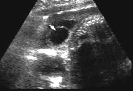 I was examined in a private clinic, and at the 32nd week of pregnancy I was told that my child had spina bifida.
The doctor could not explain anything, except that it was a severe form of disability, and did not give any predictions. There was no information in Russian on the Internet, and in Russia there was not a single foundation where one could get support and answers to questions about spina bifida. I applied to various Russian NGOs, but everywhere I got the same answer: sorry, our organization helps children with Down syndrome, or children with heart disease, or children with autism. No one helped children with spina bifida. nine0003
I felt as if my life was collapsing and everything was falling into the abyss: it is impossible to prepare a pregnant woman who already has a healthy child to have a son with a severe disability. It always happens very unexpectedly, and you do not understand how to continue to live.
Then one of my friends recommended one of the most famous and best maternity hospitals in Moscow to me. The birth was scheduled for June 14, 2012, and on June 12, an anonymous call was received from the maternity hospital with a request not to come. nine0003
I was told: “You have a difficult pregnancy, there is a possibility of a fatal outcome in a child during childbirth. We don't want to spoil the statistics."
So, just a couple of days before the birth, I had to independently look for a maternity hospital through my acquaintances and friends, where, as a result, I had a caesarean section.
When Matvey was born, we moved to the neurosurgery center: our son had to be operated on on the first day after birth in order to close the spinal hernia and avoid infection of the spinal cord - this could lead to meningitis and ultimately to death. nine0003
But Matvey could not be operated on for three weeks. Everyone was waiting: either when the head physician returns from vacation, or until the child gains weight, or something else. At some point, I realized that the doctors were just playing for time so as not to do the operation anymore: they thought that there would be no sense in it. Then, through friends, I found a doctor in Israel, and my family and I went to Tel Aviv to operate on my son there. When his condition became stable, we returned to Russia. nine0003
Time passed, Matvey grew up, but it remained unclear how to live with such a diagnosis: in Russia, there was still no help for children with spina bifida. Then I decided that it was necessary to combine the efforts of doctors, public figures and the parent community and create a system from scratch. So, four years after the birth of my son, I founded the Spina Bifida Foundation.
/readers-and-charity/
6 reasons to start doing charity work right now
Matvey in the first months after birth. At that time, practically nothing was known about spina bifida in Russia. Therefore, I did not understand how to live with such a diagnosis and whether it was even possibleWhat options do parents of children with spina bifida have
Parents who are expecting a child with spina bifida now have four options.
Terminate pregnancy. If a woman finds out about the child's illness before the 12th week of pregnancy, she can have an abortion if she wishes. But most often the defect can be seen on a simple ultrasound after this period. Then you can terminate the pregnancy for medical reasons by the decision of a council of doctors.
Some doctors still scare parents with back bifida, and under pressure they decide to have an abortion. The psychologists of our partner fund "Light in Hands" support such parents and help them recover from the loss. nine0003
Perform intrauterine surgery. At 24-26 weeks of pregnancy, intrauterine surgery can be performed to minimize the chance of orthopedic, urological and neurosurgical complications in the baby. In Russia, since 2016, such an operation has been carried out free of charge. Studies show that it has a better effect on the health of the child than the operation after birth, the one that we did to Matvey.
Thanks to intrauterine surgery, excess fluid is removed from the brain, the formed cavities in the spinal cord dissolve and the pressure between the cranial cavity and the spinal column normalizes. There are many children who have no disability at all after the operation, they live a normal life: they do not have a hernia, problems with the genitourinary system, they walk with their own feet. nine0003
/pregnancy/
How much does it cost to carry a baby? Another intrauterine operation for children with spina bifida is performed at the Lapino private hospital in Krasnogorsk.
We are trying to disseminate information about intrauterine operations, because not all doctors in the regions still know about them. Our foundation helps families with travel, accommodation and related expenses. As of October 2021, more than 50 families in Russia decided to have such an operation. nine0003 Dasha became one of 50 children who were operated on in utero.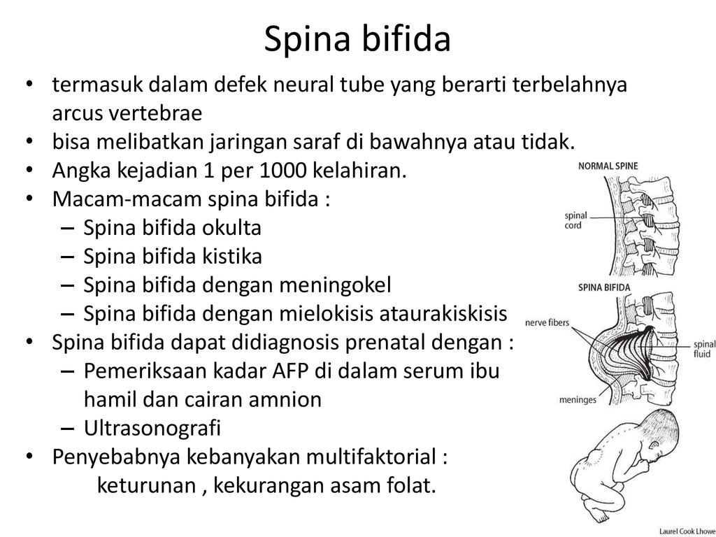 So far, she is too small to say how the operation has affected her life, but the girl's condition is now stable
Perform the operation on the first day after the birth of the child. Some women have contraindications for intrauterine surgery: for example, twin pregnancy, intrauterine infection, diabetes mellitus or tachycardia. In addition, in Russia there is a problem with the diagnosis of spina bifida: a defect is noticed when an intrauterine operation can no longer be performed. So it was with me. nine0003
Previously, more than half of spina bifida cases were detected after the 30th week of pregnancy at the third screening. But from the beginning of 2021, by order of the Ministry of Health, it was canceled. Then the parents could at least somehow prepare: find a maternity hospital with intensive care, find out everything about the diagnosis, contact the foundation, come to Moscow for childbirth. Now they don't have that opportunity either.
/platnie-rodi/
How much does it cost to give birth in Moscow? In Moscow, the leading expert of our foundation, neurosurgeon Dmitry Zinenko, performs more than a hundred such operations a year. nine0003
In general, surgery after birth can be done in any city, because it is conditionally called classical, but not all doctors have the necessary experience. Therefore, our foundation highly recommends mothers to contact us and come to Moscow for childbirth and postpartum surgery.
Anya was operated on after she was born. Now her condition is stableAbandon the child. Many parents are not ready for the birth of a child with spina bifida, including because of the frightening stories of doctors. Therefore, some are forced to abandon their children. nine0003
Since the foundation was established, the number of children with spina bifida in orphanages has decreased. It makes me very happy. Now there are more than 130 children in our orphanage program. Of these, only four children were born in 2017-2021. I attribute this to increased awareness of the diagnosis.
What problems do families face after the birth of a child with spina bifida
Life with a child with spina bifida is a quest.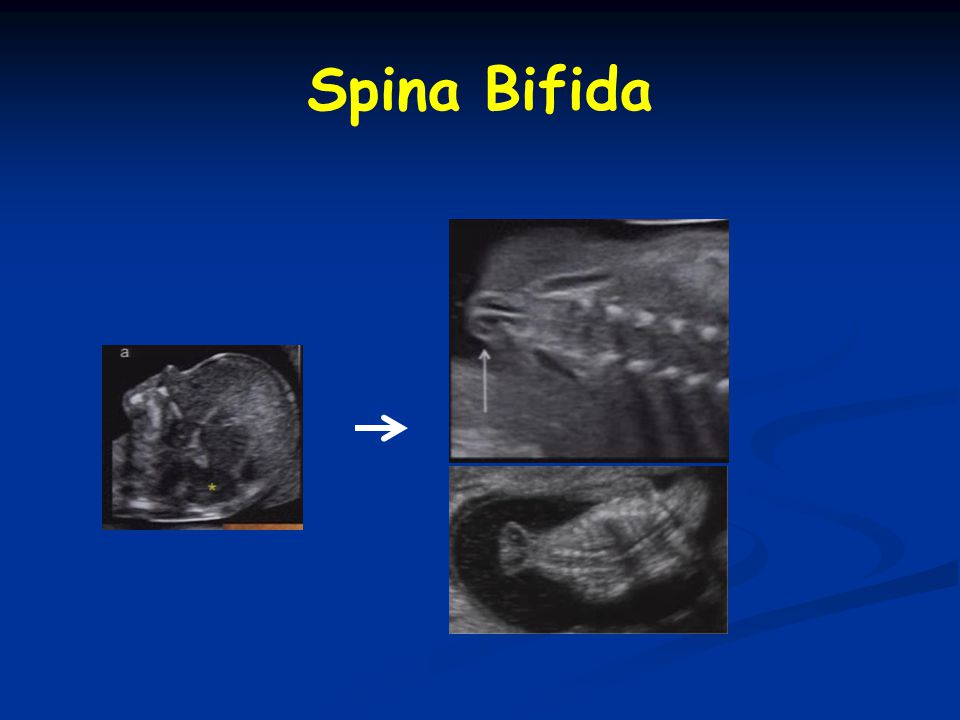 It's like a game where mice jump out of the circles and you need to hit them with a hammer so that they hide. nine0003
A child with spina bifida constantly has something unknown: problems with the kidneys, bedsores, osteoporosis or lack of calcium. This is a constant "game", in which you need to constantly monitor the child's condition, prevent complications and be in touch with all doctors.
Hydrocephalus. Spina bifida is often accompanied by hydrocephalus, since the paths through which the liquor can get from the skull into the spinal canal are pinched. Due to an excess of cerebrospinal fluid, squeezing of the brain occurs - this disrupts its work. Violation can be noticeable visually - a person will have a big head. nine0003
To eliminate the disease, surgeons install a special tube - a shunt - from the spina bifida to the patient. It drains excess fluid into the abdominal cavity, and thanks to this, hydrocephalus is compensated - the child does not have problems with the brain. It can live for many years if you monitor the operation of the shunt: change and reinstall it in time.
Unfortunately, shunts are often not of good quality, it happens that they are installed by inexperienced doctors. In 2021, two wards of the foundation died, whose bypass systems became unusable. nine0003
What to do? 17.02.20
How to insure against medical errors before surgery?
Diseases of the kidneys. In addition to hydrocephalus, children develop urinary and fecal incontinence in almost 99% of cases. Therefore, they are recommended regular catheterization. Children are given a urethral catheter - a special tube that removes stagnant urine from the bladder. The lack of timely examinations causes a high risk of kidney disease, so they must be constantly monitored. The kidneys are a life-supporting organ that must work. nine0003
Many parents often think about putting their child from dorsal bifida to his feet. They throw all their strength into it, but forget about the brain and spinal cord and about the genitourinary system. The legs are not a life-threatening or life-supporting system: a child in a wheelchair with broken legs can live to be 100 years old if their kidneys, brain, and spinal cord are functioning.
In 2021, we began to cooperate with clinics that conduct neuro-urological examinations in Yekaterinburg, Kazan and St. Petersburg. This includes a comprehensive urodynamic study, ultrasound of the bladder, cystoscopy, cystography, consultations of a neurourologist. Now we can examine 100 children a year in these cities and prevent the risk of kidney inflammation, dialysis, and death from kidney failure. nine0003
Regular inspections. All children with spina bifida should undergo regular magnetic resonance imaging, MRI, and electroneuromyography, ENMG to avoid recurrent complications. An MRI can reveal how correctly the shunt works, the state of the brain and spinal cord, and whether there is fixation - tension - of the spinal cord. If there is fixation, a second operation is necessary to free the spinal cord. ENMG is needed to understand how the dynamics change when impulses are conducted through the nerves of the legs and pelvis. We send all wards for regular check-ups to Dr. Dmitry Zinenko. nine0003
Also, patients with spina bifida should undergo annual examinations with a physical therapist, occupational therapist, neurourologist, neurosurgeon and orthopedist.
/list/pediatr-deti/
11 important questions to pediatrician Sergei Butriy
In Russia, there is still no single center for people with spina bifida. That is, a family cannot just go to one place and go through all the examinations at once. I have to fly to Kurgan to treat the spine, to St. Petersburg - legs, to Moscow - for hydrocephalus surgery, and so on. nine0003
For example, I had to spend a year with Matvey in Kurgan to install a metal structure in his spine: his son has a severely curved spine, and the metal structure corrects and aligns his back.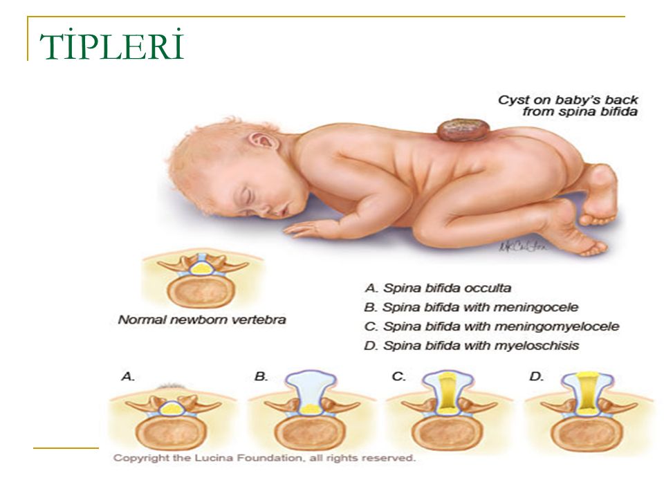 Unfortunately, the design did not take root in Matvey: osteomyelitis began, and it had to be removed. The curvature of the spine returned again.
Our foundation has a dream to initiate the creation of a center where all doctors will be gathered in one place. This will make life much easier for both parents and children with spina bifida. nine0003
How our foundation helps families
The Spina Bifida Charitable Foundation has been operating for the sixth year, and now we have two main programs. In the first one, we help 1000 wards from spina bifida. Among them are pregnant women, children left without parental care, and children in families. We help 100 children with spina bifida who were taken from orphanages. We provide legal and targeted assistance to pregnant women, help with medical examinations.
In addition, we are doing a lot of outreach work, in which we share our experience. On September 1, we launched the Spina Bifida Institute online project. The first nine courses are devoted to the basics of working with children with spina bifida for early intervention specialists, occupational therapists, physical therapists, rehabilitation therapists, sports coaches, psychologists, as well as for parents of children with such a diagnosis. Training will start in 2022. nine0003
Our specialists work all over the country. If we find a child with spina bifida in some city or village, then the specialist comes to this family right at home, looks at the child and the conditions in which he lives. After that, the specialist draws up an individual plan to help the child and family for the next year.
For example, we provide legal or psychological assistance to a mother or child, help organize an accessible environment for the patient. We agree to take the child to school and install a ramp in it. Then there will be no need for external rehabilitation: the child will be able to live a quality life in his home, in his family, go to school, go to the pool and ride in a wheelchair. nine0003
/rehabcenter/
How to choose a rehab center
How to reduce the risk of spina bifida in a child
The US Centers for Disease Control and Prevention - CDC - conducted a study that showed that folic acid intake before pregnancy reduces the risk of neural tube defects . It is they that lead to spina bifida and other diseases. Folic acid is prescribed not only for the prevention of spina bifida, but also for heart disease and even autism.
It often happens in Russia like this: a mother finds out about spina bifida in a child during pregnancy, comes to the doctor, and he prescribes folic acid to her. But a neural tube defect or spina bifida is formed in the first weeks of a child's life. Therefore, it is important to take folic acid at a dosage of 400 mg per day just before pregnancy, preferably from the age of 18.
Recommendations for the use of folic acid to reduce the incidence of spina bifida and other neural tube defects - CDC
If a family member is known to have spina bifida, the woman should take 10 times more folic acid. For example, my daughter needs it, because I gave birth to a son with such a disease. nine0003
To date, there are no other proven ways to prevent spina bifida.
In some countries, the program for the prevention of this disease has been brought to the state level.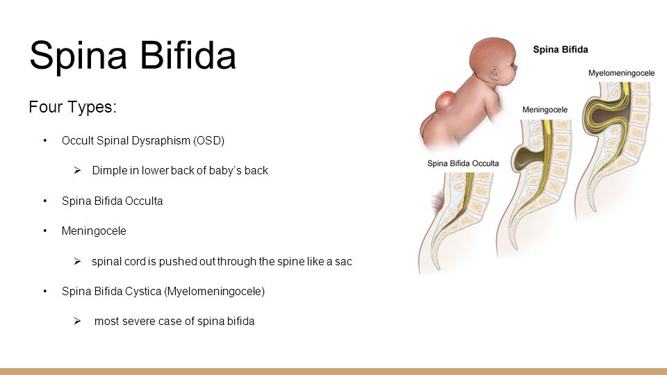 For example, in the UK and many other countries, all bread is fortified with folic acid. In Africa, corn is enriched with it as a popular food product. We hope that at some point we will be able to lobby for this program in Russia as well. We would like bread to be fortified with folic acid at the very least. nine0003
/pay-for-childbirth/
“I duplicated any tests if there were doubts”: how much does it cost to bear a child
In other countries, there have long been no such severe forms of spina bifida as in Russia. When foreign rehabilitation specialists came to our conference and saw my child, they said that they had not met children with this form for decades. I think this is due to the fact that early diagnosis of the disease is more developed there, intrauterine operations were started earlier, and there is also prevention of spina bifida. nine0003
How my son lives now
Now Matvey is nine years old, he is in the third grade.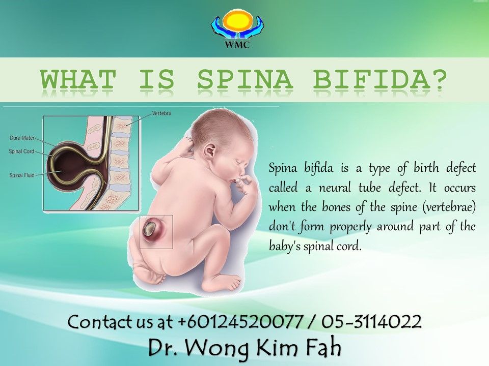 He does not feel his legs and cannot stand on them, so he moves in a wheelchair. Matvey and I often travel, he loves to swim and dreams of becoming a swimming coach for children with spina bifida.
Like me, Matvey has a motto: delegate or die.
He has learned to ask others for help, so he can live completely on his own. That is, he knows how to organize the environment around him so that someone helps him if necessary. That is why I am calm for him and I am sure that at the conditional 18 years old, Matvey will be able to leave home and live on his own. The world around always responds. nine0003 My son loves the sea and wants to connect his future profession with it
During the pandemic, our foundation went online and started a small community of people from spina bifida. This is how we met Paralympic champions, athletes, world champions in dance, tennis, and people who skydive in wheelchairs.
Among them was Nursina Galiyeva in a wheelchair from Kazan, who at the age of 17 just got on a train alone, went to Moscow, entered an institute and began to live in a hostel. Now she is a world champion in wheelchair dancing and works for the Downside Up Charitable Foundation. nine0003
/list/make-life-brighter/
Find a friend and go on stage: 5 projects for people with disabilities
We also met Riyana, who at the age of 12 became the champion of Tatarstan in tennisI realized that absolutely everything is possible . The task of the parents of children with spina bifida is to teach the child independence so that he can cook his own food and clean up after himself. This will probably be the main contribution to his life. After all, when the parent is gone, the child should be able to live on his own. Therefore, my son spends a lot of time without me: at school, in society, with friends, traveling. nine0003
It's great that children with spina bifida are intellectually and emotionally intact and develop absolutely normally. This means that they have great potential and can do a lot for society.
They feel the world a little differently because they are in a slightly vulnerable position themselves. Therefore, they can bring a lot of good and make the world a better place, so that society becomes more tolerant, kind and open.
How to help children with spina bifida and their parents
Since 2016, the Spina Bifida Charitable Foundation has been helping people with this diagnosis so that they can live a full life. You can support the work of the foundation and make a regular donation in its favor:
- on the website of the Spina Bifida Foundation;
- in the Dobro Mail.ru service;
- on the Need Help platform;
- through the Tinkoff mobile app and the Tinkoff Cashback for Good service.
Herniated disc - protrusion of the nucleus pulposus of the disc.
A herniated disc (intervertebral hernia) is a protrusion of the nucleus pulposus of the disc consisting of gel-like cartilage tissue into the spinal canal - a cavity in the spinal column formed by arches of 5 large vertebrae.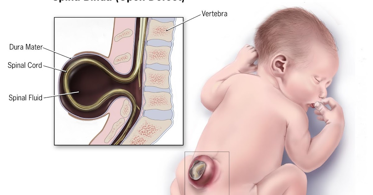
The development of pathology is accompanied by 4 stages:
- Prolapse. At this stage, changes occur in the structure of the intervertebral disc, but there are no violations of the integrity of the fibrous ring. Moisture dries up, metabolic processes are disturbed, cartilaginous tissues become thinner. The rate of changes depends on metabolism, chronic diseases - it significantly accelerates the prolapse of heavy loads on the spine (hernias are frequent companions of loaders), metabolic disorders, osteochondrosis, ankylosing spondylitis, rheumatoid arthritis, lack of vitamins, minerals. If timely treatment is started, the changes are reversible. If the treatment is ignored and delayed, cracks and micro-ruptures will begin to appear on the fibrous ring of the spine. nine0238
- Protrusion. There is a bulging of the intervertebral disc into the spinal canal (beyond the physiological boundaries), but the rupture of the fibrous ring is not observed.
 Herniation can be pronounced (the most common variant with a hernia of the lumbar spine), or visually subtle (a common variant with a cervical hernia).
 - Prolapse. The fibrous ring of the disc is strongly stretched. A person experiences severe pain, and not only direct - in the vertebrae, but also reflected, for example, in the legs. nine0238
- Direct hernia. The disc nucleus slips out and the annulus fibrosus is damaged.
The "borders" between advanced protrusion, prolapse and hernia itself can be blurred. Indeed, with a strong protrusion, bulging of the disc, there is a high risk of rupture of the fibrous membrane and displacement of the pulpous nucleus outward.
It is important to distinguish Schmorl's hernias from typical herniated discs. The nature of this pathology differs significantly. Punching occurs in the spongy bone, and not in the spinal canal. And the treatment is quite different. nine0003
Causes and frequency of intervertebral hernia
Causes that most often lead to hernia:
- Poorly developed back muscles.
- Recent injuries. Bruises are especially dangerous.
- Static overload associated with work in one position (work on the conveyor, machine tool).
- Dynamic overload associated with heavy lifting or vibration. At risk are loaders, riggers, engravers, grinders, riveters, slingers, pressers, masons, punchers. nine0238
- Anatomical anomalies in the area of the endplates on the vertebrae.
- Incorrect carrying of bags.
- Super-intense sports activities (especially those that line up with a violation of the correct technique).
- Osteochondrosis, in which micro-ruptures appear on the fibrous tissue, and over time, the damaged area is completely replaced by inelastic scar tissue.
- Scoliosis. The spinal column with this pathology is severely deformed. Scoliosis promotes herniation in the thoracic spine. nine0238
- Overweight. The spinal column begins to be constantly subjected to overloads and becomes vulnerable.
- Unbalanced or weight loss diet.
 Rapid weight loss is especially dangerous. This is a serious blow to cartilage and muscle tissue. As a result, degenerative-dystrophic processes begin in the intervertebral discs.
 - Some congenital anatomical features of the structure of the endplates on the vertebrae. Especially often this is the cause of a hernia of the lumbar and a hernia between the 11th and 12th thoracic vertebrae. nine0238
Vulnerability of the intervertebral discs is due to the fact that nutrients are supplied in a diffuse way, and not directly from the bloodstream.
That is, first - into the circulatory system, then to the end plates and from there to the discs.
If insufficient nutrients reach the discs, the nucleus pulposus loses its elasticity, dystrophic and degenerative processes develop.
The risk of herniation is higher in patients aged 45 to 55 years. At the same time, women experience pathology more often than men. nine0003
There is also a pattern that more often herniation affects the mobile parts of the spine. The lumbosacral and cervical regions are more commonly affected.
Symptoms
Symptoms differ from the area of herniation, the nature of the course of the disease, but among the most common symptoms, the following can be distinguished:
- knee numbness of the leg (sciatica). nine0238
- Sharp pain in the lower back (especially when lifting loads or vibration).
- Impaired bowel control.
- Numbness in the perineum.
- Violation of the vestibular apparatus and gait.
- Headaches. As a rule, with the defeat of the cervical, thoracic.
- Pain in the chest, which is aggravated by moving the arms, inhaling.
- "Shooting" in the shoulder (in case of pathology in the upper spine).
Herniated discs in the thoracic region often cause false sensations. It seems that there is a problem with the lungs or the heart. "It hurts" in the chest, there is pain in the shoulder blades, there is a feeling of lack of oxygen, shortness of breath occurs.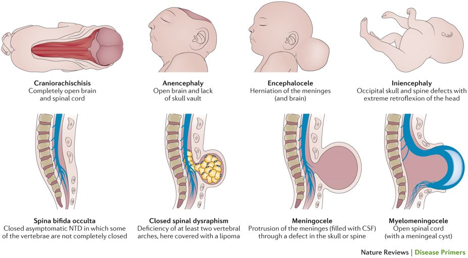 nine0003
In case of problems with the intervertebral discs of the cervical region, the first symptom is often not a problem with the spine itself, but constantly high blood pressure.
Types of hernias
Hernias are classified according to three basic parameters:
- Location of the problem area.
- Syndromes.
- Degrees of pathology development (stages).
Localization
Location of hernias can be as follows:
- To the side of the spinal canal or on one side of the vertebrae, towards the outside of the spine. Posterolateral and lateral prolapse.
- Towards the center of the vertebral bodies. Median protrusion towards the spinal canal.
- Outward facing. Protrusion - in the direction of the anterior ligament of the spine and spinous processes (anterolateral prolapse).
The location can be cervical, thoracic, sacral, lumbar. The most common variants of herniation in the lumbar region are between the 4th and 5th vertebrae and the 5th lumbar vertebra and the sacrum (it is customary to count the vertebrae from top to bottom.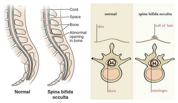
Depending on the syndrome where the pathology is reflected - on the nerves, blood vessels, the spine itself, radicular, reflex and vertebral hernia are isolated. What is the peculiarity of each of them?
- Spine. The hernia affects the nerve fibers. Part of the reflexes disappears or weakens, sensitivity is disturbed. As a result, muscle weakness appears, goosebumps begin to run along the spine, tingling, pain appear.
- Vertebrate. In the paravertebral muscles, the tone changes, muscle tension occurs. Very often the torso warps, it becomes difficult for a person to get up and sit down. nine0238
- Reflex. It usually starts with pain in the lower back or leg. Often failures can affect the intestines, pelvic organs. As a result, there are problems with stool, urination. Many have impaired coordination and impaired gait.
Unlike the more well-known horizontal hernias of the intervertebral discs in the spinal canal or nerve foramen, Schmorl's hernia is associated with pushing (falling through) of the cartilaginous tissue of the end plates into the spongy bone, inside the body of the upper or lower vertebra.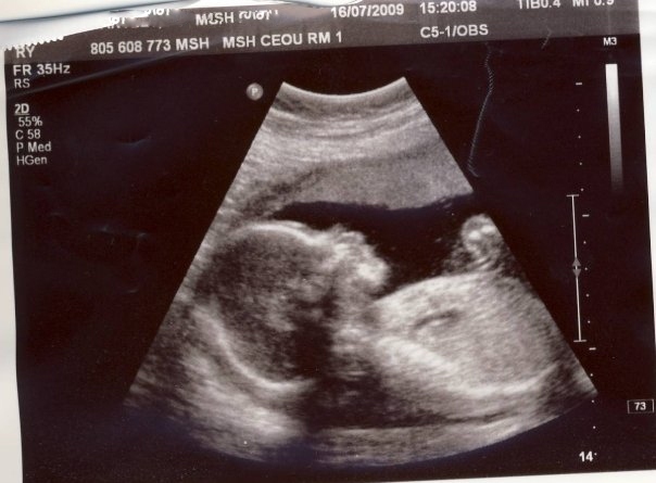 nine0003
Stages
- Stage 1. The discs do not extend beyond the vertebrae, the displacement does not exceed 2 mm.
- Stage 2. The nucleus is within the vertebra, but the protrusion of the hernia can reach up to 1.5 centimeters, the nucleus remains within the vertebral body.
- Stage 3. The nucleus is outside the vertebral body.
- Stage 4. The nucleus pulposus hangs completely outwards.
Also, a complicated hernia - stenosis - is isolated separately. In this case, we are talking not just about the hanging of the nucleus pulposus, but also about multifactorial compression - narrowing of the spinal canal. nine0003
Diagnosis of a hernia
The following measures are effective in diagnosing:
- Visual examination with an assessment of both the condition of the spine and the overall picture as a whole. The doctor pays special attention to the localization, the nature of the pain, the characteristics of the muscles (weakening, increased tone), the sensitivity of the spine and lower extremities, the reflex reactions of the tendons, body weight, concomitant diseases.
 Already at this stage, the doctor makes a decision - laboratory, functional diagnostics will be aimed specifically at identifying intervertebral hernia, or it is important to exclude another main root of the problem or pathology that accompanies herniated intervertebral discs (myositis, osteochondrosis, spondylarthrosis). nine0238
 - CT or MRI. These diagnostic tools allow you to visualize the protrusion, evaluate the width of the spinal canal, and examine the soft surrounding structures. MRI for examination of the intervertebral discs is the preferred method over CT. The latter diagnostic option is appropriate only if the pathology is the result of an injury, and bone structures also need to be examined.
- Contrast myelography. The technique is based on the introduction of a needle into the subarachnoid space surrounding the spinal cord with a contrast agent and image acquisition by means of X-ray (a cheaper, but less informative method) or MRI. nine0238
Conventional MRI is relevant for any herniation. Myelography with a contrast agent - with a radicular form (with suspicion of damage to the nerve roots of the spine). Also, this diagnostic option is relevant when there is a suspicion of a violation in the work of the vessels of the spinal column, it is necessary to determine the pressure on the spinal cord and nerve endings as accurately as possible.
In some cases, ultrasound with vascular Doppler may be additionally prescribed. This practice is relevant, for example, if you suspect a protrusion in the cervical, lumbar, sacral zone. Such a diagnosis is especially valuable if other research methods signal fiberization, the presence of cracks, and vascular problems. nine0003
If there is any doubt that this is a degenerative lesion of the spine or a pathology of a rheumatoid nature, blood tests are mandatory. Important indicators of ESR and C-reactive protein.
Methods of treatment of intervertebral hernia
Several types of treatment are used for hernia formation:
- Medication.
 In the form of injections, tablets and ointments.
 - Orthopedic. Wearing a corset.
- Surgical.
Medical treatment
If the intervertebral discs are slightly affected, there is no rupture of the fibrous tissue, complex medical treatment can help. What drugs are used?
- Painkillers. Non-steroidal drugs are used to relieve pain. With the 1st stage of a hernia, relief is possible already in the first days of treatment. The best result is achieved if you start with intensive therapy and injections, and then move on to tablets and ointments. If you start with ointments and tablets, treatment may require more time. In more advanced stages, or if the person is unresponsive to non-steroidal drugs, stronger corticosteroid drugs are used. They can be in the form of injections, tablets, gels, ointments. But doctors recommend using cortico-containing substances only in extreme cases. Most of them have a fairly strong side effect. In addition, addiction may occur.
 nine0238
- Muscle relaxants. Relieve spasms, clamps. Tone muscle fibers. Improve the level of mobility of the spine. Effective when combined with injections and tablets.
- Chondroprotectors. They are effective for patients with severe cartilage degeneration. They nourish the intervertebral discs with chondroetin.
- Increased doses of vitamin B. Important for increasing nerve conduction. The best effect is achieved by injection.
Wearing orthoses
Orthoses help to successfully solve the following tasks:
- Fix motor segments in the correct physiological position.
- Provide reflex pendulum movements in static.
- Distribute the load on the spine.
- Reduce pain.
- Relieve stress on the vertebrae.
Nuance! But the proper effect is possible only in the initial stages of the disease. If the disease is advanced, wearing an orthosis must necessarily be combined with medical and physiotherapeutic treatment, or even more serious surgical treatment is needed. nine0003
In addition, the orthosis may only be worn under regular medical supervision. Otherwise, instead of solving the problem, you can get a new one - atrophy.
Physiotherapy
Popular physiotherapy methods include massage, laser therapy, and acupuncture.
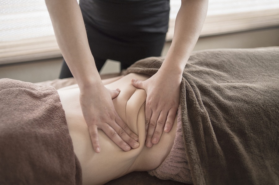
- Massage . It is used if the patient has stable blood pressure, there are no inflammatory processes. The most effective and safest massage is prescribed for hernias in the lumbosacral spine. nine0238
- Laser therapy . It is most effective after surgical treatment in persons with the presence of compression-vascular syndrome. Do not confuse classical laser therapy with vaporization of the patient - removal of a hernia. Vaporization is no longer physical therapy, but a surgical procedure using laser technology. The goal of laser therapy is to stop the pain, reduce the size of the hernia, and the goal of laser surgery is to excise it.
- Acupuncture.
 An effective method for small hernias accompanied by mobility restrictions, constipation, urination disorders, potency problems. Well removes tension from spasmodic muscles, eliminates pinched nerve fibers. Despite the fact that acupuncture seems to be the safest way to solve the problem, unfortunately, the method is contraindicated if there are serious diseases of the kidneys and heart. Also, before starting the procedures, it is important to be sure that everything is in order with blood clotting. nine0238
Surgical treatment
Surgical treatment is prescribed if the hernia is advanced, the annulus fibrosus is torn, drug therapy and corset treatment did not give any effect.
The urgency of the operation is especially important if there is paresis and muscle tissue atrophy progresses. An indication for surgery is also a pinched hernia.
The uniting moment for all operations is that they are focused on the direct removal of the hernia. But other tasks during surgery may differ. For some patients, it is enough to stabilize the intervertebral discs, for others to replace them (perform arthroplasty). nine0003
Hernia removal can be done in several ways:
- Method 1. Discectomy . Open operation with the removal of the intervertebral discs themselves, the disc or part of it. It is performed under general anesthesia. Previously practiced for excision of most hernias, now mainly in situations where the size of the intervertebral hernia does not exceed 0.6 mm or surgery is urgently required.
- Method 2. Decompression laminectomy. Effective for problems in the cervical, thoracic and lumbar regions. It is the removal of the vertebral arch (or arches) to reduce the pressure of bone tissue on the spinal cord. It is prescribed if the affected area is large and the problem cannot be solved by endoscopic surgery, and at the same time, according to the diagnostic results, contraindications for discectomy have been identified.
 It is also an effective method of surgical intervention, if, along with excision of a hernia, it is necessary to simultaneously stabilize the structure of the spine. nine0238
 - Method 4. Minimally invasive endoscopic removal of a prolapsed fragment of the nucleus of the intervertebral disc and release from compression of the spinal root (decompression). Formation of access is carried out through a puncture in the skin. Stabilizing structures (muscles, ligaments) expand without being injured. Removal of a hernia is carried out through a probe. When the nerve fibers are completely free, stitching is performed. The operation can be performed under local anesthesia or sedation. nine0238
Herniated disc prevention
- To avoid the problem, it is important to follow preventive measures:
Avoid staying in a static position for a long time.
If diagnosed with scoliosis, osteochondrosis, other diseases of the spine, treat them in a timely manner.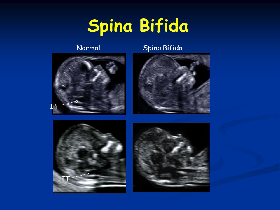
- Avoid sudden jerks when lifting heavy objects. When lifting weights, make sure that your legs are bent, not your back. To do this, when taking the load, be sure to squat slightly, press the object to yourself, and then get up. If you need to move a heavy object forward, give preference to pushing. This will significantly reduce the load on the spine. If you need to lift loads above head level, then it is better to use a stepladder, a chair. nine0238
- Stretch. It promotes increased blood flow, regeneration of cartilage tissue in the interdiscal space. Especially for intervertebral discs, regular stretching on an inclined board is beneficial.
- Swim regularly. This is especially good prevention of hernia formation in the thoracic region.
- Give preference to a child's rather than a soft mattress.
- Avoid carrying heavy bags on your shoulder. It is better to replace them with bags, suitcases on wheels. Last resort - on a backpack. nine0238
- Be smart about your diet.
