Pubic bone anatomy female
Anatomy, Function of Bones, Muscles, Ligaments
What is the female pelvis?
The pelvis is the lower part of the torso. It’s located between the abdomen and the legs. This area provides support for the intestines and also contains the bladder and reproductive organs.
There are some structural differences between the female and the male pelvis. Most of these differences involve providing enough space for a baby to develop and pass through the birth canal of the female pelvis. As a result, the female pelvis is generally broader and wider than the male pelvis.
Below, learn more about the bones, muscles, and organs of the female pelvis.
Female pelvis anatomy and function
Female pelvis bones
Hip bones
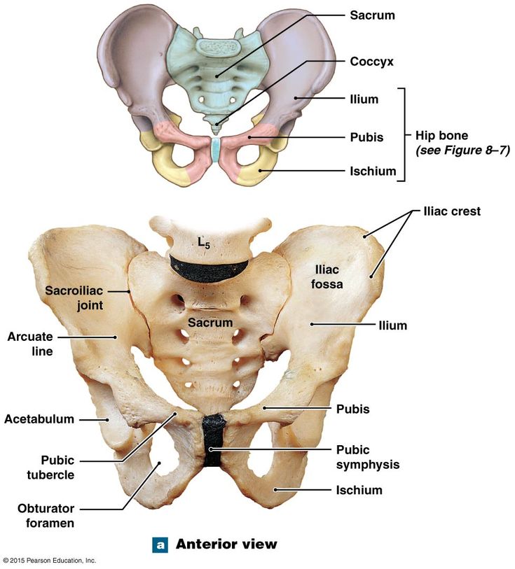
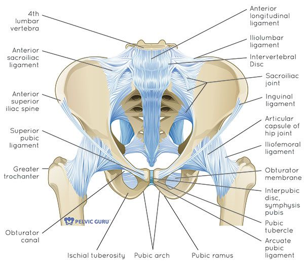
There are two hip bones, one on the left side of the body and the other on the right. Together, they form the part of the pelvis called the pelvic girdle.
The hip bones join to the upper part of the skeleton through attachment at the sacrum. Each hip bone is made of three smaller bones that fuse together during adolescence:
- Ilium. The largest part of the hip bone, the ilium, is broad and fan-shaped. You can feel the arches of these bones when you put your hands on your hips.
- Pubis. The pubis bone of each hip bone connects to the other at a joint called the pubis symphysis.
- Ischium. When you sit down, most of your body weight falls on these bones. This is why they’re sometimes called sit bones.
The ilium, pubis, and ischium of each hip bone come together to form the acetabulum, where the head of the thigh bone (femur) attaches.
Sacrum
The sacrum is connected to the lower part of the vertebrae. It’s actually made up of five vertebrae that have fused together. The sacrum is quite thick and helps to support body weight.
Coccyx
The coccyx is sometimes called the tailbone. It’s connected to the bottom of the sacrum supported by several ligaments.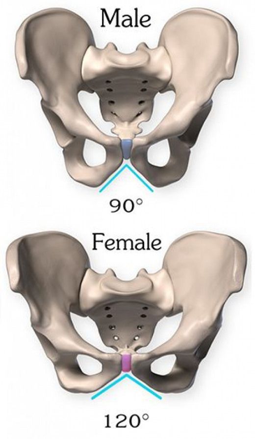
The coccyx is made up of four vertebrae that have fused into a triangle-like shape.
Female pelvis muscles
Levator ani muscles
The levator ani muscles are the largest group of muscles in the pelvis. They have several functions, including helping to support the pelvic organs.
The levator ani muscles consist of three separate muscles:
- Puborectalis. This muscle is responsible for holding in urine and feces. It relaxes when you urinate or have a bowel movement.
- Pubococcygeus. This muscle makes up most of the levator ani muscles. It originates at the pubis bone and connects to the coccyx.
- Iliococcygeus. The iliococcygeus has thinner fibers and serves to lift the pelvic floor as well as the anal canal.
Coccygeus
This small pelvic floor muscle originates at the ischium and connects to the sacrum and coccyx.
Female pelvis organs
Uterus
The uterus is a thick-walled, hollow organ where a baby develops during pregnancy.
During the reproductive years, the lining of the uterus sheds every month during menstruation if you don’t become pregnant.
Ovaries
There are two ovaries located on either side of the uterus. The ovaries produce eggs and also release hormones, such estrogen and progesterone.
Fallopian tubes
The fallopian tubes connect each ovary to the uterus. Specialized cells in the fallopian tubes use hair-like structures called cilia to help direct eggs from the ovaries toward the uterus.
Cervix
The cervix connects the uterus to the vagina. It’s able to widen, allowing sperm to pass into the uterus.
In addition, thick mucus produced in the cervix can help to prevent bacteria from reaching the uterus.
Vagina
The vagina connects the cervix to the exterior female genitalia. It’s also called the birth canal, as the baby passes through the vagina during delivery.
Rectum
The rectum is the lowest part of the large intestine.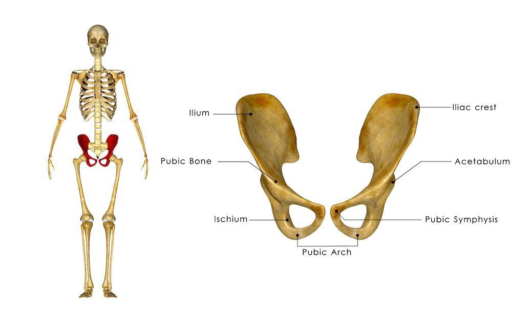 Feces collects here until exiting through the anus.
Bladder
The bladder is the organ that collects and stores urine until it’s released. Urine reaches the bladder through tubes called ureters that connect to the kidneys.
Urethra
The urethra is the tube that urine travels through to exit the body from the bladder. The female urethra is much shorter than the male urethra.
Female pelvis ligaments
Broad ligament
The broad ligament supports the uterus, fallopian tubes, and ovaries. It extends to both sides of the pelvic wall.
The broad ligament can be further divided into three components that are linked to different parts of the female reproductive organs:
- mesometrium, which supports the uterus
- mesovarium, which supports the ovaries
- mesosalpinx, which supports the fallopian tubes
Uterine ligaments
Uterine ligaments provide additional support for the uterus. Some of the main uterine ligaments include:
- the round ligament
- cardinal ligaments
- pubocervical ligaments
- uterosacral ligaments
Ovarian ligaments
The ovarian ligaments support the ovaries. There are two main ovarian ligaments:
- the ovarian ligament
- the suspensory ligament of the ovary
Female pelvis diagram
Explore this interactive 3-D diagram to learn more about the female pelvis:
Female pelvis conditions
The pelvis contains a large number of organs, bones, muscles, and ligaments, so many conditions can affect the entire pelvis or parts within it.
Some conditions that can affect the female pelvis as a whole include:
- Pelvic inflammatory disease (PID). PID is an infection that occurs in the female reproductive system. While it’s often caused by a sexually transmitted infection, other infections can also cause PID. Untreated PID can lead to complications, such as infertility or ectopic pregnancy.
- Pelvic organ prolapse. Pelvic organ prolapse occurs when the muscles in the pelvis can no longer support its organs, such as the bladder, uterus, or rectum. This can cause one or more of these organs to press down on the vagina. In some cases, this can cause a bulge to form outside of the vagina.
- Endometriosis. Endometriosis occurs when the tissue that lines the inside walls of the uterus (endometrium) begins to grow outside of the uterus. The ovaries, fallopian tubes, and other tissues in the pelvis are typically affected by the condition. Endometriosis can lead to complications, including infertility or ovarian cancer.
Symptoms of a pelvic condition
Some common symptoms of a pelvic condition can include:
- pain in the lower abdomen or pelvis
- a feeling of pressure or fullness in the pelvis
- unusual or foul-smelling vaginal discharge
- pain during sex
- bleeding in between periods
- painful cramping during or before periods
- pain during bowel movements or when urinating
- a burning feeling when urinating
Tips for a healthy pelvis
The female pelvis is a complex, important part of the body. Follow these tips to keep it in good health:
Stay on top of your reproductive health
See a gynecologist for a yearly health screen. Things like pelvic exams and Pap smears can aid in identifying pelvic conditions or infections early.
You can get a free or low-cost pelvic exam at your local Planned Parenthood clinic.
Practice safe sex
Use barriers — such as condoms or dental dams — during sexual activity, especially with a new partner, to avoid infections that could lead to PID.
Try pelvic floor exercises
These types of exercises can help to strengthen the muscles in the pelvis, including those around the bladder and vagina.
Stronger pelvic floor muscles can aid in preventing things like incontinence or organ prolapse. Here’s how to get started.
Never ignore unusual symptoms
If you’re experiencing anything unusual in your pelvic area, such as bleeding between periods or unexplained pelvic pain, make an appointment with your doctor. Left untreated, some pelvic conditions can have lasting impacts on your health and fertility.
Anatomy, Function of Bones, Muscles, Ligaments
What is the female pelvis?
The pelvis is the lower part of the torso. It’s located between the abdomen and the legs. This area provides support for the intestines and also contains the bladder and reproductive organs.
There are some structural differences between the female and the male pelvis. Most of these differences involve providing enough space for a baby to develop and pass through the birth canal of the female pelvis. As a result, the female pelvis is generally broader and wider than the male pelvis.
Below, learn more about the bones, muscles, and organs of the female pelvis.
Female pelvis anatomy and function
Female pelvis bones
Hip bones
There are two hip bones, one on the left side of the body and the other on the right. Together, they form the part of the pelvis called the pelvic girdle.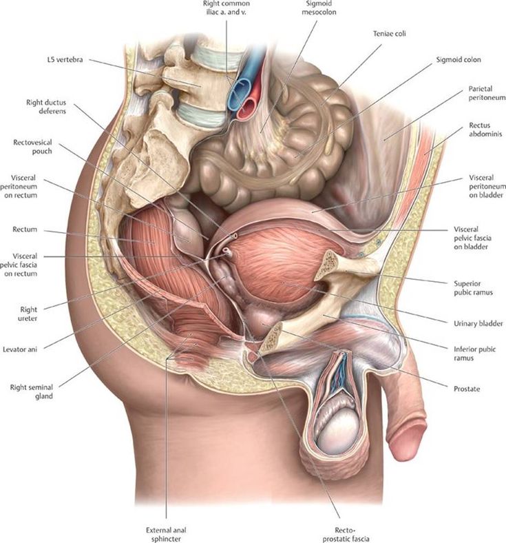
The hip bones join to the upper part of the skeleton through attachment at the sacrum. Each hip bone is made of three smaller bones that fuse together during adolescence:
- Ilium. The largest part of the hip bone, the ilium, is broad and fan-shaped. You can feel the arches of these bones when you put your hands on your hips.
- Pubis. The pubis bone of each hip bone connects to the other at a joint called the pubis symphysis.
- Ischium. When you sit down, most of your body weight falls on these bones. This is why they’re sometimes called sit bones.
The ilium, pubis, and ischium of each hip bone come together to form the acetabulum, where the head of the thigh bone (femur) attaches.
Sacrum
The sacrum is connected to the lower part of the vertebrae. It’s actually made up of five vertebrae that have fused together. The sacrum is quite thick and helps to support body weight.
Coccyx
The coccyx is sometimes called the tailbone. It’s connected to the bottom of the sacrum supported by several ligaments.
The coccyx is made up of four vertebrae that have fused into a triangle-like shape.
Female pelvis muscles
Levator ani muscles
The levator ani muscles are the largest group of muscles in the pelvis. They have several functions, including helping to support the pelvic organs.
The levator ani muscles consist of three separate muscles:
- Puborectalis. This muscle is responsible for holding in urine and feces. It relaxes when you urinate or have a bowel movement.
- Pubococcygeus. This muscle makes up most of the levator ani muscles. It originates at the pubis bone and connects to the coccyx.
- Iliococcygeus. The iliococcygeus has thinner fibers and serves to lift the pelvic floor as well as the anal canal.
Coccygeus
This small pelvic floor muscle originates at the ischium and connects to the sacrum and coccyx.
Female pelvis organs
Uterus
The uterus is a thick-walled, hollow organ where a baby develops during pregnancy.
During the reproductive years, the lining of the uterus sheds every month during menstruation if you don’t become pregnant.
Ovaries
There are two ovaries located on either side of the uterus. The ovaries produce eggs and also release hormones, such estrogen and progesterone.
Fallopian tubes
The fallopian tubes connect each ovary to the uterus. Specialized cells in the fallopian tubes use hair-like structures called cilia to help direct eggs from the ovaries toward the uterus.
Cervix
The cervix connects the uterus to the vagina. It’s able to widen, allowing sperm to pass into the uterus.
In addition, thick mucus produced in the cervix can help to prevent bacteria from reaching the uterus.
Vagina
The vagina connects the cervix to the exterior female genitalia. It’s also called the birth canal, as the baby passes through the vagina during delivery.
Rectum
The rectum is the lowest part of the large intestine. Feces collects here until exiting through the anus.
Bladder
The bladder is the organ that collects and stores urine until it’s released. Urine reaches the bladder through tubes called ureters that connect to the kidneys.
Urethra
The urethra is the tube that urine travels through to exit the body from the bladder. The female urethra is much shorter than the male urethra.
Female pelvis ligaments
Broad ligament
The broad ligament supports the uterus, fallopian tubes, and ovaries. It extends to both sides of the pelvic wall.
The broad ligament can be further divided into three components that are linked to different parts of the female reproductive organs:
- mesometrium, which supports the uterus
- mesovarium, which supports the ovaries
- mesosalpinx, which supports the fallopian tubes
Uterine ligaments
Uterine ligaments provide additional support for the uterus.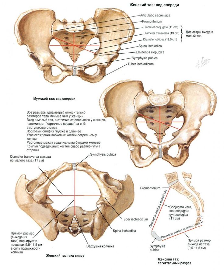 Some of the main uterine ligaments include:
- the round ligament
- cardinal ligaments
- pubocervical ligaments
- uterosacral ligaments
Ovarian ligaments
The ovarian ligaments support the ovaries. There are two main ovarian ligaments:
- the ovarian ligament
- the suspensory ligament of the ovary
Female pelvis diagram
Explore this interactive 3-D diagram to learn more about the female pelvis:
Female pelvis conditions
The pelvis contains a large number of organs, bones, muscles, and ligaments, so many conditions can affect the entire pelvis or parts within it.
Some conditions that can affect the female pelvis as a whole include:
- Pelvic inflammatory disease (PID). PID is an infection that occurs in the female reproductive system. While it’s often caused by a sexually transmitted infection, other infections can also cause PID. Untreated PID can lead to complications, such as infertility or ectopic pregnancy.
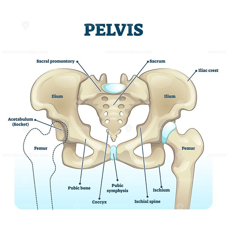
- Pelvic organ prolapse. Pelvic organ prolapse occurs when the muscles in the pelvis can no longer support its organs, such as the bladder, uterus, or rectum. This can cause one or more of these organs to press down on the vagina. In some cases, this can cause a bulge to form outside of the vagina.
- Endometriosis. Endometriosis occurs when the tissue that lines the inside walls of the uterus (endometrium) begins to grow outside of the uterus. The ovaries, fallopian tubes, and other tissues in the pelvis are typically affected by the condition. Endometriosis can lead to complications, including infertility or ovarian cancer.
Symptoms of a pelvic condition
Some common symptoms of a pelvic condition can include:
- pain in the lower abdomen or pelvis
- a feeling of pressure or fullness in the pelvis
- unusual or foul-smelling vaginal discharge
- pain during sex
- bleeding in between periods
- painful cramping during or before periods
- pain during bowel movements or when urinating
- a burning feeling when urinating
Tips for a healthy pelvis
The female pelvis is a complex, important part of the body. Follow these tips to keep it in good health:
Stay on top of your reproductive health
See a gynecologist for a yearly health screen. Things like pelvic exams and Pap smears can aid in identifying pelvic conditions or infections early.
You can get a free or low-cost pelvic exam at your local Planned Parenthood clinic.
Practice safe sex
Use barriers — such as condoms or dental dams — during sexual activity, especially with a new partner, to avoid infections that could lead to PID.
Try pelvic floor exercises
These types of exercises can help to strengthen the muscles in the pelvis, including those around the bladder and vagina.
Stronger pelvic floor muscles can aid in preventing things like incontinence or organ prolapse. Here’s how to get started.
Never ignore unusual symptoms
If you’re experiencing anything unusual in your pelvic area, such as bleeding between periods or unexplained pelvic pain, make an appointment with your doctor.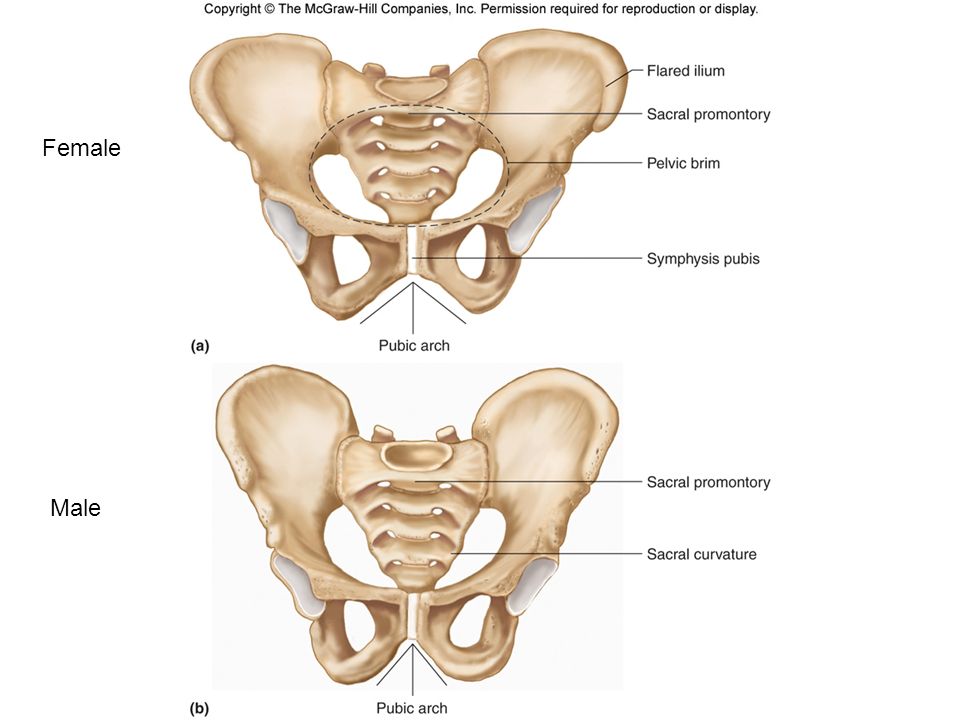 Left untreated, some pelvic conditions can have lasting impacts on your health and fertility.
Inferior ramus of pubis - e-Anatomy
SUBSCRIBE
SUBSCRIBE
Definition
English Portugues
No definition for this anatomical structure yet
Definition for:
English Portugues
I consent to the assignment of the rights associated with my participation in the project, in accordance with the Terms and Conditions of Use of the site.
I agree to the assignment of the rights associated with my participation in the project, in accordance with the Terms and conditions of use of the site.
Gallery
Comparative anatomy of animals
- Caudal ramus of pubis
Translations
IMAIOS and certain third parties use cookies or similar technologies, in particular for audience measurement.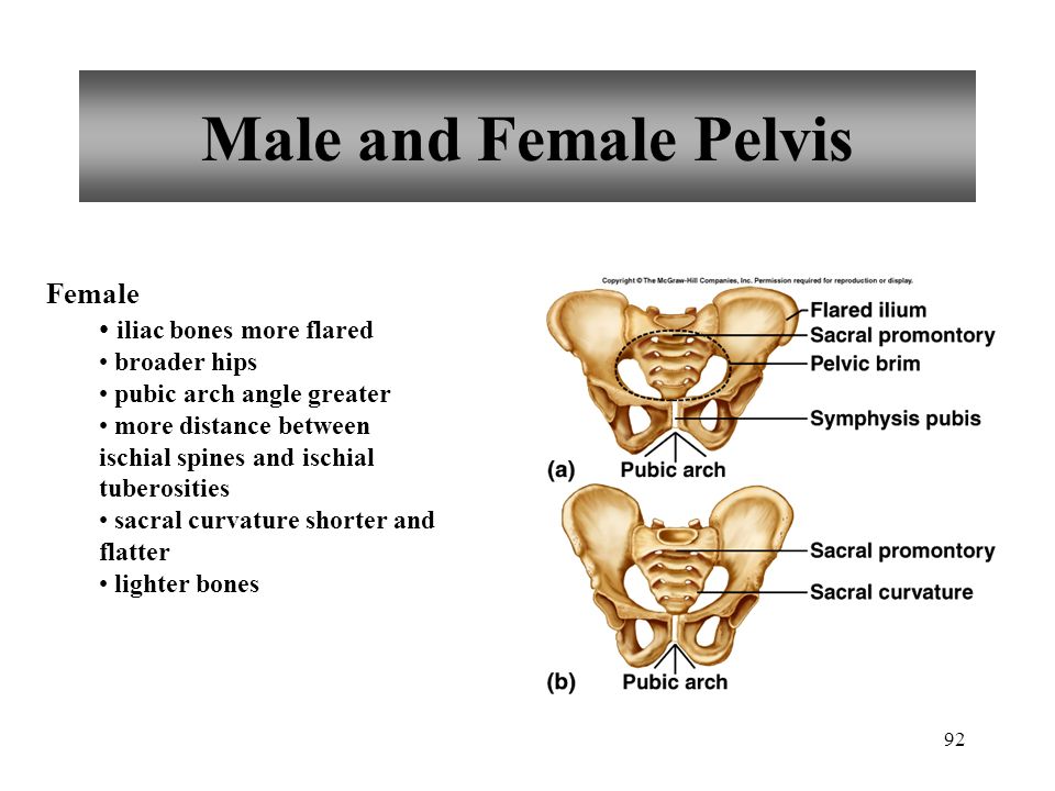 Cookies allow us to analyze and store information such as your device characteristics and certain personal data (for example, IP addresses, navigation, usage and location data, unique identifiers). This data is processed for the following purposes: to analyze and improve the user experience and/or our content, products and services, to measure and analyze the audience, to interact with social networks, to display personalized content, to measure the performance and attractiveness of content. For more information, please see our privacy policy: privacy policy.
You can give, withdraw or withdraw your consent to data processing at any time using our cookie settings tool. If you do not agree to the use of these technologies, this will be regarded as a refusal of the legitimate interest storage of any cookies. To consent to the use of these technologies, click the "Accept all cookies" button.
Analytical cookies
These cookies are designed to measure the audience: site traffic statistics help improve the quality of its work.
- Google Analytics
The structure of the pubis and what it does
The pubis, also known as the pubis, is located in front of the pelvic girdle. Posteriorly, the ilium and ischium form a bowl-shaped pelvic girdle. The two halves of the pubic bone are connected in the middle by a piece of cartilage called the pubic symphysis.
Contents of the article
- 1 Anatomy
- 2 Anatomical variants
- 3 Function
- 4 What is the pelvis?
- 5 Pilous bones form a bowl
- 6 Pilling bottom - this is a trampoline
- 7 pelvis plays a key role when walking
- 8 The female pelvis can change the size of
- 9 Pregnancy is forever changed by pelvis
- 10 pelvis more than
- 00 9000 9000 9000 large bones in the back of the pelvic girdle are higher. These bones are located almost directly above the femur and are often seen in women and people with little body fat.
 The pubic bone is not visible from the outside of the body and joins the anterior half of the pelvic girdle.
 Anatomy
The pubis is located in front of the body, slightly below the abdomen. This area provides structure and protection to the urinary organs in humans, including the bladder, uterus, ovaries, prostate, and testes.
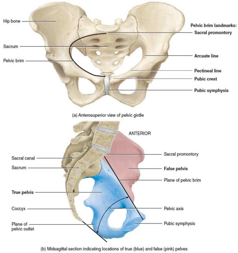
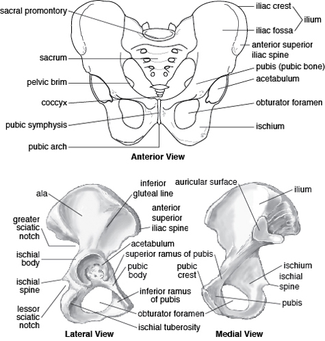
The largest part of the pubis is called the pubis body, which is located at the highest point of the pubis. Back of the pubis connects to the ilium, one of the bones at the back of the pelvis belts. The area where the ischium and pubis meet is limited obturator foramen and obturator groove. This back is where the pubic tubercle is located, to which muscles and ligaments are attached.
Pubic area where the pubic bone meets with the ilium is called the superior pubic tubercle. Between the top the pubic branch and the upper pubic area is the pectin line, which is another area where muscles and ligaments are inserted for stabilization.
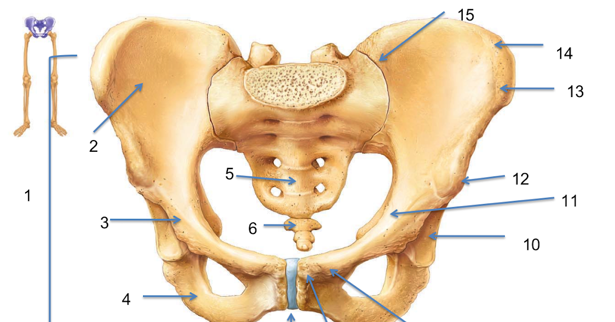 Just opposite the superior pubic ramus is the inferior pubic ramus, which points down to the lateral body of the pubis.
 The pubis is then arched downward and turns into cartilage in the middle. This arcuate region of the bone is called pubic arch, which also connects to the pubic symphysis, where they meet two ends of the pubic bone.
Variants of the anatomical structure
One of the most significant anatomical pubic bone variation is the difference in pelvic measurements. It means, that the distance between the pubic symphysis and the insertion point of the thigh can vary, as do the angles of the pubic arch, the length of the pubic symphysis and the radius recess into which the thigh is inserted. Variations in the pubic bone relative to fertility is also present.
The human pelvis is classified into types according to shape entrances:
- Gynecoid. Normal female pelvis. The most suitable for childbirth, as it has a round hole.
- Android.
 Male pelvis. The shape of the entrance is triangular.
 - Anthropoid. The hole has an oval shape, elongated along a vertical line.
- Platipelloidal. The oval shape of the entrance, located in a horizontal line.
You should know that android, anthropoid and platipelloid type of pelvic opening are an indication for caesarean section.
Function
The main function of the pubis is to protect intestines, bladder and internal genital organs. pubis also connects to the hip bones and provides support close to the body, allowing move further down the leg.
The pubis connects to the posterior bones of the pelvis belts, holding them in place and allowing the circular structure to connect the top half of the body with the lower half of the body.
The pubic bone also has several skeletal landmarks that connect and hold muscles, cartilage, ligaments and tendons. Each of these structures provides a connection between joints, bones and bodily structures.

Pubic bone has little motor function, since its main role is to stabilize pelvic girdle. The cartilaginous pubic symphysis has little movement in its free connection of the two halves of the pubic bone. However, the main purpose this cartilage is also to stabilize. All organs within the pelvic girdle have complex innervation, that is, several major nerves pass through pelvic girdle and its structures.
What is a pelvis?
"The pelvis refers to the lower abdomen as in both men and women,” says Gillogly, professor of gynecology at USA. “An important function of the pelvic region is to protect the organs used for digestion and reproduction, although all of its functions are crucial value,” she says. “It protects the bladder, both large and and the small intestine, as well as the male and female reproductive organs. Another one the key role is to support the hip joints.”
Hip bones form a bowl
Pelvis human is formed by three fused bones:
- Ilium.
 The biggest bone that expands upward.
 - Ischium. located below, has the shape of an arc. On it are the ischial tubercles, which take on assume the weight of the body when the person is sitting.
- Pubic bone. Formed by pubic bones and is located in front of the pelvis.
The pelvic floor is a trampoline
At the bottom of the pelvis is the pelvic floor. His can and should be strengthened with Kegel exercises to maintain its strength. Pelvic the bottom is like a "mini-trampoline made of solid muscle. Like a trampoline The pelvic floor is flexible and can move up and down. It is also the surface for pelvic organs: bladder, uterus and intestines. It has holes for vagina, urethra and anus.
The pelvis plays a key role in walking
Anyone who has ever broken a pelvic bone or stretched the pelvic muscle, knows how important the pelvic bone plays in functions such as walking and standing. “The pelvis also serves as a solid foundation to attach the spine and legs,” Gillogly says.
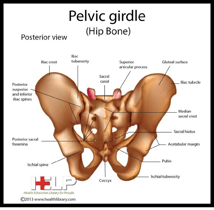 Peculiarities the structures of the pelvis endowed man with upright posture. It provides balance distributes the load. Smoothness of movement is provided by numerous ligaments, especially the iliac-femoral.
 The female pelvis can change size
Gillogly says that: “The female pelvis has tendency to be larger and wider than the male in order to fit the child into during pregnancy and ensure natural childbirth. However, the female pelvis narrows with age, when there is no need for gestation. It is believed that the change the shape of the pelvis is a key part of our evolutionary history because it changed when we started walking straight.
Pregnancy changes the pelvis forever
During pregnancy, the body releases a hormone known as relaxin to help the body adjust to the growing baby and soften the cervix. What happens, however, is: “The joints between the pelvic bones do loosen and pull apart slightly during pregnancy and childbirth,” Gillogly says.
Sometimes, however, relaxin can make the joints too loose, causing a painful syndrome known as pubic symphysis dysfunction (SPD), causing the pelvic joint to become unstable, causing pain and weakness in the pelvis, perineum, and even upper thighs during walking and other activities.
