Is transvaginal ultrasound safe
Is it painful, purpose, and results
A transvaginal, or endovaginal, ultrasound is a safe, straightforward way for doctors to examine the internal organs of the female pelvic region.
An ultrasound uses high-frequency sound waves to produce detailed images of internal organs.
Unlike X-rays, ultrasound scanning does not involve radiation, which means that it has no known side effects and is very safe.
In this article, we explain the uses of transvaginal ultrasounds and how to prepare for one. We also describe what to expect during the scan.
A healthcare professional uses an ultrasound to create images of the inside of the body. High-frequency sound waves bounce off the internal organs and form these images.
There are two ways to perform an ultrasound — abdominally and transvaginally.
A transvaginal ultrasound is an internal scan of the female reproductive organs. It involves inserting a small ultrasound probe, called a transducer, into the vagina to produce incredibly detailed images of the organs in the pelvic region.
It may be necessary to use a transvaginal ultrasound to examine the:
- vagina
- cervix
- uterus
- fallopian tubes
- ovaries
- bladder
Transvaginal ultrasounds can check for:
- the shape, position, and size of the ovaries and uterus
- the thickness and length of the cervix
- blood flow through the organs in the pelvis
- the shape of the bladder and any changes
- the thickness and presence of fluids near the bladder or in the:
- fallopian tubes
- myometrium, the muscle tissue of the uterus
- endometrium
Doctors may request a transvaginal ultrasound for a variety of reasons. For example, it might be necessary to identify the cause of:
- pelvic pain
- unexplained vaginal bleeding
- infertility
- abnormal results of a pelvic or abdominal exam
These scans can also help diagnose:
- benign growths, such as fibroids, cysts, and masses
- pelvic inflammatory disease
- endometriosis
- postmenopausal bleeding
In addition, it can check for the presence of an intrauterine contraceptive device.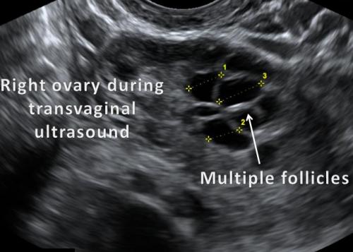
Doctors may request a transvaginal ultrasound during pregnancy, as it can help:
- check the heartbeat of the fetus
- confirm the date of delivery
- assess the condition of the placenta
- check for an ectopic pregnancy
- monitor pregnancies with a higher risk of pregnancy loss
A transvaginal ultrasound is not typically painful, but the insertion of the probe may be uncomfortable.
The healthcare professional performing the scan first covers the probe in a sheath and lubricating gel before inserting it slowly into the vagina to a depth of around 5–8 centimeters (cm). There may be mild pressure or discomfort at this stage.
There are no after-effects of a transvaginal ultrasound, and a person can return to their regular activities afterward.
A pelvic ultrasound is a noninvasive exam that produces images of the internal reproductive organs to help healthcare professionals diagnose certain conditions.
Doctors may use the term “pelvic ultrasound” to describe both a transvaginal and a transabdominal ultrasound.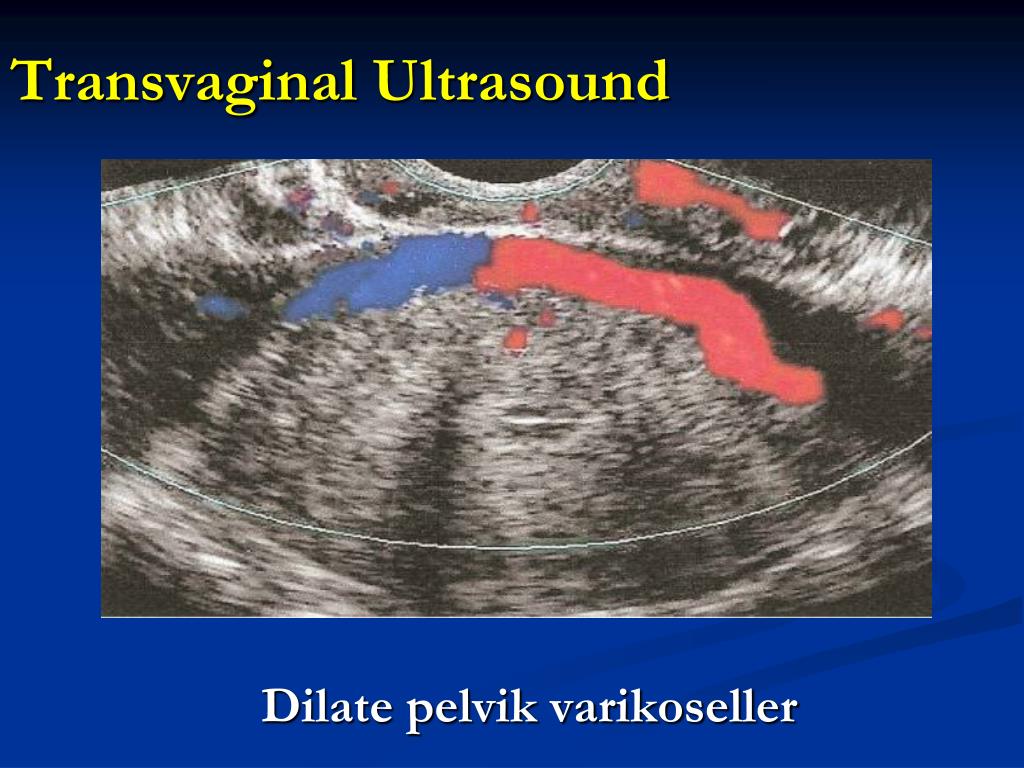 During a transabdominal ultrasound, the person performing the scan uses the probe outside the abdomen.
Another name for a transabdominal ultrasound is an external pelvic ultrasound. A person lies on their back on an examination table, and the healthcare professional applies a warm gel to the lower section of the person’s abdomen. Then, they move a probe over the area.
The person might feel slight pressure on their abdomen, but the scan is not painful. The probe uses sound waves to form an image of the internal organs and structures of the pelvic area.
By comparison, a transvaginal ultrasound can provide more close-up images of the internal organs than an external pelvic ultrasound.
A transvaginal ultrasound is a simple, painless scan that requires very little preparation. A person should be able to eat and drink normally before the appointment.
At the appointment, a healthcare professional will describe any necessary steps. A person needs to empty their bladder right before the scan, and anyone using a tampon should remove it.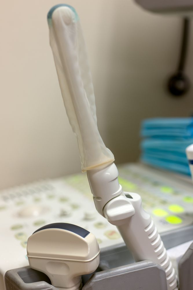
A doctor or a specially trained technician, called a sonographer, performs most transvaginal ultrasounds.
A person needs to undress from the waist down and put on a hospital gown. Next, they lie on an examination table with their knees bent. The healthcare professional uses a sheet to cover the person’s lower body.
The transducer resembles a wand and is slightly larger than a tampon. The sonographer or doctor covers the transducer with a sheath and lubricating gel before inserting it 5–8 cm into the vagina.
Once the transducer is in place, it produces sound waves that bounce off of the internal organs and relay information. To create a complete picture and bring different areas into focus, the sonographer or doctor rotates the transducer. This tool transmits the information directly to a screen.
The images display immediately on the screen, making it possible for the person and the healthcare professional to monitor the scan in real-time.
The whole process may last 15–30 minutes.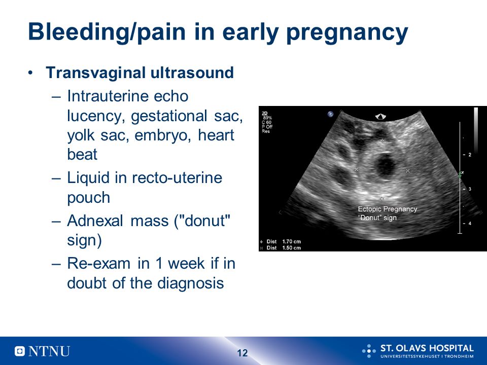
Unlike a traditional X-ray, a transvaginal ultrasound does not use radiation. As a result, it is very safe — there are no known risks.
It is also safe to perform transvaginal ultrasounds during pregnancy — there is no risk to the fetus.
During the insertion of the transducer, there may be some pressure and minimal discomfort. This feeling should go away after the scan.
It is essential to let the sonographer or doctor right away if anything feels particularly uncomfortable.
As the Miscarriage Association confirms, there is no evidence that a transvaginal ultrasound can harm a fetus or cause pregnancy loss.
If a person notices bleeding after a transvaginal ultrasound, this may be because blood has collected higher up in the vagina, and the transducer may have dislodged it.
People may have a transvaginal ultrasound in the first 11–12 weeks of pregnancy. After 11–12 weeks, it may be more common to have a transabdominal ultrasound. However, in some cases, doctors may still request a transvaginal ultrasound, which is a more accurate and detailed scan.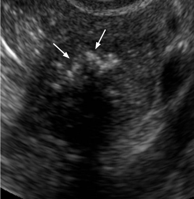
Transvaginal ultrasounds are safe up until a person’s water breaks.
If a specialist is present at the ultrasound, the person may hear the results immediately. If a sonographer does the scan, they send the images to a radiologist for analysis. The radiologist then sends a written report of the results to the person’s doctor.
Either way, it is important to discuss the results with a doctor, who can provide more information. This discussion can take place in person or over the phone.
A transvaginal ultrasound is a safe scan, with no known risks. People might experience some light discomfort during it, but this should go away afterward.
The ultrasound may take 15–30 minutes, and the results may be available immediately or within a few days, depending on whether a doctor was present during the scan.
If a person experiences extreme discomfort during the ultrasound, the doctor or sonographer may perform a transabdominal ultrasound instead. This type does not involve inserting the scanning tool into the vagina.
Vaginal ultrasound | Pregnancy Birth and Baby
beginning of content3-minute read
Listen
A vaginal ultrasound is an ultrasound scan taken by a probe inserted into the vagina. It gives a clear picture of the fetus, cervix and placenta. It is also called an internal ultrasound or a transvaginal ultrasound.
What does a vaginal ultrasound test for?
A vaginal ultrasound lets the doctor or ultrasound technologist (sonographer) see and measure the fetus. You might even discover if you are pregnant with twins or triplets.
It also allows the doctor or sonographer to look at your vagina, placenta, cervix, fallopian tubes, uterus and ovaries.
A vaginal ultrasound can be used as well as, or instead of, the standard abdominal ultrasound in which the probe is put on your belly.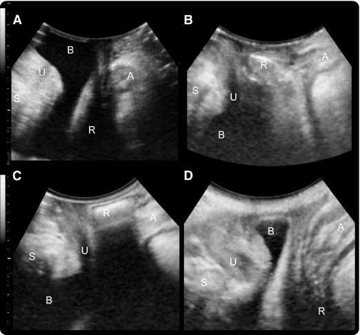
Why is a vaginal ultrasound recommended?
A vaginal ultrasound will be recommended when your doctor or midwife wants a clearer diagnostic image than the one they can get through a standard abdominal ultrasound.
When is it used during pregnancy?
A vaginal ultrasound can confirm you are pregnant as it can detect the heartbeat very early in your pregnancy. It can also record the location and size of the fetus and determine if you are pregnant with 1 baby or more.
A vaginal ultrasound can also be used to diagnose problems or potential problems, including to:
- detect an ectopic pregnancy
- measure the cervix to determine the risk of a premature birth, which will allow any necessary interventions
- detect abnormalities in the placenta or cervix
- to determine the source of any bleeding
How is a vaginal ultrasound done?
A vaginal ultrasound is done by your doctor or sonographer in a hospital, clinic or consulting room. It uses a probe that is slightly wider than your finger.
You’ll probably be asked to empty your bladder (pee) before the ultrasound starts. If you’re using a tampon, you’ll need to take it out.
You’ll be asked to take off the lower half of your clothing and lie back on the examination table with your knees bent. There might be stirrups, or your hips might be slightly raised.
The probe is covered by a sheath, which is then covered in lubricating gel. It will be inserted slowly for about 5 to 8cm into your vagina. That usually doesn’t hurt, but you will feel pressure and it can be uncomfortable.
The probe will be moved around to get the best view of what is being examined. The examination usually takes 15 to 30 minutes.
If you are not comfortable having a male perform the examination, you can ask for a female sonographer. You can also ask for a female health worker to accompany you for support, or have a family member with you.
If you live in Western Australia, you must give written consent to a vaginal ultrasound.
When will I get the results?
Often you can see the ultrasound images on a monitor while you have your scan. If your specialist is there, they might discuss the results with you straight away.
If a specialist isn’t there, the sonographer is usually not allowed to discuss what they see with you. Your doctor or midwife will see the images after they have been processed. It usually takes a day or two to get the results.
Are there any risks involved?
Vaginal ultrasounds are safe for you and your baby, as long as your waters haven’t broken.
If you are allergic to latex, let the sonographer know so they can use a latex-free sheath on the probe.
There are no after-effects of the procedure, so you can get back to your normal activities, including driving yourself home if you wish.
Sources:
Australasian Society for Ultrasound in Medicine (Guidelines for the performance of first trimester ultrasound), Inside Radiology (Transvaginal ultrasound), Royal Australian and New Zealand College of Obstetricians and Gynaecologists (Screening in early pregnancy for adverse perinatal outcomes), The Royal Women's Hospital (Bleeding in early pregnancy)Learn more here about the development and quality assurance of healthdirect content.
Last reviewed: June 2020
Back To Top
Related pages
- Pregnancy checkups, screenings and scans
- Ultrasound scans during pregnancy
This information is for your general information and use only and is not intended to be used as medical advice and should not be used to diagnose, treat, cure or prevent any medical condition, nor should it be used for therapeutic purposes.
The information is not a substitute for independent professional advice and should not be used as an alternative to professional health care. If you have a particular medical problem, please consult a healthcare professional.
Except as permitted under the Copyright Act 1968, this publication or any part of it may not be reproduced, altered, adapted, stored and/or distributed in any form or by any means without the prior written permission of Healthdirect Australia.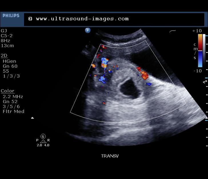
Support this browser is being discontinued for Pregnancy, Birth and Baby
Support for this browser is being discontinued for this site
- Internet Explorer 11 and lower
We currently support Microsoft Edge, Chrome, Firefox and Safari. For more information, please visit the links below:
- Chrome by Google
- Firefox by Mozilla
- Microsoft Edge
- Safari by Apple
You are welcome to continue browsing this site with this browser. Some features, tools or interaction may not work correctly.
vaginal and transvaginal - MEDSI
What is vaginal ultrasound?
Vaginal (transvaginal) pelvic ultrasound is performed by inserting a special device equipped with a transducer into the vagina.
The device is a rod with a handle, which is made of plastic, about 10-12 centimeters long and up to three centimeters in diameter. A special groove can be built into it to insert a needle for taking biopsy material.
Examination allows to determine the presence of pathologies, neoplasms or diseases in the following female genital organs:
- Uterus
- Fallopian tubes
- Ovaries
- Cervix
It is considered the most effective for the study of these parts of the reproductive system, as it allows you to identify various health problems in the patient at an early stage. Ultrasound of the small pelvis with a sensor is able to show the presence of deviations already at a time when other studies do not show any problem areas.
How is the procedure?
The examination is organized as follows:
- The patient must remove clothing from the lower part of the body (from the waist down)
- She settles down on a special couch in the same way as during a regular gynecological examination
- The doctor prepares the transducer: puts an individual condom on it, lubricates it with a special gel for the procedure
- The physician then inserts the device shallowly into the patient's vagina
- To get a complete picture of the state of the organs, he can move the sensor from side to side
- All data is recorded and processed by the doctor
The gel is needed to facilitate the penetration of the transducer (and thereby reduce the likelihood of negative sensations) and enhance the ultrasonic effect by increasing the conductivity.
This type of examination lasts no more than 10 minutes. It is painless and gives the most complete picture even when an abdominal ultrasound shows nothing or cannot be performed.
When is a pelvic ultrasound probe needed?
There are symptoms in which the doctor must refer the patient to a transvaginal examination:
- Pain in the lower abdomen (not related to the menstrual cycle)
- Neoplasm suspected
- Too short or too long period of menstrual bleeding or lack thereof
- Impossibility of pregnancy
- Bleeding other than menstruation
- The presence of violations of the patency of the fallopian tubes
- Nausea, vomiting and weakness with bleeding from the vagina
Doctors recommend using this type of examination for preventive purposes, since not every ailment can have symptoms at an early stage, just as pregnancy in the first trimester may not manifest itself with classic symptoms (nausea, etc.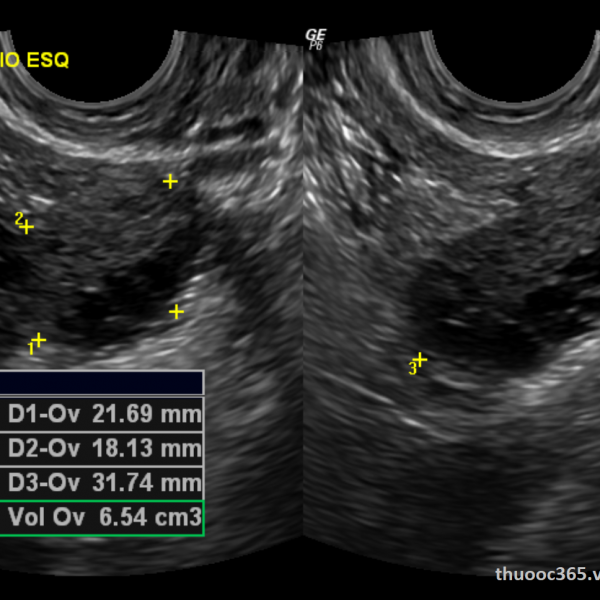 ).
).
In this case, vaginal ultrasound is used for:
- Infertility diagnostics
- The need to determine the presence of changes in the size of the ovaries and uterus
- Pregnancy diagnostics
- Pregnancy control (first trimester only)
- General supervision of the uterus, fallopian tubes and ovaries
A pelvic ultrasound with two probes can be performed at the same time. In this case, an abdominal ultrasound examination is performed first, and then a transvaginal one. The use of two types of analysis at once is necessary to detect violations in the highly located organs of the small pelvis.
What does a vaginal ultrasound show?
This examination allows to evaluate the following parameters of the organs of the reproductive system:
- The size of the uterus. In normal condition, it should be about seven centimeters long, six wide and 4.2 in diameter. If it is significantly less or more, then this indicates the presence of pathology
- Echogenicity.
 The structure of the organs should be homogeneous, uniform, have clearly defined, well-visible edges
 - General picture of the internal organs. The uterus should be slightly tilted forward. And the fallopian tubes may be slightly visible, but should not be clearly visible without the use of contrast agent
Diagnosable diseases
Transvaginal ultrasound can detect a number of diseases and problems in the reproductive system at an early stage. It allows you to detect:
- Fluid and pus in the uterus and fallopian tubes. The cause of their appearance may be infections, viruses, mechanical damage
- Endomentriosis - excessive growth of cells of the inner layer of uterine tissues into other layers and organs. It can occur due to inflammatory processes, damage (surgery, abortion), the appearance of neoplasms, disruption of the endocrine system, too frequent use of certain drugs and substances
- Myoma is a benign neoplasm in the tissues of the uterus or its cervix.
 May occur due to chronic diseases, frequent abortions, hormonal disorders, constant stress, pathologies, overweight, with hereditary predisposition
 - Cysts and polycystic ovaries are tumors filled with fluid. Occur in endocrine disorders, chronic diseases of the genitourinary system
- A variety of polyps on the walls of the uterus - benign formations in the endometrium of the organ. They can reach several centimeters in diameter. Their appearance may be associated with polycystic disease, chronic diseases, mastopathy, fibroma
- Inflammation and enlargement of organs can occur due to both infection and trauma
- Cystic skid - appears instead of a full-fledged embryo in the process of conception, filled with fluid. It occurs due to duplication of male chromosomes with the loss of female chromosomes, sometimes due to the fertilization of an egg that does not contain a nucleus. This disease is rare
- Fetal development disorders during pregnancy
- Malformations and pathologies in the development of the fallopian tubes: obstruction, spiral or too long tubes, blind passages, duplication of organs
- An ectopic pregnancy occurs when an egg, after fertilization, attaches itself outside the tissues of the uterus.
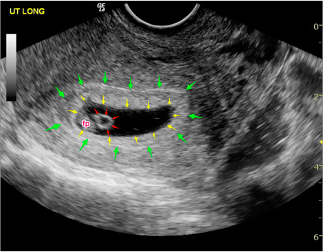 Occurs due to blockage of the fallopian tubes, congenital anomalies in them, as well as after the inflammatory process, abortion
 - Cancer - a malignant tumor in various organs:
- Uterus
- Ovaries
- Cervix
- Chorionepithelioma is a malignant neoplasm that occurs during or after pregnancy from chorion cells (the membrane of the embryo attached to the wall of the uterus)
Steps to prepare for the exam
No special preparation is required for a pelvic ultrasound with a transducer, but there are several mandatory requirements:
Doctors also recommend using such a study on certain days of the cycle, depending on which organ and for what purpose you need to diagnose:0012
It is important to remember about personal hygiene before the examination, use wet and other wipes.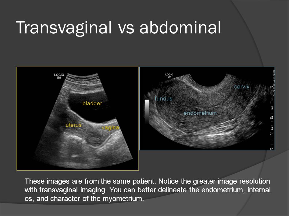
If you plan to perform a pelvic ultrasound with two sensors, then you should pay attention to the preparation for the abdominal examination.
This includes:
- Diet at least three days prior to the examination to reduce the likelihood of symptoms of flatulence and bloating
- The last meal must be finished by six o'clock in the evening on the eve of the analysis
- It is recommended to take an enema after a meal
- If there is still a risk of flatulence, special drugs should be used to reduce gas formation
- Drink at least 400 ml of water one hour before the examination
The diet involves the exclusion of a number of products from the diet:
- Sweets
- Flour (bread, biscuits and other)
- Legumes
- Cabbage
- Milk and dairy products
- Uncooked vegetables and fruits
- Coffee and strong tea
- Carbonated drinks
- Fast food
- Fatty foods (meat, fish, oils)
You can eat cereals cooked with water, low-fat boiled beef, poultry and fish, hard cheeses.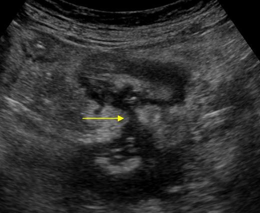 It is recommended to drink loosely brewed lightly sweetened tea.
It must be remembered that since fluids are required before the abdominal examination, the bladder must be emptied before the transvaginal examination.
Contraindications
Vaginal ultrasound has a small number of contraindications:
- It is never performed if the patient is a virgin, so as not to violate the integrity of the hymen. In this case, it is possible to perform a transrectal examination for such a patient, in which the probe is inserted into the rectum
- Examination should not be performed during the second or third trimester of pregnancy because it may cause preterm labor or uterine contractions before the expected date of delivery
- This test is not used if the patient is allergic to latex
- If the patient has epilepsy because the examination requires her to lie still
Do not delay treatment, see a doctor right now:
- Ultrasound
- Ultrasound of the pelvic organs for a child
- Hospitalization and transportation
How is a vaginal ultrasound performed during pregnancy and why is an examination necessary?
Transvaginal ultrasound is performed to obtain a more detailed picture and visualization of the smallest structures. However, there are cases when it is contraindicated.
How is a vaginal ultrasound done?
Vaginal ultrasound, which can be performed to examine the female reproductive system, allows you to get a lot of information. In order to start the procedure, the patient needs to remove clothes below the waist and lie down on the couch. In this case, the legs must be bent at the knees and spread apart. The doctor puts a condom on a special device sensor, which is otherwise called a "transducer", and lubricates it with gel. The gel should eliminate air between the transducer and the organs and also serve as a lubricant for easier insertion. The sensor is then gently inserted into the vagina, and the doctor examines the image that is displayed on the screen.
Vaginal ultrasound appointment
Ultrasound with a vaginal probe is performed to obtain information about the state of the woman's pelvic organs. Such a study allows you to determine even the slightest deviations from the norm and determine the appropriate treatment tactics for the patient's condition. A gynecologist or surgeon can refer a patient for examination in case of pain, delays in the onset of the next menstruation, discharge of an unhealthy color, in the absence of a planned pregnancy, and in many other cases.
What is transvaginal ultrasound?
By itself, vaginal ultrasound is a completely safe, diagnostic method. Such a study is based on ultrasonic waves, which are reflected differently from tissues, depending on their structure. Based on how exactly the waves were reflected from certain organs and their parts, the doctor draws up a conclusion about the patient's state of health. A feature of the transvaginal examination method is that during the procedure it is necessary to insert the probe into the woman's vagina, and not just examine the organs through the abdominal wall.
Indications for vaginal ultrasound
An examination of the pelvic organs with a vaginal ultrasound probe can be prescribed by a surgeon or gynecologist. Such a study is necessary in a number of cases:
- spotting appeared, which is not related to menstruation;
- pregnancy does not occur within six months of active sexual activity;
- in addition to menstrual pain, a woman is worried about other pain;
- too long or short menstruation - beyond 3-7 days;
- for prophylactic purposes under certain conditions.
In these cases, the procedure allows you to obtain valuable information about the state of health of the woman and determine the nature of the unpleasant symptoms. The undoubted advantage of ultrasound is its complete safety, due to which the procedure can be prescribed even for pregnant women.
Preparation for transvaginal ultrasound
If you do not know what a vaginal ultrasound is and how this procedure is done, it is worth knowing that preparatory measures are not needed. The only thing to do before the examination is to empty the bladder. The doctor may even ask the patient about it if the last time the woman urinated more than an hour ago. The day before the procedure, you should not eat foods that cause increased gas formation. In addition, no preparation is required for the vaginal ultrasound .
How is the procedure?
The examination itself does not take more than 10 minutes. After the doctor examines the image displayed on the screen, he writes a conclusion. During this time, the patient may be asked to wait outside the office. The conclusion is handed out - with it you can go to the attending physician. It is worth knowing that such a procedure is not prescribed for girls who do not have sex. In this situation, the examination is performed rectally or with the help of abdominal ultrasound.
On what day of the cycle is a vaginal ultrasound done?
If you do not know what a vaginal ultrasound is, when it is best to do it, you should find out that the most suitable time for examination is the 5-8th day of the cycle, that is, immediately after menstruation.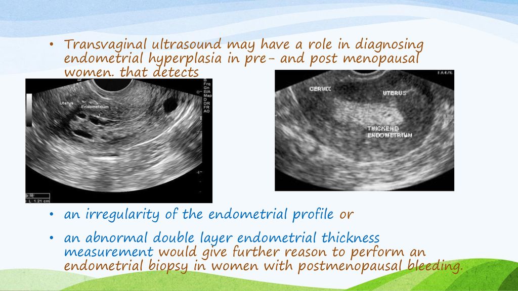 However, this is only suitable if a scheduled inspection is performed. If the gynecologist suspects endometriosis of the uterus, the examination is postponed to the second half of the cycle. Several studies in one cycle allow to follow the dynamics of the disease. An emergency procedure can also be performed on any day of the cycle if bleeding has begun that is not related to menstruation.
Interpretation of results
You can find out the course of a vaginal ultrasound, which the procedure shows and get information about the state of the pelvic organs, from the conclusion drawn up by the doctor. As a rule, this is done by an ultrasound specialist who gives the result of the diagnosis to the patient after the procedure.
What does a vaginal ultrasound show?
Vaginal ultrasound may be needed during or without pregnancy, with the help of such a study, you can detect a lot of deviations from the norm, as well as confirm or refute the diagnosis. During the procedure, you may find:
- cyst, polycystic ovaries;
- endometriosis;
- ectopic or uterine pregnancy;
- inflammation;
- pathological fluid in the fallopian tubes;
- tumor of any kind;
- pelvic fluid;
- and more.

This diagnostic method allows you to determine the state of each organ and identify the presence of malfunctions in the functioning of the female reproductive system. Thanks to this, adequate and timely treatment is prescribed.
Ultrasound of the uterus and appendages transvaginally
Many women do not know if a vaginal ultrasound can be done during pregnancy. They should learn that such a procedure can be used both to determine pregnancy, even at the earliest stages, and to control its course. Also, the procedure allows you to detect any pathological processes in the reproductive system.
With the help of such a study, you can find out the condition of the body and cervix, their size. It will be visible the location of the uterus, its size, the condition and location of the ovaries, their structure. You will see the presence of free fluid in the peritoneal cavity, the doctor also notes the number and size of follicles.


