How big is a normal uterus
Average Uterus Size & How Fibroids Change It
Thursday, May 19th, 2022
Average Uterus SizeIf your doctor has talked to you about having an average uterus size or discussed the concern of an enlarged uterus, you may wonder about the definition of “average.” It may help you better understand your uterus and any health concerns if you realize how this organ is measured and what makes it average.
The uterus measurements are found in three ways. First, it is measured by the length, from the fundus of the uterus to the outside opening. The fundus is the part of the uterus farthest from the opening. It may also be measured by the width of the uterus, which is measured across the fundus. The third measurement is the thickness of the uterus.
The uterus grows at puberty when it becomes pear-shaped. At this point, it has reached its full size, but the volume will continue to change during a woman’s reproductive years. It changes throughout the menstrual cycle, ranging from 75 ccs to 200 ccs.
The normal size of the uterus is about three inches in length and two inches at the widest part. The uterus can expand up to five times its normal size during pregnancy. Compare this change in the uterus as if it were going from a lemon to the size of a watermelon.
If your uterus has changed size and you experience other symptoms of uterine fibroids, you can seek treatment with a fibroid specialist through Uterine Fibroid Embolization (UFE). UFE is a minimally invasive procedure which can help your uterus go back to its original size and shape.
Schedule a consultation
Uterus Size with FibroidsUterine fibroids are benign tumors that can grow inside or on the uterus. The fibroids in your uterus can continue to grow and change the shape and size of the average uterus size. How much the fibroids increase the size of the uterus will depend on how many fibroids are present, where they are located, and how big they grow.
Fibroids can have a wide range of sizes, with the ability to change the average uterus size by causing it to expand.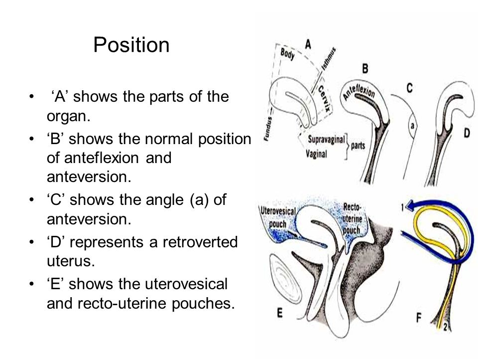 They can start small, about the size of a small seed, and grow to be quite large, tor the size of a grapefruit. Large fibroids are classified as those that range from a grapefruit to a watermelon or more than 10 cm in diameter. Even medium fibroids can be large enough to increase the size of the uterus. These fibroids may range from the size of a plum to an orange.
If you have multiple fibroids, they can cause the average size uterus to expand and make you look like you’re pregnant. They can cause the uterus to expand to the point that it reaches the rib cage. As the fibroids grow, they can cause discomfort. You may be concerned about how they will impact your uterus.
What Do Fibroids Do to Your Uterus?Fibroids can cause the average size of the uterus to change shape and expand as they grow. They can also cause the uterus to press on other organs, such as the bladder, making it difficult to empty the bladder or cause you to feel the need to urinate more often.
Fibroids often cause heavy bleeding because the pressure makes the endometrial tissue bleed more than normal. They may prevent the uterus from contracting properly to stop menstrual bleeding as expected with a normal period.
Submucosal Fibroids
Submucosal fibroids are one type of uterine fibroid located in the uterus’s inner lining, also known as the endometrium. Submucosal fibroids can grow alone or in a cluster. They often cause heavy menstrual bleeding, which can lead to anemia. They may cause pain in the lower back and fatigue and dizziness, which is often attributed to anemia.
Subserosal Fibroids
Subserosal fibroids are another type of growth which may not change the average uterus size because they are found on the outside of the organ. They can impact nearby organs, such as the bladder. These fibroids can vary in size and location in different areas around the uterus. They may cause abdominal pain, cramping, feeling full or heavy, and frequent urination.
Fibroid symptoms can vary based on size and location in a normal size uterus. They can also be similar to other conditions that affect the reproductive system, so it is important to discuss symptoms with your doctor.
Typical symptoms of fibroids include:
- Heavy menstrual bleeding and longer periods
- Bleeding between periods
- Severe menstrual cramping
- Pain in the pelvis or lower back
- Frequent urination
- Stomach swelling
- Pain during sex
- Anemia
- Bloating and/or constipation
If you experience these symptoms, you should see your doctor. They may diagnose fibroids during a regular pelvic exam or use other tests to reach a diagnosis. They may recommend:
- Ultrasound
- MRI
- Hysterosonography
- Hysteroscopy
These tests can confirm a diagnosis of uterine fibroids.
Fibroids can cause complications with pregnancy. They may grow in size during pregnancy because of the increase in progesterone. They can outgrow their blood supply, which may lead to severe pain and hospitalization to monitor the mother and baby’s conditions.
Fibroids can affect the baby’s position in the uterus, increasing the risk of miscarriage. They may also increase the risk for preterm delivery and the need for a cesarean section delivery.
Uterine Fibroid TreatmentUSA Fibroid Centers offers a minimally invasive treatment for uterine fibroids to help reduce their size and accompanying symptoms. UFE treats conditions to reduce or eliminate symptoms while keeping the uterus intact, unlike a hysterectomy.
When UFE is performed, a fibroid specialist will use ultrasound technology to locate the fibroid. They will insert a tiny catheter into the thigh or wrist and inject embolic materials into the artery feeding the fibroid.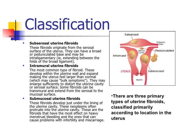 When the fibroid can no longer receive nutrients, it shrinks and dies.
If you are concerned about fibroids, how they can affect an average size uterus, or the symptoms that negatively impact your life, you can get help with USA Fibroid Centers. Our specialists will work with you to determine the right treatment plan. Schedule a free consultation online or call us at 855.615.2555 to visit one of our facilities.
Our Locations
Related Posts
Does a Uterus Really Double in Size During Menstruation?
A viral post that claims the ballooning size of a uterus during a period is to blame for a “heavy” feeling many women experience isn’t backed by science.
A viral photo making its way around Facebook claims to explain the reason many women feel “heavy” during their periods.
The photo compares two uterus models side-by-side. One represents a smaller, nonmenstruating uterus. The other model is a dark-colored menstruating uterus almost double in size.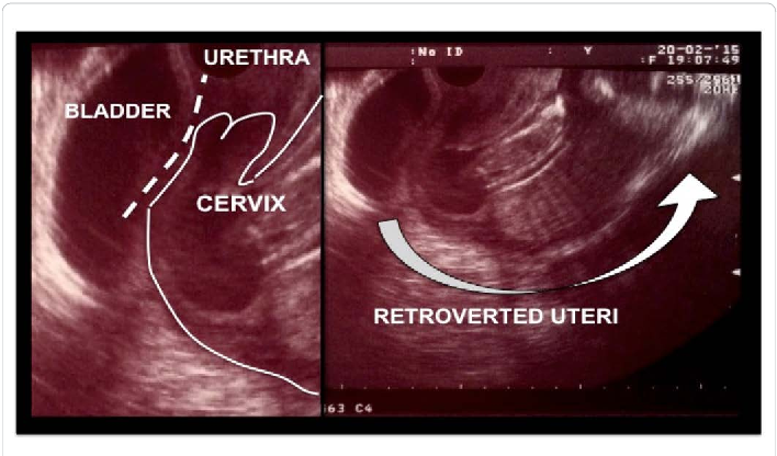
The claim comes from Apples and Ovaries, an account run by a holistic nutritional therapist and natural fertility health consultant.
“This is why we feel so heavy at the beginning of our bleed. Why it can feel like our uterus is about to drop out and hit the pavement… And why we need to take things slooooooow,” Apples and Ovaries wrote as a caption for the photo.
Nearly 25,000 people have shared the Facebook post and 19,000 have liked it. The resounding response in more than 6,000 comments is, “Well that explains it.”
One woman wrote, “Ahhh, that is a wonderful confirmation of the discomfort. All makes more sense now.”
Another woman wrote, “WOW good to know cause I do feel so bloated and heavy on the 2nd day.”
But two gynecologists say the models aren’t grounded in science.
Healthline asked Dr. Safrir Neuwirth, an OB-GYN at CentraState Healthcare System, if a woman’s uterus nearly doubles in size monthly during her period.
“The simple answer is no,” Neuwirth responded. “In my 20 years of practice, I’ve never noticed much of a change in the size of the uterus during the period.”
Another OB-GYN, Dr. Kimberly Gecsi, who practices at University Hospitals Cleveland Medical Center, had a similar reaction. “I’ve never heard anything about a uterus increasing in size during a woman’s period,” she said.
“There are certain things that happen physiologically during a woman’s period that do increase the volume of the uterus and make it slightly more swollen,” Neuwirth noted. “But does the uterus double in size like this picture? No way.”
Neuwirth explained that there’s increased blood flow to the uterus during that time of the month, driven by a surge in hormones.
The lining of the uterus, which is what sheds during a period, also thickens by about half a centimeter leading up to the first day of menstruation.
“The combination of the two factors might slightly increase the volume of the uterus by as much as 10 to 15 percent,” Neuwirth told Healthline.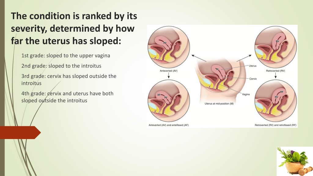
Megan Assaf, a licensed massage therapist and the founder of Wombs for Wisdom, created these uterus models. She said the two sizes are “based on a unique combination of medical science data, medical illustrations, cadaver observation, over a decade of manual palpation of living wombs, and concepts from traditional folk healing.”
Assaf told Healthline that the medical basis she cited comes from a Maya Abdominal Therapy book written by Rosita Arvigo, DN.
According to Assaf, the book states that in a woman who isn’t pregnant, the uterus weighs 4 ounces. During menstruation, the uterus can weigh as much as 8 ounces.
However, changes in weight and size aren’t the same thing.
The typical size of a uterus is approximately 7 centimeters long, 5 centimeters wide, and 4 centimeters thick.
But the size varies considerably, emphasized Neuwirth.
“The uterus models in the picture are both totally normal uterine sizes,” Gecsi clarified. “The smaller one looks like a uterus you could see in someone who’s never had children or is postmenopausal, and the other is pretty representative of a uterus in a woman who’s had a few children. ”
”
Neuwirth further explained that having babies increases the size of the uterus, or womb. Most of the time after childbirth, the uterus will remain larger, he said.
There are a few other reasons for the variety in uterus size. About 20 to 80 percent of women will develop uterine fibroids by the time they’re 50. Fibroids are noncancerous growths on the side of the uterus wall. They vary in size and can enlarge the uterus overall.
Genetics are also at play in uterus size.
“It’s important know that if a woman’s uterus is enlarged, it could be a sign of a different problem” and to consult a doctor, Gecsi warned.
She pointed out that since the uterus is set deep into the pelvis, a woman wouldn’t be able to notice the change in size herself.
It’s easy for things to go viral without being fact checked, and these models certainly look compelling.
But the lack of scientific evidence to back up the change in uterine size during menstruation highlights the need to research medical information you read on social media if it’s not from a trusted source.
Since feeling “heavy” during a period is common, the picture of the uterus models resonated with thousands of women.
While women might experience a heavier feeling because of the increased volume of blood in the uterus, Neuwirth said, the main factor behind that full, uncomfortable feeling comes from a combination of bloating, extra water retention, and excess gas.
You can blame those symptoms on the plummeting levels of the hormone progesterone during a period — not a ballooning uterus.
Ultrasound of the uterus - Ultrasound of the cervix and appendages in Nizhny Novgorod, transvaginal ultrasound
Ultrasound of the uterus is one of the most important and common methods for examining this organ. For a better understanding of the process of conducting ultrasound of the uterus and appendages, consider the uterus as an organ of the female reproductive system.
The uterus is a hollow muscular organ consisting of three layers: endometrium (inner), myometrium (middle), perimetrium (outer), and is located in the pelvic cavity, between the bladder and rectum.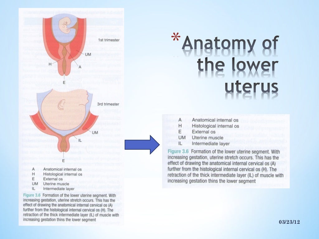 The normal anatomical position of the uterus in the small pelvis is provided by the supporting muscular pelvic floor and the suspension ligamentous apparatus of the uterus. The uterus has a fundus (upper part of the uterus), a body, and a cervix. The cervix connects the uterus to the vagina. Therefore, the vaginal and supravaginal parts of the uterus are isolated.
The function of the uterus is to provide a place and conditions for the implantation of the embryo and for the further development of the fetus.
The fallopian tubes and ovaries are the so-called appendages of the uterus. The ovaries are paired sex glands that work alternately. They contain many different degrees of development of follicles in which eggs mature. The fallopian tubes run from the uterus to the ovaries. They are where fertilization takes place.
Therefore, in order to enjoy the happiness of motherhood, it is necessary to undergo prophylactic Ultrasound of the uterus and appendages and timely diagnosis and treatment of the uterus and other genital organs of a woman.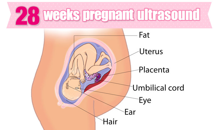
Ultrasound of the uterus and appendages a gynecologist can prescribe if there are any symptoms of a disease of the female genital organs:
- Various pains in the lower abdomen, pain during menstruation
- Irregular menses
- Uterine bleeding
- Anomalies in the development of the uterus
- Bleeding during menopause
As well as ultrasound of the uterus and appendages is used for:
- Clarification of the presence and location of the intrauterine contraceptive (spiral)
- Establishing pregnancy
- Neoplasm diagnostics
For the study of the pelvic organs in a sexually active woman, transvaginal ultrasound of uterus is performed. To prepare for a transvaginal ultrasound of the uterus, 2 days before the examination, do not eat foods that cause gas formation, and, if necessary, make an enema.
Transvaginal ultrasound of the uterus is not performed for girls before the onset of sexual activity.
To do this, use the transabdominal method of conducting ultrasound of the uterus. It is necessary before the study to try to fill the bladder as much as possible - drink about 1 liter of water or tea. This is necessary in order to visualize the uterus well on ultrasound.
The most important condition for transvaginal ultrasound and transabdominal ultrasound of the uterus and appendages is its 5-7 day of the menstrual cycle or during the first 2 days after the end of menstruation.
With an ultrasound of the uterus, the doctor determines the position, size and structure of the uterus, and also assesses the condition of the cervix. It is also taken into account when conducting an ultrasound of the uterus, that before childbirth, the size of the uterus in a girl is minimal, and in a woman who has given birth, it is maximum.
Normal ultrasound findings of the uterus in reproductive age (from 20 to 35 years):
- Uterine length 30-40 mm, cervix 40-50 mm, anterior-posterior size 28-45 mm, width 35-50 mm.
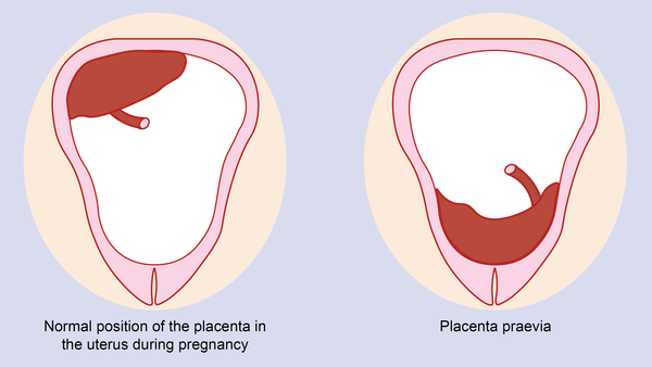
- Position of the uterus in anteflexio - inclination towards the bladder. Bend of the uterus to the rectum (retroflexio) is a normal variant, but less common
- The thickness of the endometrium changes during the menstrual cycle
By the 5-7th day of the menstrual cycle, the endometrium is clearly defined, its thickness is up to 6-9 mm. - Myometrium homogeneous in structure and without formations
Ultrasound of the uterus in the early reproductive period (from the onset of menstruation to 20 years) shows the smallest size of the uterus. With ultrasound of the uterus in postmenopause (begins at the age of 45 to 55 years), the uterus decreases and becomes homogeneous in echostructure. The endometrium may not be visualized.
Ultrasound of the uterus helps to detect the following diseases:
- Anomalies of development (unicornuate, bicornuate, bifid, with a septum)
- Endometriosis
- Adenomyosis
- Metroendometritis
- Endometritis
- Uterine polyps
- Tumors (benign and malignant)
In addition to ultrasound of the uterus, an ultrasound of the cervix is also performed:
During ultrasound of the cervix, its shape, structure and thickness of the cervical canal, the structure of the muscular layer of the cervix are assessed. On a normal picture Ultrasound of the cervix the contours are clear and even, the dimensions are variable, compared to the uterus, the echogenicity is greater, the structure is homogeneous.
When performing ultrasound of the cervix, special attention should be paid to it, due to the high incidence of cervical fibroids.
Ultrasound of the cervix can detect:
- Cervicitis - inflammation of the cervix
- Polyp
- Kistu
- Endometriosis
- Cervical atresia, narrowing of the cervical canal
- Scars
- Tumors
Ultrasound of the uterus in Nizhny Novgorod
The TONUS PREMIUM Center for Radiological Diagnostics provides the best conditions for Ultrasound of the uterus in Nizhny Novgorod . For uterine ultrasound, doctors with extensive experience use expert-class ultrasound machines, and the entire uterine ultrasound procedure takes place in a cozy and relaxing environment of the center.
You can sign up for an ultrasound by calling 8 (831) 411-13-13
Ultrasound of the uterus and appendages
How the study is carried out
If the examination is conducted through the abdominal wall, it is enough for a woman to provide access to the abdomen, to expose its bottom so that there are no obstacles when the transducer sensor moves. To facilitate its movements on the skin, a profile gel-like composition is used, which after the procedure is easily removed with a damp cloth, leaving no trace.
If the doctor needs to obtain cervical data, then a transvaginal examination option is used. Simultaneously with the assessment of the organ under study, the doctor also notes the position of other structures of the small pelvis. For carrying out, a special elongated sensor is used, on which a condom is put on for sterility, after which the instrument is inserted into the vagina.
For rectal administration, a thinner transducer is used, also with a protective agent.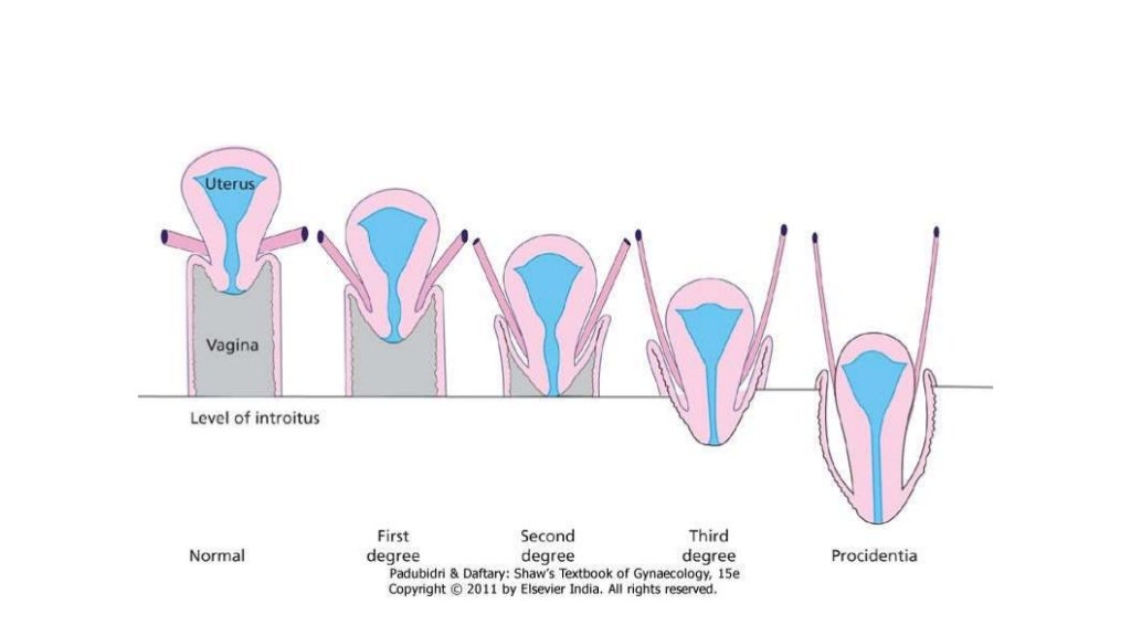 The study is carried out in the supine position. Before you lie down on the couch, you should remove your underwear, providing access to the genitals.
Explanation of ultrasound results
Before dwelling on the decoding, you should refresh your knowledge of the physiology of the female reproductive system. The uterus is a pear-shaped organ made up of muscle tissue. In an ideal anatomical structure, the uterus is characterized by a slight forward bend. In the opposite situation, problems with conception and pregnancy often occur, bowel function suffers.
Normally, the contour lines are distinguished by an even and smooth bend. If bumps are present, this indicates the presence of pathologies. Among them may be benign and malignant tumors, signs of inflammatory processes.
Ovarian and uterus size norms
When discussing the normal size of the ovaries and uterus, reference is made to the position of the woman and her age. In the absence of pregnancy, the length of the uterus should be 4. 5-7 cm (45-70 mm), the width of the organ should not exceed 4.6-6.4 cm (46-64 mm). The norm for thickness is 4.2 cm (42mm).
Twenty years after the onset of menopause, these dimensions are reduced to 42 mm, 44 mm, 30 mm - (length, width, thickness - respectively). If the doctor diagnoses a large organ during scanning, the result may indicate the presence of uterine fibroids or pregnancy, as a result of which the volume of the organ has increased.
As for the ovaries, in the normal position they are 30-41, 20-31, 14-22 mm - length, width, thickness. The volume of one ovary is in the range from 2 to 8 cubic meters. see If the doctor says that the ultrasound showed the corpus luteum, this indicates the appearance of a temporary gland in the organ, which is an indicator of the ovulation period in the cycle under study.
Ultrasound contraindications
Experts confidently say that ultrasound is a safe non-invasive method. Evidence confirming the negative impact of ultrasound on well-being and health is not officially registered, no matter how many studies on this topic have been conducted.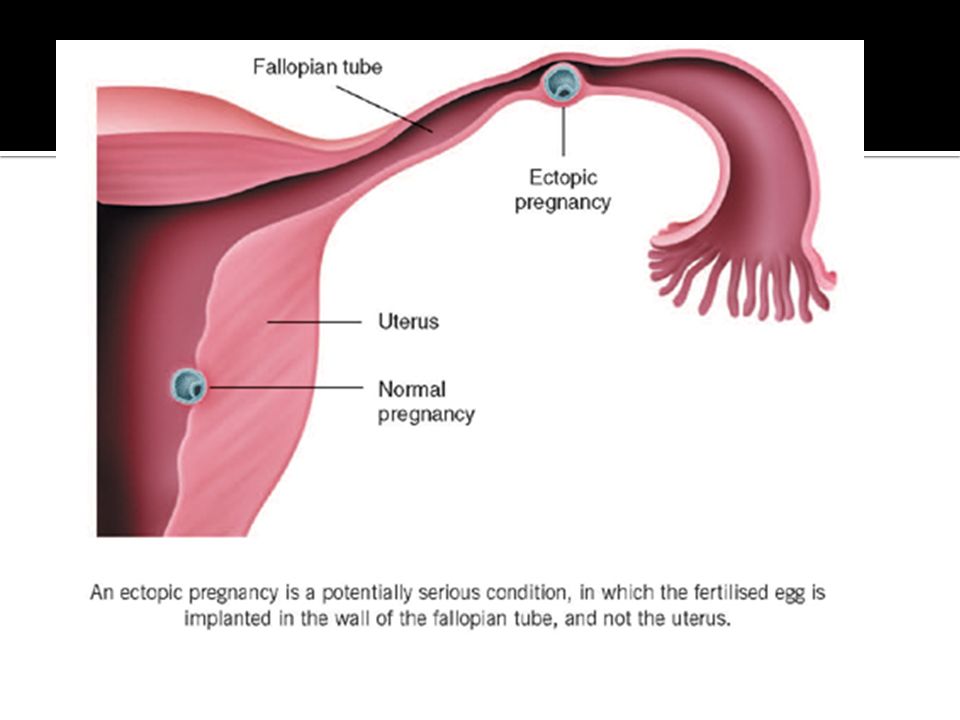
In accordance with this, ultrasound has no contraindications to conduct. The only thing that should be stipulated is the ban on the use of the transvaginal method for virgins. In order to preserve the hymen, it is not recommended to insert the transducer through the vagina.
Where to get an ultrasound of the uterus and appendages in Kazan
The question of where to do an ultrasound of the uterus and appendages is not as simple as it seems at first glance. The technology does not tolerate the poor quality of the research, since we are talking about the health of the woman, and during pregnancy and the correct, harmonious development of the fetus in the womb.
The medical center "AM Medica" performs all types of ultrasound on modern equipment, which gives a clear visualization of the objects under study. We offer reasonable prices for all types of diagnostics. Make an appointment to undergo the study recommended to you, and, if necessary, get a consultation with a specialist, taking into account the results you have received.