How accurate is a ultrasound
Accuracy of Ultrasound in Diagnosing Abdominal Masses | JAMA Surgery
Accuracy of Ultrasound in Diagnosing Abdominal Masses | JAMA Surgery | JAMA Network [Skip to Navigation]This Issue
- Download PDF
- Full Text
-
Share
Twitter Facebook Email LinkedIn
- Cite This
- Permissions
Article
August 1975
Robert Richardson, MD; Lawrence W. Norton, MD; John Eule, MD; et al
Ben Eiseman, MD
Author Affiliations
From the Department of Surgery, Denver General Hospital and the University of Colorado School of Medicine, Denver.
Arch Surg. 1975;110(8):933-939. doi:10.1001/archsurg.1975.01360140077016
Full Text
Abstract
B-mode ultrasonography was performed in 246 patients with suspected abdominal masses over a seven-year period. In 105 (43%), the accuracy of ultrasonic diagnosis was evaluated surgically. Sonography was proven correct in 60 (57%) patients who had undergone operation. Among 141 patients who had not undergone operation and whose diagnoses were established by other means, ultrasonography agreed with the clinical diagnosis in 69 (31%).
Ultrasound accuracy, as confirmed by operation, was highest for splenic masses (100%) and for aortic aneurysm (88%). Liver masses were correctly identified in 56% of patients and gallbladder lesions in 38%. While only a 48% accuracy was obtained in diagnosing pancreatic disease, 64% of all pseudocysts were localized. Ultrasonography correlated positively with operative findings in 56% of renal masses. Intraperitoneal abscess was accurately diagnosed in 61% of patients but retroperitoneal adenopathy in only 33%.
Abdominal ultrasonography, while accurately diagnosing splenic and aortic masses, failed to identify approximately half of other mass lesions. Improved techniques hold promise of improving this diagnostic accuracy.
Full Text
Add or change institution
- Academic Medicine
- Acid Base, Electrolytes, Fluids
- Allergy and Clinical Immunology
- Anesthesiology
- Anticoagulation
- Art and Images in Psychiatry
- Bleeding and Transfusion
- Cardiology
- Caring for the Critically Ill Patient
- Challenges in Clinical Electrocardiography
- Clinical Challenge
- Clinical Decision Support
- Clinical Implications of Basic Neuroscience
- Clinical Pharmacy and Pharmacology
- Complementary and Alternative Medicine
- Consensus Statements
- Coronavirus (COVID-19)
- Critical Care Medicine
- Cultural Competency
- Dental Medicine
- Dermatology
- Diabetes and Endocrinology
- Diagnostic Test Interpretation
- Drug Development
- Electronic Health Records
- Emergency Medicine
- End of Life
- Environmental Health
- Equity, Diversity, and Inclusion
- Ethics
- Facial Plastic Surgery
- Gastroenterology and Hepatology
- Genetics and Genomics
- Genomics and Precision Health
- Geriatrics
- Global Health
- Guide to Statistics and Methods
- Guidelines
- Hair Disorders
- Health Care Delivery Models
- Health Care Economics, Insurance, Payment
- Health Care Quality
- Health Care Reform
- Health Care Safety
- Health Care Workforce
- Health Disparities
- Health Inequities
- Health Informatics
- Health Policy
- Hematology
- History of Medicine
- Humanities
- Hypertension
- Images in Neurology
- Implementation Science
- Infectious Diseases
- Innovations in Health Care Delivery
- JAMA Infographic
- Law and Medicine
- Leading Change
- Less is More
- LGBTQIA Medicine
- Lifestyle Behaviors
- Medical Coding
- Medical Devices and Equipment
- Medical Education
- Medical Education and Training
- Medical Journals and Publishing
- Melanoma
- Mobile Health and Telemedicine
- Narrative Medicine
- Nephrology
- Neurology
- Neuroscience and Psychiatry
- Notable Notes
- Nursing
- Nutrition
- Nutrition, Obesity, Exercise
- Obesity
- Obstetrics and Gynecology
- Occupational Health
- Oncology
- Ophthalmic Images
- Ophthalmology
- Orthopedics
- Otolaryngology
- Pain Medicine
- Pathology and Laboratory Medicine
- Patient Care
- Patient Information
- Pediatrics
- Performance Improvement
- Performance Measures
- Perioperative Care and Consultation
- Pharmacoeconomics
- Pharmacoepidemiology
- Pharmacogenetics
- Pharmacy and Clinical Pharmacology
- Physical Medicine and Rehabilitation
- Physical Therapy
- Physician Leadership
- Poetry
- Population Health
- Preventive Medicine
- Professional Well-being
- Professionalism
- Psychiatry and Behavioral Health
- Public Health
- Pulmonary Medicine
- Radiology
- Regulatory Agencies
- Research, Methods, Statistics
- Resuscitation
- Rheumatology
- Risk Management
- Scientific Discovery and the Future of Medicine
- Shared Decision Making and Communication
- Sleep Medicine
- Sports Medicine
- Stem Cell Transplantation
- Substance Use and Addiction Medicine
- Surgery
- Surgical Innovation
- Surgical Pearls
- Teachable Moment
- Technology and Finance
- The Art of JAMA
- The Arts and Medicine
- The Rational Clinical Examination
- Tobacco and e-Cigarettes
- Toxicology
- Trauma and Injury
- Treatment Adherence
- Ultrasonography
- Urology
- Users' Guide to the Medical Literature
- Vaccination
- Venous Thromboembolism
- Veterans Health
- Violence
- Women's Health
- Workflow and Process
- Wound Care, Infection, Healing
Save Preferences
Privacy Policy | Terms of Use
Ultrasound Accuracy for Pregnancy | MXR
MXR Imaging acquired Conquest Imaging in December 2018 |
by J.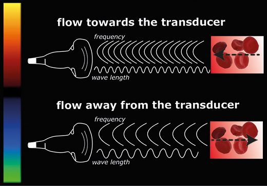 Guerra on 2.26.2020
Expectant first-time mothers have all sorts of questions running through their heads. As the baby develops, the mother wonders about its health, if she will be a good mother, what the future will hold for the family, and if they will be one and done or have a big family. Medical professionals and doctors can only answer so much. They are smart people, but they don’t know everything. They aren’t able to predict the future or know what’s in the minds of others. What they can do is monitor the health and development of the baby and make educated predictions about the future. It’s kind of like predicting the future, but they use skill, science, and data to make those predictions. One of the best tools at their disposal is the ultrasound machine.
Prenatal care is the treatment most often associated with ultrasound machines. Ultrasound images of babies in utero are used in marketing campaigns, in movies and television, and are quite pervasive in the public consciousness. Long before the average person becomes a parent, they have some semblance of what an ultrasound image is and how it’s obtained. The ultrasound machine is a versatile machine that has many uses, while new uses are developing all the time.
The main technology of the ultrasound is not new; it’s based on sonar, sending and receiving and interpreting sound waves. While the basics are rudimentary, the ancillary technology in ultrasounds is cutting edge. Advances in 4D imaging and AI make ultrasounds among the most advanced medical equipment in the world. How reliable is ultrasound accuracy for pregnancy? Let’s find out.
HOW ACCURATE ARE THEY FOR PREDICTING DUE DATE?
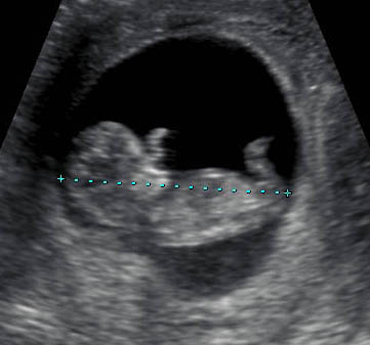
This is a top-of-mind question for all expecting parents. They can gauge it on their own with a slight degree of accuracy, but a doctor can get a lot closer. Evidence suggests that the ultrasound is more accurate than using the last menstrual period for predicting when the baby is due. The ultrasound is more accurate during the first trimester, and early in the second trimester, it is accurate to within a week. Later in the second and into the third trimesters, the ultrasound will be less accurate in predicting. Keep in mind that this is only an estimate. No doctor can predict the exact day that a baby will be born, unless scheduling a cesarean section.
DETECTING FETAL HEARTBEAT
In detecting a fetal heartbeat, an ultrasound should be 100 percent accurate. If there is one, the ultrasound will detect it with ease. The heartbeat is viewable during the gestational period beyond seven to eight weeks. In the early part of the first trimester, it is difficult to differentiate between an earlier-than-estimated pregnancy and an overlooked miscarriage. It usually takes two ultrasounds with several days in between to confirm or rule out a miscarriage at this point. An abdominal ultrasound will be able to detect the heartbeat after eight weeks. If the pregnancy has a gestational age of less than eight weeks, then a transvaginal ultrasound is needed for an accurate reading.
DIAGNOSING A MISCARRIAGE
As mentioned above, in the first trimester it can be hard to differentiate between a pregnancy and a miscarriage based on the results of one ultrasound exam. Doctors will order a second one if they aren’t sure about what they are seeing. There are times when one ultrasound is able to determine pregnancy, so there is not a defined number of ultrasounds that will detect a miscarriage. It all depends on the skill of the technician, the doctor, and the machine doing the ultrasound. Tests like checking the woman’s hCG level are used in conjunction with an ultrasound to get an accurate diagnosis.
DIAGNOSING BIRTH DEFECTS
Doctors can use ultrasounds for diagnosing birth defects, but there are questions about the accuracy. It is believed that a second-trimester ultrasound performed between 16–20 weeks can detect three of four major birth defects. But it is not uncommon for a woman to have an ultrasound give a false positive for birth defects. It’s important for pregnant women to have this information before the exam is done so they can make informed decisions based on the results. Second-trimester ultrasounds have a higher success rate at finding anomalies in the fetus than in the first trimester. First-trimester exams can still provide important information. A study published in 2016 found that first-trimester ultrasounds were capable of finding anomalies. The researchers were successful in finding birth defects in 30 percent of women at low risk and 60 percent of women at high risk of having babies with anomalies. In other cases, like Down syndrome, ultrasounds can’t give a definite diagnosis, only show markers that indicate an elevated risk of certain conditions.
DETERMINING THE BABY’S GENDER
Midway through the pregnancy, an ultrasound can find out the gender of the baby, if you choose to know. For many mothers this is the one and only ultrasound they will have if everything is progressing normally. Ultrasound imaging is good enough to find the telltale signs of gender in the womb. The only limiting factors are the position of the baby and the skill of the ultrasound technician. If the baby isn’t cooperating and is in the wrong position, getting a clear image will be difficult.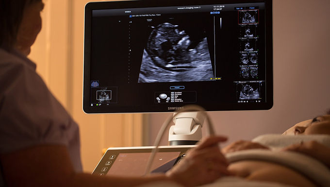 There are specific structures specific to males and females, beyond fully formed reproductive organs, that the technician will look for to determine the sex.
PREDICTING THE BABY’S SIZE
This is an area in which the ultrasound isn’t very reliable. Ultrasounds can be off by multiple pounds in predicting the weight and overall size of a baby. The best an ultrasound exam can do is estimate weight. The difficulty goes both ways, low and high. It is accepted that the exam is not good for predicting low birth weight, and estimates vary on assessing babies that are oversize. If the doctor is worried about low birth weight, there are other tools available that will give a better diagnosis.
MXR Imaging has portable Philips ultrasound machines for sale. Keep your staff adaptable and mobile with this versatile machine. Contact us today for availability.
J. Guerra |
How accurately can ultrasound show myocardial infarction?
Myocardial infarction is one of the main causes of death among the working population.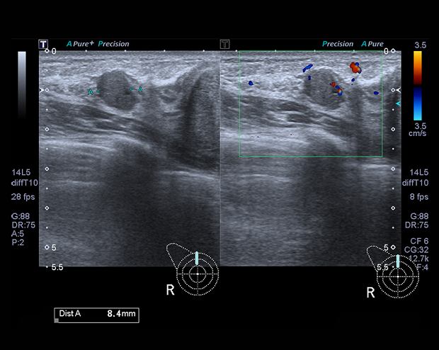 Pathology does not differ in specific clinical signs that would allow an accurate diagnosis without additional research. Ultrasound of the heart is one of the diagnostic methods, which shows changes in the heart muscle.
The essence of ultrasound diagnostics
The method is based on the application of ultrasonic waves with certain characteristics. Ultrasound is not audible to the human ear. The apparatus generates a stream of waves which are directed by the transducer to the desired area of study. Having reached the tissues, they are reflected and returned back.
Based on the received signals, the ultrasound machine generates an image of the organ, which is studied by the doctor of ultrasound diagnostics. The study has no contraindications, age restrictions . Ultrasonic waves do not give radiation exposure to the body , so the number of procedures is not limited. Special training is not required.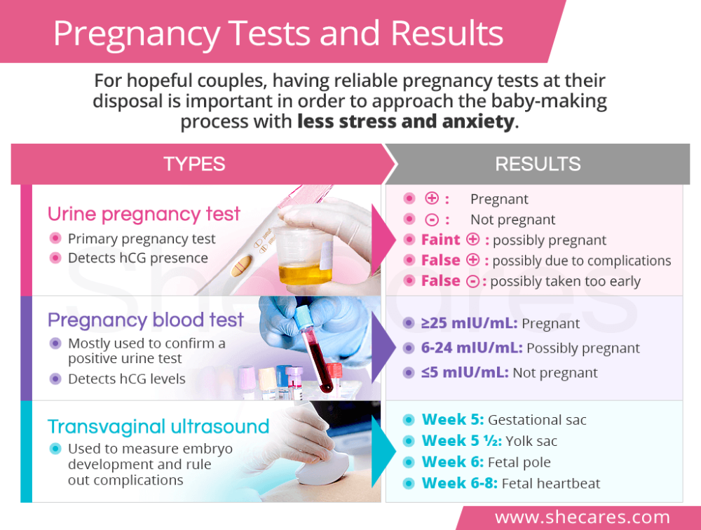
What does an ultrasound of the heart show?
During the examination, the specialist evaluates:
- organ dimensions, integrity, structure, thickness of its walls;
- valve operation;
- contractility;
- pressure level in the pulmonary artery;
- cardiac blood flow rate;
- location of pathology, degree of damage, presence of complications.
ECG and ultrasound of the heart are not interchangeable studies, as they reflect different aspects of the work of the muscular organ. In the first case, the function of the conducting system and its violations are studied.
ECG can indirectly judge the size of the cardiac cavities. It does not provide information about the structural features of the heart - the operation of valves, the presence of inflammation, blood clots, malformations, etc. Ultrasound diagnostics allows you to evaluate all layers of the heart, chambers, identify defects, aneurysms after a heart attack and other changes .
Ultrasound as a preventive measure for myocardial infarction
Prophylactic testing is recommended for people who are at risk for the disease:
- persons over 45 years of age;
- history of atherosclerosis;
- suffering from hypertension, obesity;
- having bad habits;
- persons with a genetic predisposition.
Consult a doctor for advice, diagnostics if:
- suffocation;
- cough;
- chest pains radiating to left arm, ear, jaw, neck;
- arrhythmias.
A heart attack may be asymptomatic , if it is suspected, ultrasound is done several times. The first one is for making a diagnosis, the next ones are during the course of treatment in order to monitor its effectiveness, to identify the likelihood of complications.
How is an ultrasound of the heart performed?
If the person is taking any medications, the doctor must be informed before the start of the study , as they may affect the results. If there are results of previous ultrasounds, you must bring them with you so that the doctor can see the process in dynamics.
The patient undresses to the waist, lies on the couch on his back or on his side. A special gel is applied to the skin in the chest area for good glide of the sensor, enhancing the conduction of ultrasonic waves. During the procedure, the patient does not experience discomfort . On the monitor, the specialist evaluates the condition of the heart, its walls and other characteristics. The survey takes 15-25 minutes .
After the examination, the ultrasound diagnostician draws up a conclusion, describes the resulting image, and makes a presumptive diagnosis. The results indicate the individual indicators of the patient. The final diagnosis is made by a cardiologist on the basis of the collected history, examination, research results, and then prescribes treatment .
Features of diagnosis after a heart attack
Ultrasound determines the degree of necrotic lesion according to the thickness of the scar after a heart attack.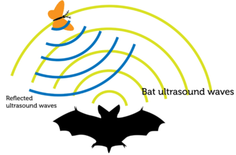 If at the time of the study it is thick, then the pathology is at the initial stage, it is possible to normalize blood circulation. The thinner the wall, the stronger the lesion and the less likely it is to restore the work of the myocardium.
When the blood flow is disturbed, parts of the heart muscle begin to be replaced by connective tissue, which cannot perform any functions. Examining the myocardium, the doctor examines vascular patency, the presence of blood clots that can clog the lumen and stop blood flow. In this case, necrotic changes will occur in 15-30 minutes. nine0003
The direction of blood flow can be used to determine the location of the area of necrosis . With inflammation of the pericardium in the post-infarction period, uncharacteristic movements and adhesions can be seen on the image. With ultrasound, the index of contractility of the walls of the left ventricle is determined. In a healthy person, the indicator is equal to one, with a heart attack it is much higher.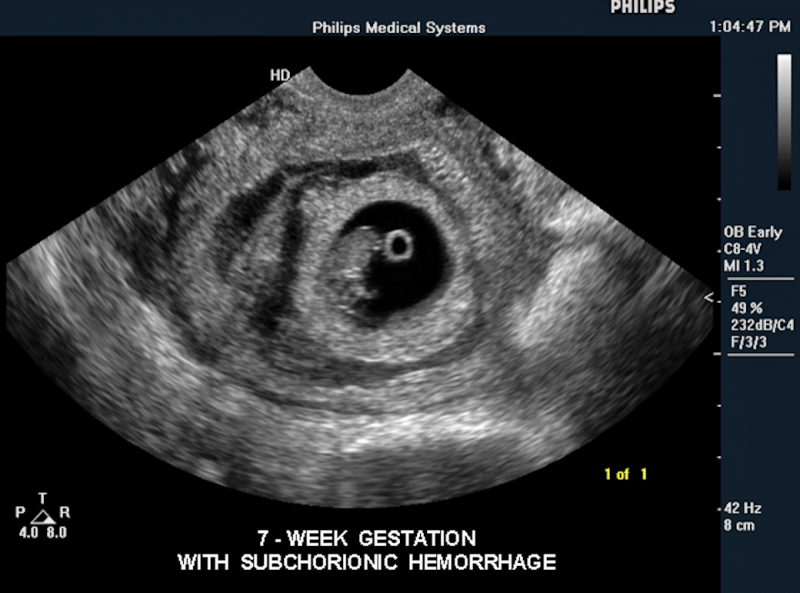
Examination in the diagnostic center "DonMed"
Ultrasound of the heart and other types of examination can be done in the multidisciplinary medical center "DonMed" . The reception is carried out by qualified specialists, the examination takes place on modern expert-class equipment that provides high-quality images - the “ Philips Affiniti 70 ” system, for more information about which you can learn at link . Studies can be completed without a doctor's appointment, and after receiving the results, the patient can make an appointment with a specialized specialist for treatment, and, if necessary, further diagnostics.
Ultrasound is a safe and highly informative diagnostic method , which helps to evaluate the work of the heart visually in dynamics. A timely appeal to the specialists of the center will help to identify cardiovascular pathologies in the early stages of development, to avoid complications.
Deciphering the results takes 20-60 minutes, in complex clinical cases - a day. The center employs experienced gynecologists, phlebologists, therapists, gastroenterologists and other specialists. To make an appointment with a doctor, take tests, undergo diagnostics at the DonMed center, call the phone number indicated on the website or leave a request for feedback on the website. nine0003
Find out prices for ultrasound of the heart in the clinic "DonMed" You can find out by link .
The staff of MC “DonMed” applies high quality standards in medicine now in Lyubertsy (MOSCOW) , clinic “Horizont”.
The clinic has modern devices that allow accurate, high-quality diagnostics.
The Philips Affiniti 70 Expert Ultrasound is an innovative ultrasound system designed for use in Cardiology, Pediatrics, General and Women's Health, enabling scanning of technical patients with high penetration transducers.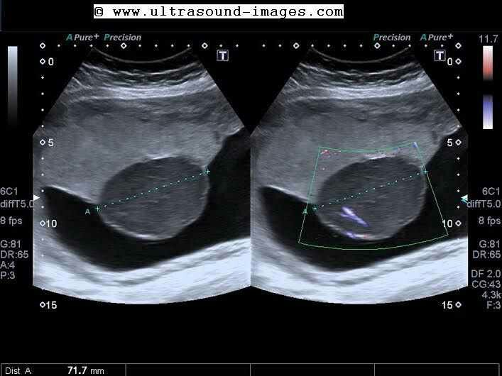 nine0003
Samsung Medison W10 Expert Class Ultrasound System is a premium ultrasound scanner that enhances diagnostic capabilities with high-tech computing system, accurate analysis tools.
Go to the website MC Horizon
vaginal and transvaginal - MEDSI
What is vaginal ultrasound?
Vaginal (transvaginal) pelvic ultrasound is performed by inserting a special device equipped with a transducer into the vagina. nine0003
The device is a rod with a handle, which is made of plastic, about 10-12 centimeters long and up to three centimeters in diameter. A special groove can be built into it to insert a needle for taking biopsy material.
Examination allows to determine the presence of pathologies, neoplasms or diseases in the following female genital organs:
- Uterus
- Fallopian tubes
- Ovaries nine0026 Cervix
It is considered the most effective for the study of these parts of the reproductive system, as it allows you to identify various health problems in the patient at an early stage. Ultrasound of the small pelvis with a sensor is able to show the presence of deviations already at a time when other studies do not show any problem areas.
How is the procedure?
The examination is organized as follows:
- The patient must remove clothing from the lower part of the body (from the waist down)
- She settles down on a special couch in the same way as during a regular gynecological examination
- The doctor prepares the transducer: puts an individual condom on it, lubricates it with a special gel for the procedure
- The physician then inserts the device shallowly into the patient's vagina
- To get a complete picture of the state of the organs, he can move the sensor from side to side nine0026 All data is recorded and processed by the doctor
The gel is needed to facilitate the penetration of the transducer (and thereby reduce the likelihood of negative sensations) and enhance the ultrasonic effect by increasing the conductivity.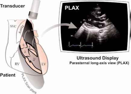
This type of examination lasts no more than 10 minutes. It is painless and gives the most complete picture even when an abdominal ultrasound shows nothing or cannot be performed.
When is a pelvic ultrasound probe needed? nine0009
There are symptoms in which the doctor must refer the patient for a transvaginal examination:
- Pain in the lower abdomen (not related to the menstrual cycle)
- Neoplasm suspected
- Too short or too long period of menstrual bleeding or its absence
- Impossibility of pregnancy
- Non-menstrual bleeding
- The presence of violations of the patency of the fallopian tubes
- Nausea, vomiting and weakness with bleeding from the vagina
Doctors recommend using this type of examination for preventive purposes, since not every ailment may have symptoms at an early stage, just as pregnancy in the first trimester may not manifest itself with classic symptoms (nausea, etc.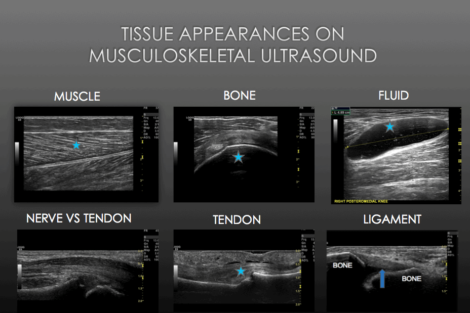 ).
).
In this case, vaginal ultrasound is used for:
- Infertility diagnostics
- The need to determine the presence of changes in the size of the ovaries and uterus
- Pregnancy diagnostics
- Pregnancy monitoring (first trimester only)
- General supervision of the uterus, fallopian tubes and ovaries
A pelvic ultrasound with two probes can be performed at the same time. In this case, an abdominal ultrasound examination is performed first, and then a transvaginal one. The use of two types of analysis at once is necessary to detect violations in the highly located organs of the small pelvis. nine0003
What does a vaginal ultrasound show?
This examination allows to evaluate the following parameters of the organs of the reproductive system:
- Size of the uterus. In normal condition, it should be about seven centimeters long, six wide and 4.2 in diameter. If it is significantly less or more, then this indicates the presence of pathology
- Echogenicity.
 The structure of the organs should be homogeneous, uniform, have clearly defined, well-visible edges
 - General picture of internal organs. The uterus should be slightly tilted forward. And the fallopian tubes may be slightly visible, but should not be clearly visible without the use of contrast agent
Diagnosable diseases
Transvaginal ultrasound can detect a number of diseases and problems in the reproductive system at an early stage. It allows you to detect:
- Fluid and pus in the uterus and fallopian tubes. The cause of their appearance may be infections, viruses, mechanical damage
- Endomentriosis - excessive growth of cells of the inner layer of uterine tissues into other layers and organs. It can occur due to inflammatory processes, damage (surgery, abortion), the appearance of neoplasms, disorders in the endocrine system, too frequent use of certain drugs and substances
- Myoma is a benign neoplasm in the tissues of the uterus or its cervix.
 May occur due to chronic diseases, frequent abortions, hormonal disorders, constant stress, pathologies, overweight, with hereditary predisposition
 - Cysts and polycystic ovaries are tumors filled with fluid. Occur in endocrine disorders, chronic diseases of the genitourinary system
- A variety of polyps on the walls of the uterus - benign formations in the endometrium of the organ. They can reach several centimeters in diameter. Their appearance may be associated with polycystic disease, chronic diseases, mastopathy, fibroma
- Inflammation and enlargement of organs can occur both due to infection and trauma
- Vesical skid - appears instead of a full-fledged embryo in the process of conception, filled with liquid. It occurs due to duplication of male chromosomes with the loss of female chromosomes, sometimes due to the fertilization of an egg that does not contain a nucleus. This disease is rare
- Fetal development disorders during pregnancy
- Malformations and pathologies in the development of the fallopian tubes: obstruction, spiral or too long tubes, blind passages, duplication of organs
- An ectopic pregnancy occurs when an egg, after fertilization, attaches itself outside the tissues of the uterus.
 Occurs due to blockage of the fallopian tubes, congenital anomalies in them, as well as after the inflammatory process, abortion
 - Cancer - a malignant tumor in various organs:
- Uterus
- Ovaries
- Cervix
- Chorionepithelioma - a malignant neoplasm that occurs during or after pregnancy from chorion cells (the membrane of the embryo attached to the wall of the uterus)
Pre-exam preparation steps
No special preparation is required for a pelvic ultrasound with a transducer, but there are a few requirements:
Doctors also recommend using such a study on certain days of the cycle, depending on which organ and for what purpose you need to diagnose:0027
It is important to remember about personal hygiene before the examination, use wet and other wipes. nine0003
If you plan to perform a pelvic ultrasound with two sensors, then you should pay attention to the preparation for the abdominal examination.
This includes:
- Diet at least three days prior to the examination to reduce the likelihood of symptoms of flatulence and bloating
- The last meal must be finished by six o'clock in the evening on the eve of the analysis
- It is recommended to take an enema after a meal
- If there is still a risk of flatulence, special drugs should be used to reduce gas formation
- Drink at least 400 ml of water one hour before the test
The diet involves the exclusion of a number of products from the diet:
- Sweets
- Flour (bread, biscuits and other)
- Legumes
- Cabbage
- Milk and dairy products
- Uncooked vegetables and fruits
- Coffee and strong tea
- Carbonated drinks
- Fast food
- Fatty foods (meat, fish, oils)
You can eat cereals cooked with water, low-fat boiled beef, poultry and fish, hard cheeses.