Functions of the pelvic girdle
8.3 The Pelvic Girdle and Pelvis - Anatomy and Physiology 2e
Learning Objectives
By the end of this section, you will be able to:
- Define the pelvic girdle and describe the bones and ligaments of the pelvis
- Explain the three regions of the hip bone and identify their bony landmarks
- Describe the openings of the pelvis and the boundaries of the greater and lesser pelvis
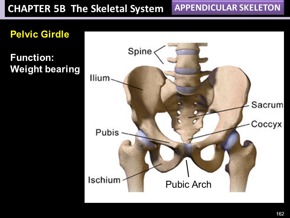
The pelvic girdle (hip girdle) is formed by a single bone, the hip bone or coxal bone (coxal = “hip”), which serves as the attachment point for each lower limb. Each hip bone, in turn, is firmly joined to the axial skeleton via its attachment to the sacrum of the vertebral column. The right and left hip bones also converge anteriorly to attach to each other. The bony pelvis is the entire structure formed by the two hip bones, the sacrum, and, attached inferiorly to the sacrum, the coccyx (Figure 8.12).
Unlike the bones of the pectoral girdle, which are highly mobile to enhance the range of upper limb movements, the bones of the pelvis are strongly united to each other to form a largely immobile, weight-bearing structure. This is important for stability because it enables the weight of the body to be easily transferred laterally from the vertebral column, through the pelvic girdle and hip joints, and into either lower limb whenever the other limb is not bearing weight. Thus, the immobility of the pelvis provides a strong foundation for the upper body as it rests on top of the mobile lower limbs.
Figure 8.12 Pelvis The pelvic girdle is formed by a single hip bone. The hip bone attaches the lower limb to the axial skeleton through its articulation with the sacrum. The right and left hip bones, plus the sacrum and the coccyx, together form the pelvis.
Hip Bone
The hip bone, or coxal bone, forms the pelvic girdle portion of the pelvis. The paired hip bones are the large, curved bones that form the lateral and anterior aspects of the pelvis. Each adult hip bone is formed by three separate bones that fuse together during the late teenage years. These bony components are the ilium, ischium, and pubis (Figure 8.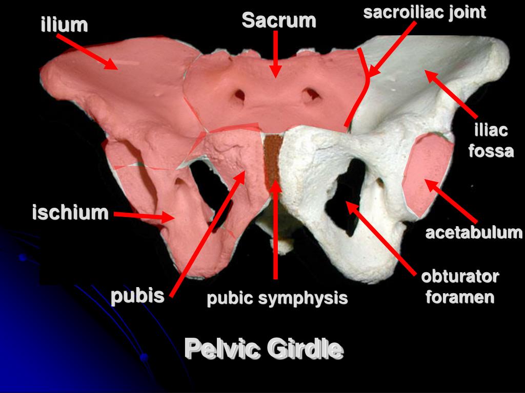 13). These names are retained and used to define the three regions of the adult hip bone.
Figure 8.13 The Hip Bone The adult hip bone consists of three regions. The ilium forms the large, fan-shaped superior portion, the ischium forms the posteroinferior portion, and the pubis forms the anteromedial portion.
The ilium is the fan-like, superior region that forms the largest part of the hip bone. It is firmly united to the sacrum at the largely immobile sacroiliac joint (see Figure 8.12). The ischium forms the posteroinferior region of each hip bone. It supports the body when sitting. The pubis forms the anterior portion of the hip bone. The pubis curves medially, where it joins to the pubis of the opposite hip bone at a specialized joint called the pubic symphysis.
Ilium
When you place your hands on your waist, you can feel the arching, superior margin of the ilium along your waistline (see Figure 8.13). This curved, superior margin of the ilium is the iliac crest.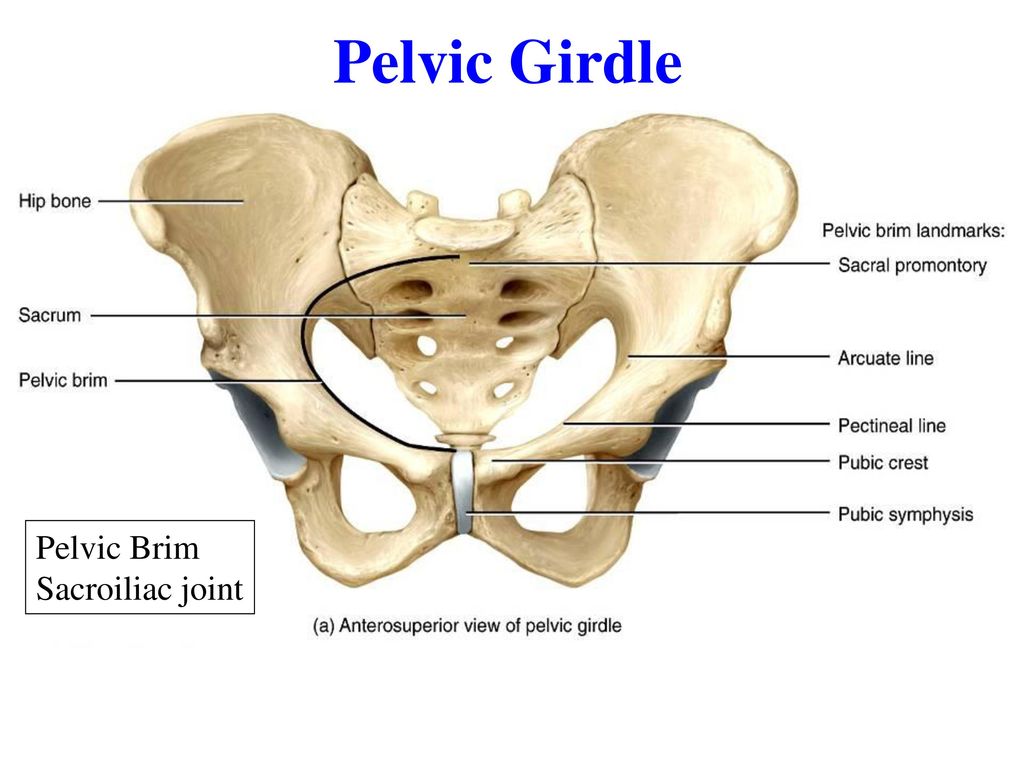 The rounded, anterior termination of the iliac crest is the anterior superior iliac spine. This important bony landmark can be felt at your anterolateral hip. Inferior to the anterior superior iliac spine is a rounded protuberance called the anterior inferior iliac spine. Both of these iliac spines serve as attachment points for muscles of the thigh. Posteriorly, the iliac crest curves downward to terminate as the posterior superior iliac spine. Muscles and ligaments surround but do not cover this bony landmark, thus sometimes producing a depression seen as a “dimple” located on the lower back. More inferiorly is the posterior inferior iliac spine. This is located at the inferior end of a large, roughened area called the auricular surface of the ilium. The auricular surface articulates with the auricular surface of the sacrum to form the sacroiliac joint. Both the posterior superior and posterior inferior iliac spines serve as attachment points for the muscles and very strong ligaments that support the sacroiliac joint.
The shallow depression located on the anteromedial (internal) surface of the upper ilium is called the iliac fossa. The inferior margin of this space is formed by the arcuate line of the ilium, the ridge formed by the pronounced change in curvature between the upper and lower portions of the ilium. The large, inverted U-shaped indentation located on the posterior margin of the lower ilium is called the greater sciatic notch.
Ischium
The ischium forms the posterolateral portion of the hip bone (see Figure 8.13). The large, roughened area of the inferior ischium is the ischial tuberosity. This serves as the attachment for the posterior thigh muscles and also carries the weight of the body when sitting. You can feel the ischial tuberosity if you wiggle your pelvis against the seat of a chair. Projecting superiorly and anteriorly from the ischial tuberosity is a narrow segment of bone called the ischial ramus. The slightly curved posterior margin of the ischium above the ischial tuberosity is the lesser sciatic notch. The bony projection separating the lesser sciatic notch and greater sciatic notch is the ischial spine.
Pubis
The pubis forms the anterior portion of the hip bone (see Figure 8.13). The enlarged medial portion of the pubis is the pubic body. Located superiorly on the pubic body is a small bump called the pubic tubercle. The superior pubic ramus is the segment of bone that passes laterally from the pubic body to join the ilium. The narrow ridge running along the superior margin of the superior pubic ramus is the pectineal line of the pubis.
The pubic body is joined to the pubic body of the opposite hip bone by the pubic symphysis. Extending downward and laterally from the body is the inferior pubic ramus. The pubic arch is the bony structure formed by the pubic symphysis, and the bodies and inferior pubic rami of the adjacent pubic bones. The inferior pubic ramus extends downward to join the ischial ramus. Together, these form the single ischiopubic ramus, which extends from the pubic body to the ischial tuberosity. The inverted V-shape formed as the ischiopubic rami from both sides come together at the pubic symphysis is called the subpubic angle.
Pelvis
The pelvis consists of four bones: the right and left hip bones, the sacrum, and the coccyx (see Figure 8.12). The pelvis has several important functions. Its primary role is to support the weight of the upper body when sitting and to transfer this weight to the lower limbs when standing. It serves as an attachment point for trunk and lower limb muscles, and also protects the internal pelvic organs. When standing in the anatomical position, the pelvis is tilted anteriorly. In this position, the anterior superior iliac spines and the pubic tubercles lie in the same vertical plane, and the anterior (internal) surface of the sacrum faces forward and downward.
The three areas of each hip bone, the ilium, pubis, and ischium, converge centrally to form a deep, cup-shaped cavity called the acetabulum. This is located on the lateral side of the hip bone and is part of the hip joint.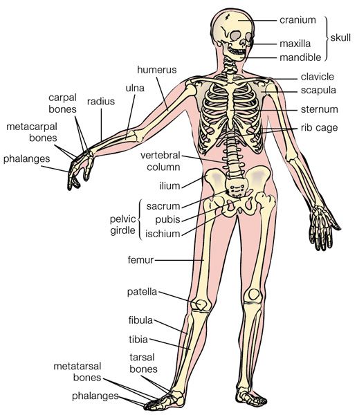 The large opening in the anteroinferior hip bone between the ischium and pubis is the obturator foramen. This space is largely filled in by a layer of connective tissue and serves for the attachment of muscles on both its internal and external surfaces.
Several ligaments unite the bones of the pelvis (Figure 8.14). The largely immobile sacroiliac joint is supported by a pair of strong ligaments that are attached between the sacrum and ilium portions of the hip bone. These are the anterior sacroiliac ligament on the anterior side of the joint and the posterior sacroiliac ligament on the posterior side. Also spanning the sacrum and hip bone are two additional ligaments. The sacrospinous ligament runs from the sacrum to the ischial spine, and the sacrotuberous ligament runs from the sacrum to the ischial tuberosity. These ligaments help to support and immobilize the sacrum as it carries the weight of the body.
Figure 8.14 Ligaments of the Pelvis The posterior sacroiliac ligament supports the sacroiliac joint. The sacrospinous ligament spans the sacrum to the ischial spine, and the sacrotuberous ligament spans the sacrum to the ischial tuberosity. The sacrospinous and sacrotuberous ligaments contribute to the formation of the greater and lesser sciatic foramina.
Interactive Link
Watch this video for a 3-D view of the pelvis and its associated ligaments. What is the large opening in the bony pelvis, located between the ischium and pubic regions, and what two parts of the pubis contribute to the formation of this opening?
The sacrospinous and sacrotuberous ligaments also help to define two openings on the posterolateral sides of the pelvis through which muscles, nerves, and blood vessels for the lower limb exit. The superior opening is the greater sciatic foramen. This large opening is formed by the greater sciatic notch of the hip bone, the sacrum, and the sacrospinous ligament. The smaller, more inferior lesser sciatic foramen is formed by the lesser sciatic notch of the hip bone, together with the sacrospinous and sacrotuberous ligaments.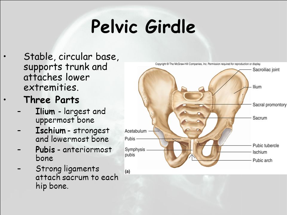
The space enclosed by the bony pelvis is divided into two regions (Figure 8.15). The broad, superior region, defined laterally by the large, fan-like portion of the upper hip bone, is called the greater pelvis (greater pelvic cavity; false pelvis). This broad area is occupied by portions of the small and large intestines, and because it is more closely associated with the abdominal cavity, it is sometimes referred to as the false pelvis. More inferiorly, the narrow, rounded space of the lesser pelvis (lesser pelvic cavity; true pelvis) contains the bladder and other pelvic organs, and thus is also known as the true pelvis. The pelvic brim (also known as the pelvic inlet) forms the superior margin of the lesser pelvis, separating it from the greater pelvis. The pelvic brim is defined by a line formed by the upper margin of the pubic symphysis anteriorly, and the pectineal line of the pubis, the arcuate line of the ilium, and the sacral promontory (the anterior margin of the superior sacrum) posteriorly.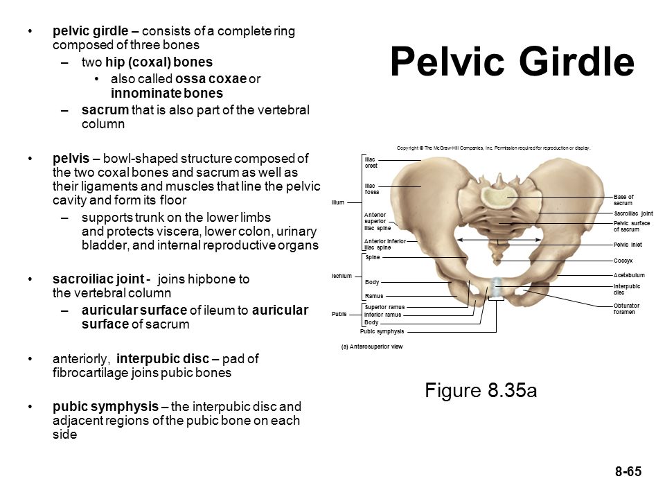 The inferior limit of the lesser pelvic cavity is called the pelvic outlet. This large opening is defined by the inferior margin of the pubic symphysis anteriorly, and the ischiopubic ramus, the ischial tuberosity, the sacrotuberous ligament, and the inferior tip of the coccyx posteriorly. Because of the anterior tilt of the pelvis, the lesser pelvis is also angled, giving it an anterosuperior (pelvic inlet) to posteroinferior (pelvic outlet) orientation.
Figure 8.15 Male and Female Pelvis The female pelvis is adapted for childbirth and is broader, with a larger subpubic angle, a rounder pelvic brim, and a wider and more shallow lesser pelvic cavity than the male pelvis.
Comparison of the Female and Male Pelvis
The differences between the adult female and male pelvis relate to function and body size. In general, the bones of the male pelvis are thicker and heavier, adapted for support of the male’s heavier physical build and stronger muscles.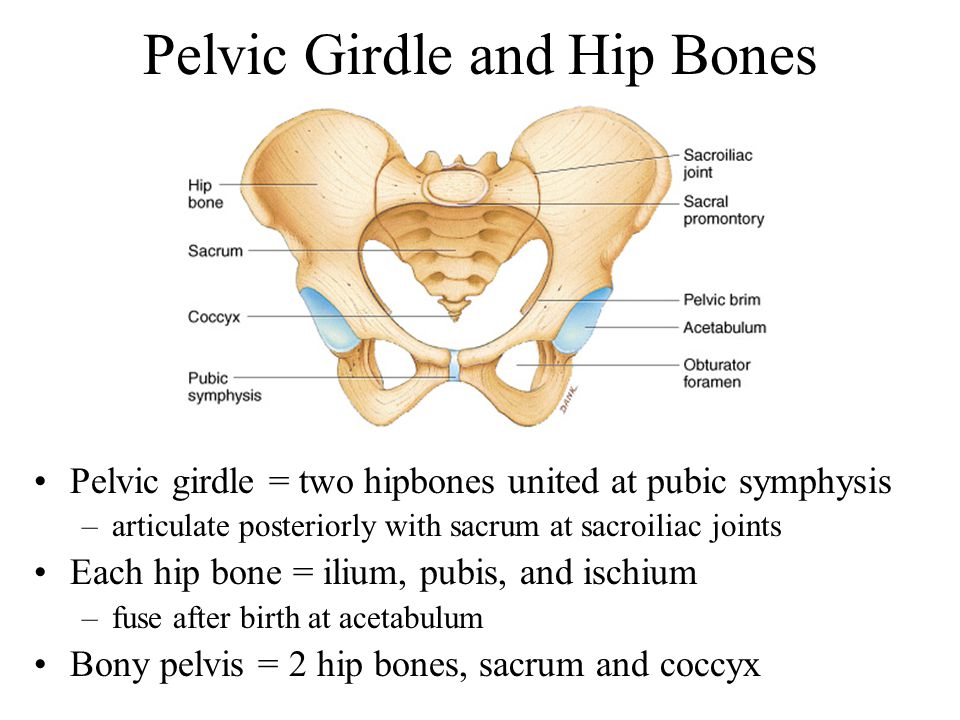 The greater sciatic notch of the male hip bone is narrower and deeper than the broader notch of females. Because the female pelvis is adapted for childbirth, it is wider than the male pelvis, as evidenced by the distance between the anterior superior iliac spines (see Figure 8.15). The ischial tuberosities of females are also farther apart, which increases the size of the pelvic outlet. Because of this increased pelvic width, the subpubic angle is larger in females (greater than 80 degrees) than it is in males (less than 70 degrees). The female sacrum is wider, shorter, and less curved, and the sacral promontory projects less into the pelvic cavity, thus giving the female pelvic inlet (pelvic brim) a more rounded or oval shape compared to males. The lesser pelvic cavity of females is also wider and more shallow than the narrower, deeper, and tapering lesser pelvis of males. Because of the obvious differences between female and male hip bones, this is the one bone of the body that allows for the most accurate sex determination.
 Table 8.1 provides an overview of the general differences between the female and male pelvis.
Overview of Differences between the Female and Male Pelvis
| Female pelvis | Male pelvis | |
|---|---|---|
| Pelvic weight | Bones of the pelvis are lighter and thinner | Bones of the pelvis are thicker and heavier |
| Pelvic inlet shape | Pelvic inlet has a round or oval shape | Pelvic inlet is heart-shaped |
| Lesser pelvic cavity shape | Lesser pelvic cavity is shorter and wider | Lesser pelvic cavity is longer and narrower |
| Subpubic angle | Subpubic angle is greater than 80 degrees | Subpubic angle is less than 70 degrees |
| Pelvic outlet shape | Pelvic outlet is rounded and larger | Pelvic outlet is smaller |
Table 8.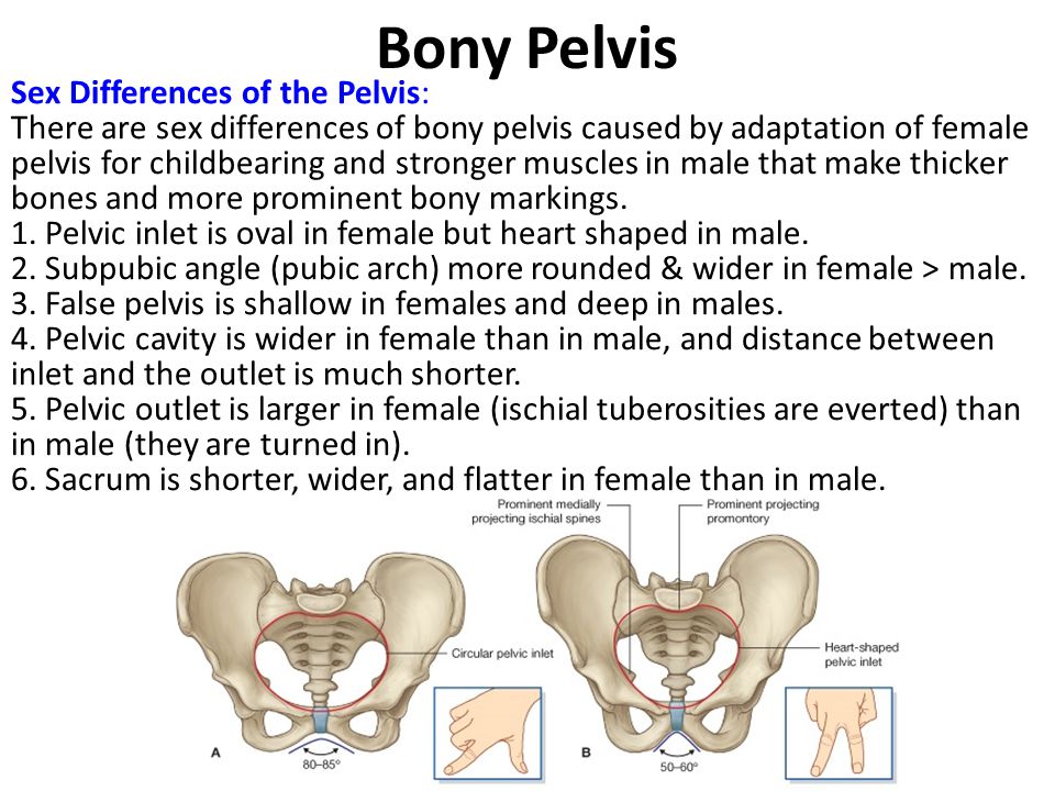 1
1
Career Connection
Forensic Pathology and Forensic Anthropology
A forensic pathologist (also known as a medical examiner) is a medically trained physician who has been specifically trained in pathology to examine the bodies of the deceased to determine the cause of death. A forensic pathologist applies understanding of disease as well as toxins, blood and DNA analysis, firearms and ballistics, and other factors to assess the cause and manner of death. At times, a forensic pathologist will be called to testify under oath in situations that involve a possible crime. Forensic pathology is a field that has received much media attention on television shows or following a high-profile death.
While forensic pathologists are responsible for determining whether the cause of someone’s death was natural, by suicide, accidental, or by homicide, there are times when uncovering the cause of death is more complex, and other skills are needed.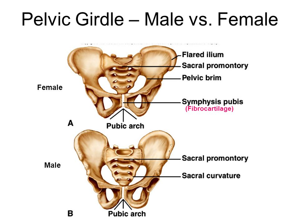 Forensic anthropology brings the tools and knowledge of physical anthropology and human osteology (the study of the skeleton) to the task of investigating a death. A forensic anthropologist assists medical and legal professionals in identifying human remains. The science behind forensic anthropology involves the study of archaeological excavation; the examination of hair; an understanding of plants, insects, and footprints; the ability to determine how much time has elapsed since the person died; the analysis of past medical history and toxicology; the ability to determine whether there are any postmortem injuries or alterations of the skeleton; and the identification of the decedent (deceased person) using skeletal and dental evidence.
Due to the extensive knowledge and understanding of excavation techniques, a forensic anthropologist is an integral and invaluable team member to have on-site when investigating a crime scene, especially when the recovery of human skeletal remains is involved.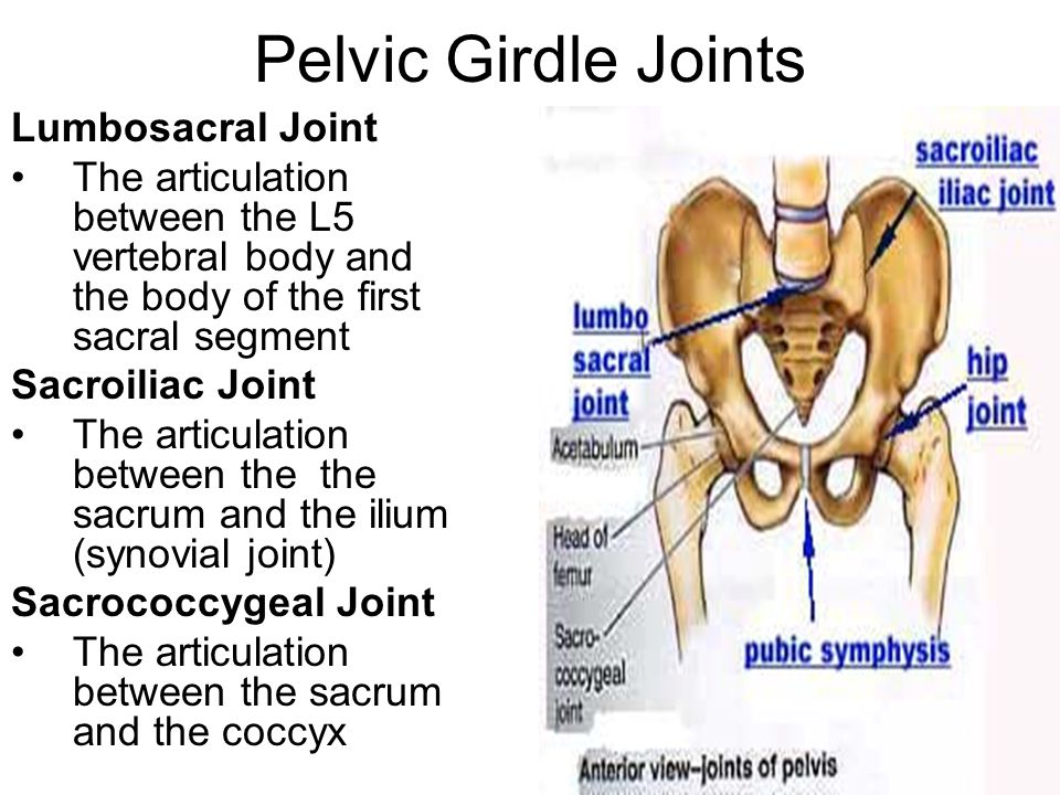 When remains are bought to a forensic anthropologist for examination, they must first determine whether the remains are in fact human. Once the remains have been identified as belonging to a person and not to an animal, the next step is to approximate the individual’s age, sex, race, and height. The forensic anthropologist does not determine the cause of death, but rather provides information to the forensic pathologist, who will use all of the data collected to make a final determination regarding the cause of death.
The Pelvic Girdle - Structure - Function - Assessment
The pelvic girdle is a ring-like bony structure, located in the lower part of the trunk. It connects the axial skeleton to the lower limbs.
In this article, we shall look at the anatomy of the pelvic girdle – its bony landmarks, functions, and its clinical relevance.
Structure of the Pelvic GirdleThe bony pelvis consists of the two hip bones (also known as innominate or pelvic bones), the sacrum and the coccyx.
There are four articulations within the pelvis:
- Sacroiliac joints (x2) – between the ilium of the hip bones, and the sacrum
- Sacrococcygeal symphysis – between the sacrum and the coccyx.
- Pubic symphysis – between the pubis bodies of the two hip bones.
Ligaments attach the lateral border of the sacrum to various bony landmarks on the bony pelvis to aid stability.
By TeachMeSeries Ltd (2023)
Fig 1 – The pelvic girdle is formed by the hip bones, sacrum and coccyx.
Functions of the PelvisThe strong and rigid pelvis is adapted to serve a number of roles in the human body. The main functions being:
- Transfer of weight from the upper axial skeleton to the lower appendicular components of the skeleton, especially during movement.
- Provides attachment for a number of muscles and ligaments used in locomotion.

- Contains and protects the abdominopelvic and pelvic viscera.
The osteology of the pelvic girdle allows the pelvic region to be divided into two:
- Greater pelvis (false pelvis) – located superiorly, it provides support of the lower abdominal viscera (such as the ileum and sigmoid colon). It has little obstetric relevance.
- Lesser pelvis (true pelvis) – located inferiorly. Within the lesser pelvis reside the pelvic cavity and pelvic viscera.
The junction between the greater and lesser pelvis is known as the pelvic inlet. The outer bony edges of the pelvic inlet are called the pelvic brim.
By TeachMeSeries Ltd (2023)
Fig 2 – The greater and lesser pelvis. The lesser pelvis is the ‘true’ pelvis and contains the pelvic cavity.
Pelvic InletThe pelvic inlet marks the boundary between the greater pelvis and lesser pelvis.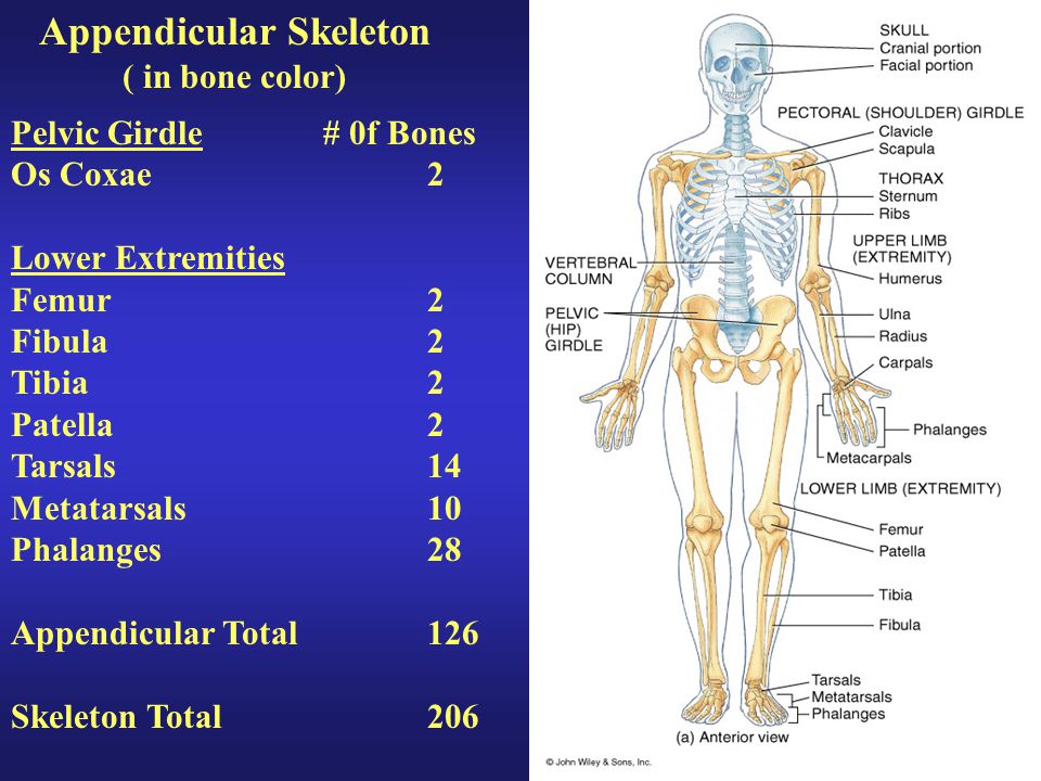 Its size is defined by its edge, the pelvic brim.
The borders of the pelvic inlet:
- Posterior – sacral promontory (the superior portion of the sacrum) and sacral wings (ala).
- Lateral – arcuate line on the inner surface of the ilium, and the pectineal line on the superior pubic ramus.
- Anterior – pubic symphysis.
The pelvic inlet determines the size and shape of the birth canal, with the prominent ridges a key site for attachment of muscle and ligaments.
Some alternative descriptive terminology can be used in describing the pelvic inlet:
- Linea terminalis – the combined pectineal line, arcuate line and sacral promontory.
- Iliopectineal line – the combined arcuate and pectineal lines. This represents the lateral border of the pelvic inlet.
By TeachMeSeries Ltd (2023)
Fig 3 – Looking down onto the pelvis, the borders of the pelvic brim.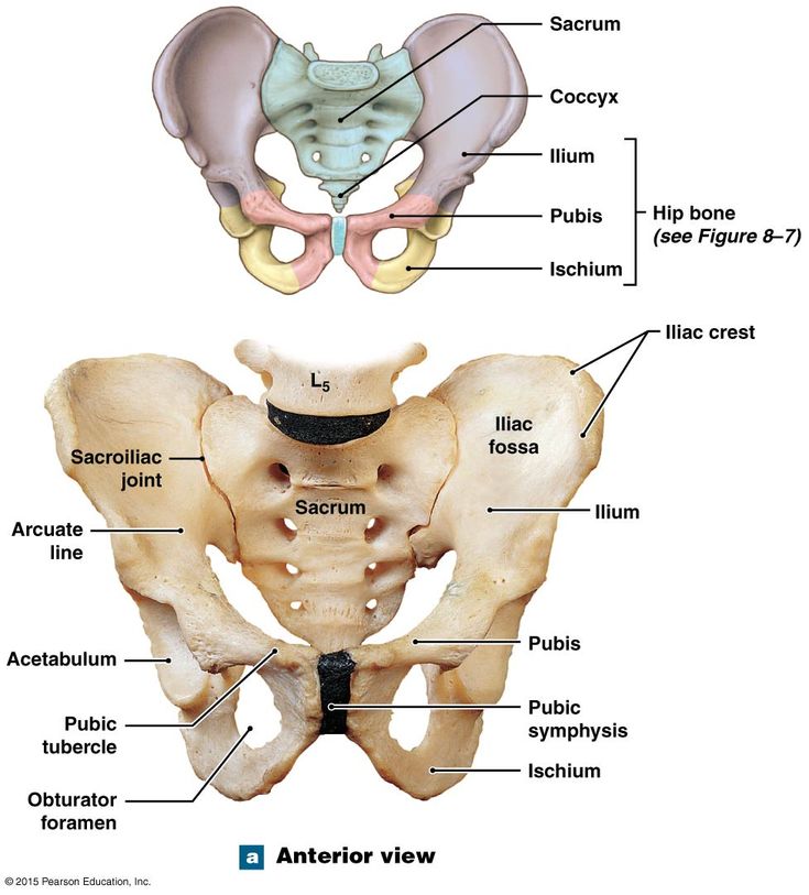
The pelvic outlet is located at the end of the lesser pelvis, and the beginning of the pelvic wall.
Its borders are:
- Posterior: The tip of the coccyx
- Lateral: The ischial tuberosities and the inferior margin of the sacrotuberous ligament
- Anterior: The pubic arch (the inferior border of the ischiopubic rami).
The angle beneath the pubic arch is known as the sub-pubic angle and is of a greater size in women.
By TeachMeSeries Ltd (2023)
Fig 4 – The borders of the pelvic outlet.
Adaptation for ChildbirthThe majority of women have a gynaecoid pelvis, as opposed to the male android pelvis. The slight differences in their structures creates a greater pelvic outlet, adapted to aid the process of childbirth. When comparing the two, the gynaecoid pelvis has:
- A wider and broader structure yet it is lighter in weight
- An oval-shaped inlet compared with the heart-shaped android pelvis.
- Less prominent ischial spines, allowing for a greater bispinous diameter
- A greater angled sub-pubic arch, more than 80-90 degrees.
- A sacrum which is shorter, more curved and with a less pronounced sacral promontory.
In addition to the bony adaptations, the sacrotuberous and sacrospinous ligaments can stretch under the influence of progesterone and increase the size of the outlet further.
By TeachMeSeries Ltd (2023)
Fig 5 – Gynaecoid pelvis vs the android pelvis.
Clinical Relevance: Assessment of the Female Bony Pelvis
The lesser pelvis is the bony canal through which the fetus has to pass during childbirth. It is therefore of great importance to determine the diameter of this canal and therefore the childbearing capacity of the mother.
The diameter can be determined by a pelvic examination or radiographically. There are two measurements that are of importance:
Obstetric ConjugateIn order to determine the narrowest fixed distance that the fetus would have to negotiate, the minimum antero-posterior diameter of the pelvic inlet is measured.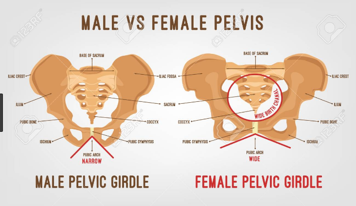
This distance is between the sacral promontory and the midpoint of the pubic symphysis (where the pubic bone is thickest) and is known as the obstetric conjugate (or true conjugate). However, this measurement cannot be assessed clinically, due to the presence of the bladder.
Diagonal Conjugate
The diagonal conjugate is the alternative, measuring from the inferior border of the pubic symphysis to the sacral promontory and can be measured manually via the vagina.
(To do this you use the tip of your middle finger to measure the sacral promontory and then using the other hand to mark the level of the inferior margin of the pubic symphysis on the examining hand. You then use the distance between the index finger and the pubic symphysis to measure the diagonal conjugate, ideally 11cm or greater)
In addition to measuring the diagonal conjugate, a mid-pelvis check is carried out. Here, the clinician is testing for straight side walls and measuring the bispinous diameter which is narrowest part of the pelvic canal.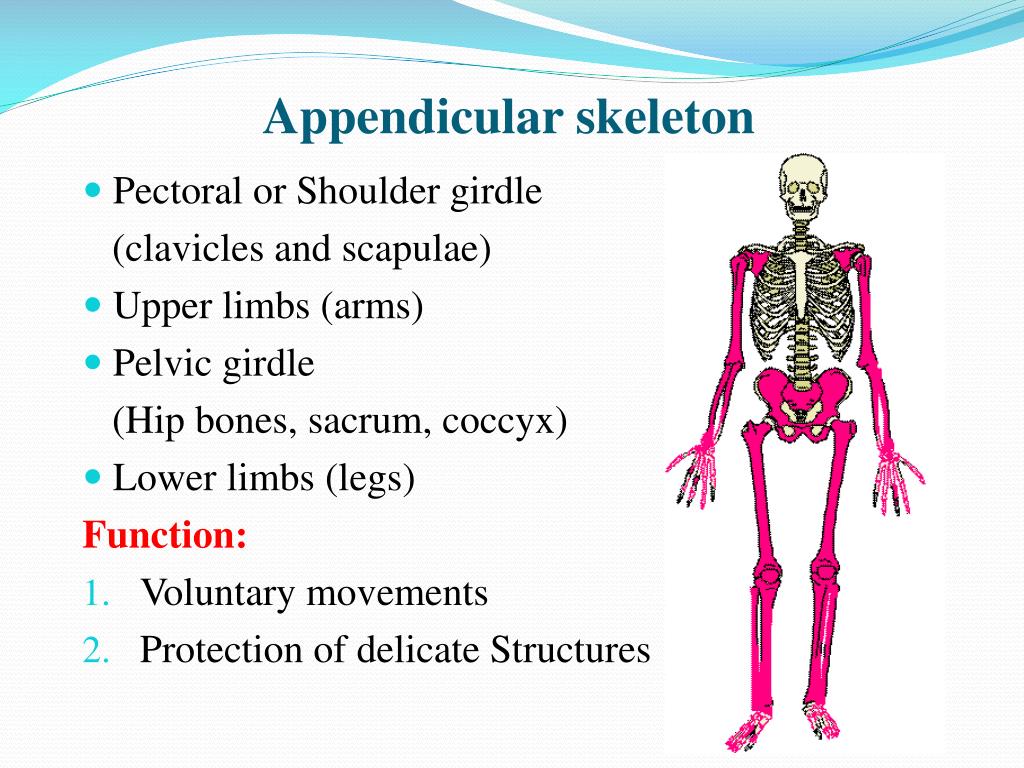 The width of the subpubic angle at the pelvic outlet can be determined by the distance between the ischial tuberosities.
By TeachMeSeries Ltd (2023)
Fig 6 – Assessment of the female pelvis, via the diagonal conjugate
printPrint this Article
Encyclopedia - Pelvic Girdle
The pelvic girdle bears the weight of the entire upper body, and the lower limbs rest against it. This part of the body experiences a lot of pressure from both sides - from below and from above, it promotes the movements of the lower limbs and protects important internal organs. The most important function of the pelvis is locomotor, contributing to the movement of the body in space.
Differences between the human pelvis and the pelvis of other mammals are related
with an upright body position. Only in humans transverse dimensions
the pelvis is more straight (anterior-posterior). Even the pelvis of the great apes
already more elongated. In the human fetus, the pelvis has the same shape,
as the pelvis of four-legged mammals. Pelvis transformations begin
under the influence of mechanical loads: gravity of the body, pressure in the hip
joint during movements, etc. Active formation of sex differences
in the structure of the pelvis occurs during puberty under the action of
hormones. Characteristically, with reduced ovarian function (female
gonads) slows down the formation of female
features - the pelvis remains relatively narrow.
Pelvic bones
The pelvic girdle, or pelvis, is a strong bone ring that is located in the lower part of the skeleton of the human torso. It is formed from almost immovably connected bones: unpaired - the sacrum and two massive, flat - right and left pelvic bones. A sacrum is wedged between the pelvic bones, to which is attached a small bone - the coccyx - a rudimentary remnant of the caudal skeleton.
In children under 16, each pelvic bone consists of 3 separate bones: the ilium, ischium and pubis, interconnected by layers of cartilage. After 16 years they grow together. In this place there is a deep fossa - the acetabulum. It includes the head of the femur, forming the hip joint.
Structure of the ischium
The ischial bone has a powerful ischial tuberosity on which the human body rests when sitting. If a person is standing, the ischial tuberosity is hidden by a thick layer of gluteal muscles and fatty tissue.
Structure of the pubic bone
The pubic bone has 2 branches connected to each other at an angle. These branches, together with the branch of the ischium, limit a large obturator opening on the pelvic bone, covered with a dense membrane. The pubic bones on the right and left are interconnected by means of cartilage - thus, a pubic symphysis (half-joint), one of the joints of the pelvic girdle, is formed. The elevation of the skin above the site of the symphysis is called the pubis.
The value of the pubic symphysis is especially great for the female body. By the time of childbirth, the cartilaginous layer between the pubic bones softens, and the gap inside it allows the bones to move apart and thereby somewhat expand the birth canal.
Structure of the ilium
The ilium consists of a body and a thin wing, which expands upward and ends in a long crest. The crest serves as an attachment site for the broad abdominal muscles. The deepening of the inner surface of the wing forms the iliac fossa. It is in this hole on the right that the cecum with the appendix (appendix) is located.
Behind the ilium there is an articular surface, shaped like an auricle. It connects tightly with exactly the same surface on the sacrum, forming a flat sacroiliac joint. This joint is strengthened on all sides by bundles of ligaments, which, by their strength, are considered the most powerful in the human body.
Pelvic angle
The pelvic bones are the attachment point for the muscles of the abdomen, back and lower extremities. In the vertical position of a person, the pelvis is tilted forward at an angle of 45-60 degrees relative to the horizontal plane. The value of the angle depends on the posture, in women it is greater than in men.
Large and small pelvis
There are large and small pelvis. The boundary line separating them runs along the inner surface of the pelvic bones from the protrusion on the spine - the cape (the junction of the last lumbar vertebra with the sacrum) to the upper edge of the pubic symphysis.
Large basin
The large pelvis is the upper part of the pelvis, formed by the deployed wings of the ilium. It is the lower wall of the abdominal cavity and serves as a support for the internal organs.
Pelvis
The small pelvis is located below the large one and is bounded behind by the sacrum and coccyx, in front and from the sides - by the ischial and pubic bones. It distinguishes between the entrance, exit and cavity.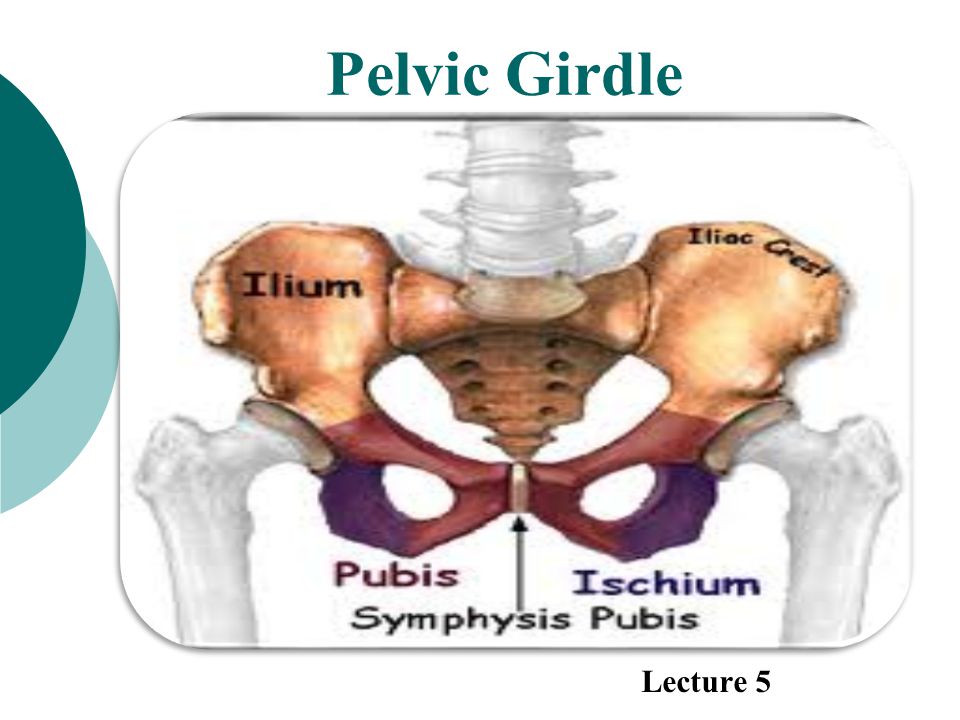 The pelvic cavity contains the bladder, rectum and internal genital organs (ovaries, fallopian tubes, uterus and vagina, prostate, seminal vesicles and vas deferens). The entrance to the small pelvis is open to the abdominal cavity and corresponds to the boundary line with the large pelvis. The exit from the pelvic cavity is closed by the muscles that form the pelvic diaphragm, the urethra and rectum pass through it in men, the urethra, rectum and vagina in women. Outside, this area of the body stands out as the perineum.
The pelvic organs differ from the abdominal organs in that they can
significantly change its volume: periodically filled
and the bladder and rectum are emptied, enlarged
and the uterus moves during pregnancy. It affects
on the work of other organs and blood supply.
Pelvis female and male
In no part of the skeleton are sex differences as pronounced as in the pelvis. Sex differences in the pelvis begin to appear in children between the ages of 8 and 10 years.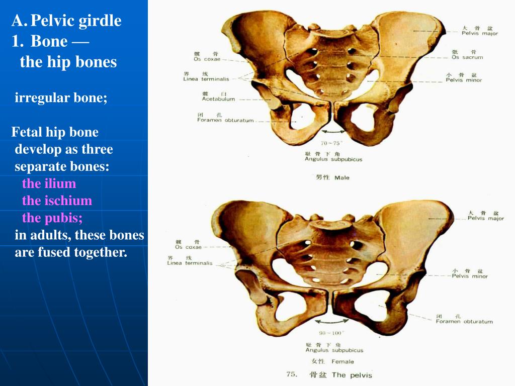 The average size of the male pelvis is about 2 cm smaller than the average size of the female pelvis. The female pelvis is wider and shorter than the male, the wings of the ilium are deployed more strongly. The angle between the lower branches of the pubic bones is rounded in the form of a pubic arch, the cape almost does not protrude into the pelvic cavity, and due to the wide, short and flat sacrum, the pelvic cavity has the shape of a cylinder.
Male pelvic structure
In men, the pelvis is already higher: the wings of the ilium are located almost vertically, the sacrum is strongly concave, and the promontory clearly protrudes into the cavity of the small pelvis, the subpubic angle is sharp. As a result, both the entrance and exit from the male pelvis are greatly narrowed, and its cavity itself has a conical shape.
The structure of the pelvis in women
The fetus moves through the small pelvis in women during childbirth, so its shape and size are of great importance for a normal birth act. The dimensions of the small pelvis are determined by indirect measurements of the large pelvis with obstetric compasses. The internal dimensions are determined during a vaginal examination and with the help of ultrasound.
For example, the distance between protruding tubercles on the iliac crest (the so-called superior anterior iliac spines) in women is normally 25–27 cm, and the distance between the most distant points of the crest on the right and left is 28–30 cm. The size of the entrance and exit from the small pelvis, which, both in direct and transverse dimensions, are about 11–13 cm in women. 1.5–2 cm due to the deviation of the tip of the coccyx posteriorly.
With violations in the development of the girl, due to rickets, spondylitis, coxitis and other diseases and poor nutrition, neglect of physical education or too much physical activity, deviations in the normal development of the pelvis are possible - a narrow pelvis. With a small degree of narrowing, childbirth is possible, but they are long and difficult. With a greater narrowing, there are obstacles to the passage of the fetus through the birth canal.
Author: Olga Gurova, Candidate of Biological Sciences, Senior Researcher, Associate Professor, Department of Human Anatomy, Peoples' Friendship University of Russia
The material uses photographs owned by shutterstock.com
Anatomy
Topic: Lower limb bones. Joints of the bones of the limbs.
Plan:
- Lower limb bones
- Pelvis as a whole, sex differences
- Bones of the free lower limb
- Lower limb bone joints
The skeleton of the lower extremities is divided into two sections: the skeleton of the girdle of the lower extremities (pelvic belt, or pelvis) and the skeleton of the free lower limbs.
Bones of the girdle of the lower extremities
The skeleton of the girdle of the lower extremities is formed by two pelvic bones and the sacrum with the coccyx.
The pelvic bone (os coxae) in children consists of three bones: the ilium, pubic and ischium, connected in the region of the acetabulum by cartilage. After the age of 16, cartilage is replaced by bone tissue and a monolithic pelvic bone is formed.
The ilium (os ilium) is the largest part of the pelvic bone, making up its upper section. It distinguishes a thickened part - the body and a flat section - the wing of the ilium, ending in a comb. On the wing in front and behind there are two protrusions: in front - upper anterior and lower anterior; iliac spines, and behind - superior posterior and inferior posterior iliac spines. The superior anterior iliac spine is well palpable. On the inner surface wing has an ilium! fossa, and on the gluteal (external) - three rough gluteal lines - front back and bottom. From these lines, the gluteal muscles begin. Rear of the wing thickened, on it is an ear-shaped (articular) surface for articulation with the sacrum.
The pubic bone (os pubis) is the anterior part of the pelvic bone. It consists of a body and two branches: top and bottom. Hai superior branch of the pubic bone is the pubic tubercle and pubic crest, passing into the arcuate line of the ilium. At the junction of the pubic bone with the ilium there is an iliac-pubic eminence.
The ischium (os ischii) forms the lower part of the pelvic bone. It consists of a body and a branch. Lower the department of the branch of the bone has a thickening - the sciatic tubercle. At the posterior edge of the body
bone is a protrusion - the ischial spine, separating the greater and lesser ischial notches.
Branches of the pubic and ischium bones form the obturator foramen. It is covered with thin connective tissue barrier membrane. In its upper part there is an obturator canal, limited by the obturator groove of the pubis. The channel serves to pass the vessels of the same name and nerve. On the outer surface of the pelvic bone, at the junction of the iliac, pubic and ischium, a significant depression is formed - the acetabulum (acetabulum),
The pelvis as a whole
The pelvis (pelvis) is formed by two pelvic bones, the sacrum and the coccyx.
Joints of the pelvic bones. The bones of the pelvis are connected to each other in front with the help of the pubic symphysis, and behind - two sacroiliac joints and numerous ligaments.
The pubic symphysis is formed by the pubic bones tightly fused with the fibrocartilaginous interpubic disc. Inside the disk there is a slit-like cavity. This symphysis strengthened by special ligaments: from above - by the upper pubic ligament and from below - arcuate ligament of the pubis. During pregnancy, the cavity of the pubic symphysis increases. Maybe also a slight expansion of the cavity of the sacroiliac joints. Due to the expansion of these cavities increase
the size of the pelvis, which is a favorable factor during childbirth.
The sacroiliac joint is flat in shape, formed by the auricular surfaces of the sacrum and iliac bone. Movement in it is extremely limited, which is facilitated by a system of powerful ventral (anterior), dorsal (posterior), and interosseous sacroiliac ligaments.
The ligaments of the pelvis include the sacrotuberous ligament - goes from the sacrum to the ischial tuberosity and sacrospinous ligament - goes from the sacrum to the ischial spine. These ligaments close the large and small sciatic notches, forming together with them a large and small sciatic foramen, through which muscles, blood vessels and nerves pass through. The posterior part of the iliac crest articulates with the transverse process of the V lumbar vertebrae by a strong iliac-lumbar ligament.
Large and small pelvis. The border line that runs along the upper edge of the pubic symphysis, crests pubic bones, the semicircular lines of the ilium and the promontory of the sacrum, the pelvis is sub-divided into two Department: large and small pelvis.
The greater pelvis is bounded by the wings of the ilium, the lesser by the ischial and pubic bones, sacrum, coccyx, sacrotuberous and sacrospinous ligaments, obturator membranes and pubic symphysis. There are two openings of the pelvic cavity: the upper - the upper aperture of the pelvis (inlet) and lower - the lower aperture of the pelvis (output). The upper aperture is limited by the boundary line, and the lower one - by the branches of the pubic and ischial bones, ischial tubercles, sacrotuberous ligaments and coccyx.
Sex differences in the pelvis. The female pelvis differs from the male pelvis in shape and size. The female pelvis is wider and smaller in height than the male. Its bones are thinner, their relief is smoothed. This due to differences in the degree of muscle development in women and men. Wings of the male pelvis located almost vertically, in women they are deployed to the sides. The pelvis is larger in women than in men. The cavity of the female pelvis is a cylindrical canal, in men it is looks like a funnel.
There are also gender differences in the subpubic angle formed by the lower branches of the pubic bones (its apex located at the lower edge of the pubic symphysis). In men, this angle is acute (about 75 °), and in women - blunt and has the shape of an arc (subpubic arc).
The upper pelvic inlet in women is wider than in men and has an elliptical shape. In men it heart-shaped due to the fact that their cape protrudes more forward. Inferior aperture of the pelvis women are also wider than men. Sex differences in the pelvis begin to emerge at older ages. 10 years.
An imaginary line connecting the midpoints of the anteroposterior dimensions of the entrance to the small pelvis, the cavity of the small pelvis and exit from the small pelvis, is the axis of the pelvis. It is also called a wire axis, or a guide. line; this is the path that the fetal head travels during childbirth. The axis of the pelvis is a curved line, its the curvature approximately corresponds to the curvature of the pelvic surface of the sacrum.
The pelvis has an anterior inclination (when the body is in a vertical position). The angle of the pelvis is formed by a line drawn through the cape and the upper edge of the pubic symphysis, and a horizontal plane. Usually it is equal 50-60°.
Bones of the free lower limb
Skeleton of the free lower limb (leg) includes the femur with the patella, the bones of the lower leg and foot bones.
The femur (femur) is the longest bone in the human body. It distinguishes the body proximal and distal ends. The spherical head at the proximal end faces the medial side. Below the head is the neck; it is located at an obtuse angle to the longitudinal axis of the bone. At the place the transition of the neck into the body of the bone there are two protrusions: the greater trochanter and the lesser trochanter (trochanter major and trochanter minor). The large trochanter lies outside and is well palpable. Between the skewers on the back The intertrochanteric ridge runs along the surface of the bone, and the intertrochanteric line runs along the anterior surface.
The body of the femur is curved, the convexity is directed anteriorly. The anterior surface of the body is smooth, along a rough line passes through the back surface. The distal end of the bone is somewhat flattened from front to back. and ends with the lateral and medial condyles. Above them from the sides rise respectively medial and lateral epicondyles. Between the latter is located behind the intercondylar fossa, in front - patella surface (for articulation with the patella). Above the intercondylar fossa there is a flat, triangular popliteal surface. The condyles of the femur have articular surfaces for connection with the tibia.
The patella (patella), or patella, is the largest sesamoid bone; it is enclosed in the tendon of the quadriceps femoris and is involved in the formation of the knee joint. On it distinguishes between an expanded upper part - the base and a narrowed, downward-facing part - top.
Leg bones: tibia, located medially, and fibula, occupies the lateral position.
The tibia (tibia) consists of a body and two ends. The proximal end is much thicker, it has two condyles: medial and lateral, articulating with the condyles of the femur. Between the condyles is the intercondylar eminence. On the outside of the lateral condyle a small peroneal articular surface is located (for connection with the head of the peroneal bones). The body of the tibia is trihedral. The anterior edge of the bone protrudes sharply, at the top it turns into roughness. At the lower end of the bone on the medial side there is a downward process - medial malleolus.
Fibula (fibula) - relatively thin, located outside of the tibia. The upper end of the fibula is thickened and is called the head. The top is isolated on the head, facing outwards and backwards. The head of the fibula articulates with the tibia. body bone has a triangular shape. The lower end of the bone is thickened, is called the lateral malleolus and is adjacent to talus outside.
The bones of the foot are divided into bones of the tarsus, metatarsals and phalanges (toes)
The bones of the tarsus are short cancellous bones. There are seven of them: talus, calcaneus, cuboid, scaphoid and three cuneiform. The talus has a body and a head. On the top of her body the block is located; together with the bones of the lower leg, it forms the ankle joint. Under the talus located calcaneus - the largest of the bones of the tarsus. On this bone are distinguished a well-defined thickening - a tubercle of the calcaneus, a process called the support of the talus, talar and cuboid articular surfaces (serve to connect with the corresponding bones).
Anterior to the calcaneus is the cuboid bone, and anterior to the head of the talus lies scaphoid. Three cuneiform bones - medial, intermediate and lateral - located distal to the scaphoid.
Five metatarsals located anterior to the cuboid and sphenoid bones. Each The metatarsal bone consists of a base, a body, and a head. Their bases articulate with the bones tarsus, and heads - with the proximal phalanges of the fingers.
The toes, like the fingers, have three phalanges, except for the first toe, which has two phalanges.
The foot skeleton has features due to its role as part of the supporting apparatus in vertical position of the body. The longitudinal axis of the foot is almost at right angles to the axis of the leg and hips. In this case, the bones of the foot do not lie in the same plane, but form a transverse and longitudinal arches, facing the concavity to the sole, and the convexity to the rear of the foot. Thanks to this, the foot rests only the tubercle of the calcaneus and the heads of the metatarsal bones. The outer edge of the foot is lower, it almost touches surface of the support and is called the supporting arch. The inner edge of the foot is raised - this is a spring vault. A similar structure of the foot ensures that it performs support and spring functions, which is associated with vertical position of the human body and upright posture.
Joints of the bones of the free lower limb
The hip joint (articulatio coxae) is formed by the acetabulum of the pelvic bone and the head femur. Along the edge of the acetabulum is the acetabular (articular) lip, which makes deeper depression. In shape, this is a kind of spherical joint - a walnut joint.
The joint is reinforced with ligaments. The strongest ilio-femoral ligament. Inside the joint cavity is the ligament of the femoral head. It goes from the bottom of the acetabulum to the fossa on the head femur. Vessels and nerves pass through it to the head of the femur; bond mechanical value slightly.
Movements in the hip joint occur around three axes: frontal - flexion and extension, sagittal - abduction and adduction, vertical - rotation inward and out. In it, as in any triaxial joint, circular movements are possible.
The knee joint (articulatio genus) is formed by three bones: the femur, tibia and patella. The medial and lateral condyles of the femur articulate with the condyles of the same name. tibia, and in front is the articular surface of the patella. Articular surfaces condyles of the tibia are slightly concave, and the articular surfaces of the condyles of the femur convex, but their curvature is not the same. The discrepancy between the articular surfaces is compensated by the medial and lateral menisci, located in the joint cavity between the condyles of the articulating bones. Menisci, being elastic formations, absorb shocks transmitted from the foot when walking, run, jump. The knee joint is a block-rotational complex joint.
Movements are carried out in the knee joint: flexion and extension of the lower leg, in addition, slight rotational movement of the lower leg around its longitudinal axis. The last movement is possible with a half-bent the position of the lower leg, when the relaxation of the collateral ligaments of the knee joint occurs.
Lower leg bone joints. The proximal ends of the bones of the lower leg are interconnected by means of tibial joint, flat in shape. Between the bodies of both bones is the interosseous membrane of the leg. The distal ends of the tibia and fibula are connected by syndesmosis (ligaments), characterized by special strength.
The ankle joint (articulatio talocruralis) is formed by both bones of the lower leg and the talus The joint is strengthened by ligaments that run from the bones of the lower leg to the talus, navicular and calcaneal bones. articular the bag is thin.
According to the shape of the articular surfaces, the joint belongs to the block-shaped. Movement goes around frontal axis: flexion and extension of the foot. Small movements to the sides (adduction and abduction) possible with strong plantar flexion.
Joints and ligaments of the foot. The bones of the foot are connected to each other through a series of joints, reinforced bundles. Among the joints of the tarsus, the talocalcaneal-navicular and calcaneocuboid joints. They are united under the general name "transverse joint tarsus" (known in surgery as the Chopard joint). This joint is strengthened on the back the surface of the foot with a bifurcated ligament - the so-called key of Chopard's joint. in the joints tarsus, supination and pronation of the foot, as well as adduction and abduction are possible.


