Early pregnancy genetic test
Prenatal Genetic Screening Tests | ACOG
Amniocentesis: A procedure in which amniotic fluid and cells are taken from the uterus for testing. The procedure uses a needle to withdraw fluid and cells from the sac that holds the fetus.
Aneuploidy: Having an abnormal number of chromosomes. Types include trisomy, in which there is an extra chromosome, or monosomy, in which a chromosome is missing. Aneuploidy can affect any chromosome, including the sex chromosomes. Down syndrome (trisomy 21) is a common aneuploidy. Others are Patau syndrome (trisomy 13) and Edwards syndrome (trisomy 18).
Carrier Screening: A test done on a person without signs or symptoms to find out whether he or she carries a gene for a genetic disorder.
Cell-Free DNA: DNA from the placenta that moves freely in a pregnant woman’s blood. Analysis of this DNA can be done as a noninvasive prenatal screening test.
Cells: The smallest units of a structure in the body. Cells are the building blocks for all parts of the body.
Chorionic Villus Sampling (CVS): A procedure in which a small sample of cells is taken from the placenta and tested.
Chromosomes: Structures that are located inside each cell in the body. They contain the genes that determine a person’s physical makeup.
Cystic Fibrosis (CF): An inherited disorder that causes problems with breathing and digestion.
Diagnostic Tests: Tests that look for a disease or cause of a disease.
DNA: The genetic material that is passed down from parent to child. DNA is packaged in structures called chromosomes.
Down Syndrome (Trisomy 21): A genetic disorder that causes abnormal features of the face and body, medical problems such as heart defects, and mental disability. Most cases of Down syndrome are caused by an extra chromosome 21 (trisomy 21).
Edwards Syndrome (Trisomy 18): A genetic condition that causes serious problems. It causes a small head, heart defects, and deafness.
Fetus: The stage of human development beyond 8 completed weeks after fertilization.
Genes: Segments of DNA that contain instructions for the development of a person’s physical traits and control of the processes in the body. The gene is the basic unit of heredity and can be passed from parent to child.
Genetic Counselor: A health care professional with special training in genetics who can provide expert advice about genetic disorders and prenatal testing.
Genetic Disorders: Disorders caused by a change in genes or chromosomes.
Inherited Disorders: Disorders caused by a change in a gene that can be passed from parents to children.
Monosomy: A condition in which there is a missing chromosome.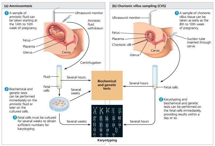
Mutations: Changes in genes that can be passed from parent to child.
Neural Tube Defects (NTDs): Birth defects that result from a problem in development of the brain, spinal cord, or their coverings.
Nuchal Translucency Screening: A test to screen for certain birth defects, such as Down syndrome, Edwards syndrome, or heart defects. The screening uses ultrasound to measure fluid at the back of the fetus’s neck.
Obstetrician: A doctor who cares for women during pregnancy and their labor.
Patau Syndrome (Trisomy 13): A genetic condition that causes serious problems. It involves the heart and brain, cleft lip and palate, and extra fingers and toes.
Placenta: An organ that provides nutrients to and takes waste away from the fetus.
Screening Tests: Tests that look for possible signs of disease in people who do not have signs or symptoms.
Sex Chromosomes: The chromosomes that determine a person’s sex. In humans, there are two sex chromosomes, X and Y. Females have two X chromosomes and males have an X and a Y chromosome.
Sickle Cell Disease: An inherited disorder in which red blood cells have a crescent shape, which causes chronic anemia and episodes of pain.
Tay–Sachs Disease: An inherited disorder that causes mental disability, blindness, seizures, and death, usually by age 5.
Trimester: A 3-month time in pregnancy. It can be first, second, or third.
Trisomy: A condition in which there is an extra chromosome.
Ultrasound Exams: Tests in which sound waves are used to examine inner parts of the body. During pregnancy, ultrasound can be used to check the fetus.
First trimester screening - Mayo Clinic
Overview
Nuchal translucency measurement
Nuchal translucency measurement
First trimester screening includes an ultrasound exam to measure the size of the clear space in the tissue at the back of a baby's neck (nuchal translucency). In Down syndrome, the nuchal translucency measurement is abnormally large — as shown on the left in the ultrasound image of an 11-week fetus. For comparison, the ultrasound image on the right shows an 11-week fetus with a normal nuchal translucency measurement.
First trimester screening is a prenatal test that offers early information about a baby's risk of certain chromosomal conditions, specifically, Down syndrome (trisomy 21) and extra sequences of chromosome 18 (trisomy 18).
First trimester screening, also called the first trimester combined test, has two steps:
- A blood test to measure levels of two pregnancy-specific substances in the mother's blood — pregnancy-associated plasma protein-A (PAPP-A) and human chorionic gonadotropin (HCG)
- An ultrasound exam to measure the size of the clear space in the tissue at the back of the baby's neck (nuchal translucency)
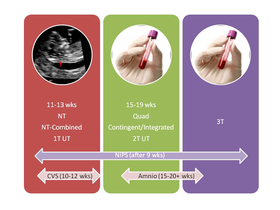
Typically, first trimester screening is done between weeks 11 and 14 of pregnancy.
Using your age and the results of the blood test and the ultrasound, your health care provider can gauge your risk of carrying a baby with Down syndrome or trisomy 18.
If results show that your risk level is moderate or high, you might choose to follow first trimester screening with another test that's more definitive.
Products & Services
- Book: Mayo Clinic Guide to a Healthy Pregnancy
- Book: Obstetricks
Why it's done
First trimester screening is done to evaluate your risk of carrying a baby with Down syndrome. The test also provides information about the risk of trisomy 18.
Down syndrome causes lifelong impairments in mental and social development, as well as various physical concerns. Trisomy 18 causes more severe delays and is often fatal by age 1.
First trimester screening doesn't evaluate the risk of neural tube defects, such as spina bifida.
Because first trimester screening can be done earlier than most other prenatal screening tests, you'll have the results early in your pregnancy. This will give you more time to make decisions about further diagnostic tests, the course of the pregnancy, medical treatment and management during and after delivery. If your baby has a higher risk of Down syndrome, you'll also have more time to prepare for the possibility of caring for a child who has special needs.
Other screening tests can be done later in pregnancy. An example is the quad screen, a blood test that's typically done between weeks 15 and 20 of pregnancy. The quad screen can evaluate your risk of carrying a baby with Down syndrome or trisomy 18, as well as neural tube defects, such as spina bifida. Some health care providers choose to combine the results of first trimester screening with the quad screen. This is called integrated screening. This can improve the detection rate of Down syndrome.
First trimester screening is optional. Test results indicate only whether you have an increased risk of carrying a baby with Down syndrome or trisomy 18, not whether your baby actually has one of these conditions.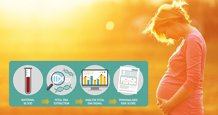
Before the screening, think about what the results will mean to you. Consider whether the screening will be worth any anxiety it might cause, or whether you'll manage your pregnancy differently depending on the results. You might also consider what level of risk would be enough for you to choose a more invasive follow-up test.
More Information
- Prenatal testing: Quick guide to common tests
Request an Appointment at Mayo Clinic
Risks
First trimester screening is a routine prenatal screening test. The screening poses no risk of miscarriage or other pregnancy complications.
How you prepare
You don't need to do anything special to prepare for first trimester screening. You can eat and drink normally before both the blood test and the ultrasound exam.
What you can expect
First trimester screening includes a blood draw and an ultrasound exam.
During the blood test, a member of your health care team takes a sample of blood by inserting a needle into a vein in your arm. The blood sample is sent to a lab for analysis. You can return to your usual activities immediately.
For the ultrasound exam, you'll lie on your back on an exam table. Your health care provider or an ultrasound technician will place a transducer — a small plastic device that sends and receives sound waves — over your abdomen. The reflected sound waves will be digitally converted into images on a monitor. Your health care provider or the technician will use these images to measure the size of the clear space in the tissue at the back of your baby's neck.
The ultrasound doesn't hurt, and you can return to your usual activities immediately.
Results
Your health care provider will use your age and the results of the blood test and ultrasound exam to gauge your risk of carrying a baby with Down syndrome or trisomy 18. Other factors — such as a prior Down syndrome pregnancy — also might affect your risk.
First trimester screening results are given as positive or negative and also as a probability, such as a 1 in 250 risk of carrying a baby with Down syndrome.
First trimester screening correctly identifies about 85 percent of women who are carrying a baby with Down syndrome. About 5 percent of women have a false-positive result, meaning that the test result is positive but the baby doesn't actually have Down syndrome.
When you consider your test results, remember that first trimester screening indicates only your overall risk of carrying a baby with Down syndrome or trisomy 18. A low-risk result doesn't guarantee that your baby won't have one of these conditions. Likewise, a high-risk result doesn't guarantee that your baby will be born with one of these conditions.
If you have a positive test result, your health care provider and a genetics professional will discuss your options, including additional testing. For example:
- Prenatal cell-free DNA (cfDNA) screening. This is a sophisticated blood test that examines fetal DNA in the maternal bloodstream to determine whether your baby is at risk of Down syndrome, extra sequences of chromosome 13 (trisomy 13) or extra sequences of chromosome 18 (trisomy 18).
 Some forms of cfDNA screening also screen for other chromosome problems and provide information about fetal sex. A normal result might eliminate the need for a more invasive prenatal diagnostic test.
 - Chorionic villus sampling (CVS). CVS can be used to diagnose chromosomal conditions, such as Down syndrome. During CVS, which is usually done during the first trimester, a sample of tissue from the placenta is removed for testing. CVS poses a small risk of miscarriage.
- Amniocentesis. Amniocentesis can be used to diagnose both chromosomal conditions, such as Down syndrome, and neural tube defects, such as spina bifida. During amniocentesis, which is usually done during the second trimester, a sample of amniotic fluid is removed from the uterus for testing. Like CVS, amniocentesis poses a small risk of miscarriage.

Your health care provider or a genetic counselor will help you understand your test results and what the results mean for your pregnancy.
By Mayo Clinic Staff
Related
Products & Services
what does genetic analysis show during pregnancy and where to do it in Moscow?
Every expectant mother wants to be 100% sure that her baby will be born healthy. But until recently, it was possible to verify this only with the help of risky research methods, which were prescribed only for vital indications. Now there is a safe way to detect genetic abnormalities in a fetus - a non-invasive DNA test. What does it show and how is it carried out? We understand the topic and answer questions.
During pregnancy, most genetic defects in the fetus can be diagnosed with a DNA test, and are often performed for this purpose.
Here are the main purposes of a DNA test for pregnant women:
Diagnosis of chromosomal abnormalities:
- Down syndrome (additional chromosome in the twenty-first pair) occurs in about one in a thousand newborns [1] .
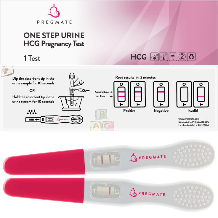 With the increase in the age of the expectant mother, the risk of giving birth to a baby with an anomaly increases. Children with Down syndrome are often born with heart defects, epilepsy, their physical development lags behind the norm, and all have more or less severe mental retardation.
 - Edwards syndrome occurs due to the presence of an extra chromosome in the eighteenth pair. This is a rare anomaly: it occurs in about one in 7000 cases [2] . Newborns with Edwards syndrome have numerous malformations: 90-95% of them die in the first months.
- Patau syndrome is trisomy of the thirteenth pair. This anomaly is even rarer, affecting approximately one in 14,000 newborns [3] . Like Edwards syndrome, this type of hereditary pathology is manifested by multiple gross malformations; life expectancy of patients rarely exceeds a year.
Important
Ultrasound screening is performed for early diagnosis of Down, Edwards and Patau syndromes.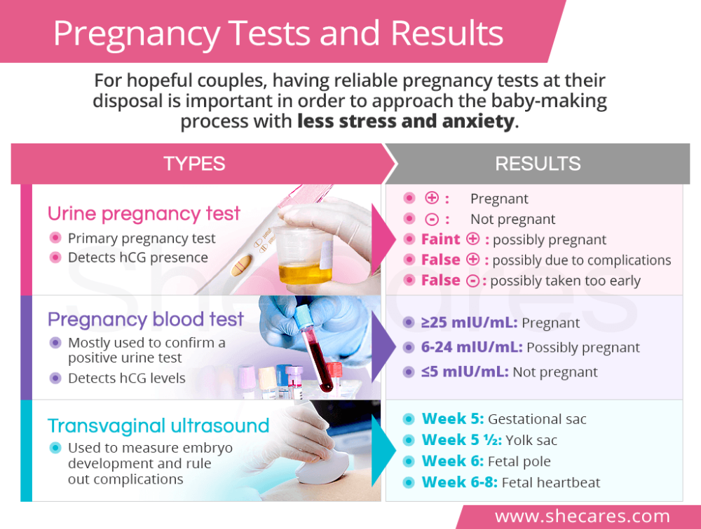 Some features of the development of the fetus allow us to suggest chromosomal abnormalities: an increased width of the collar zone, a shortened nasal bone, and others. But in the first trimester, ultrasound signs cannot be considered as reliable evidence of disorders, and later, when the screening results become more convincing, time may be lost. Meanwhile, research among 18 955 women showed that the most accurate test for the presence of chromosomal abnormalities is DNA analysis during pregnancy (in the first trimester) [4] .
- Anomalies in the number of sex chromosomes . These include Klinefelter syndrome, Shereshevsky-Turner syndrome and many other abnormalities associated with a decrease or increase in the normal number of X and Y chromosomes. Such genetic defects are often accompanied by underdevelopment of the gonads (sex glands), infertility, various changes in appearance, often mental retardation.
Diagnosis of congenital pathologies of the nervous system . For example, a DNA test early in pregnancy can detect Rett syndrome (a severe neuropsychiatric disorder that does not become apparent until several months after birth), infantile epileptic encephalopathy, and other CNS disorders.
Identification of hereditary forms of craniosynostosis (premature fusion of the bones of the skull) - Pfeiffer, Aper, Cruzon, Müncke syndromes.
Many other genetic diseases poorly diagnosed by traditional screening can be detected by DNA testing during pregnancy.
Determination of the sex of the unborn baby . Yes, you can see it on the screen of the ultrasound machine already during the second screening. But sometimes you need to get a 100% accurate result as early as possible. This is necessary primarily for the timely diagnosis of hereditary anomalies associated with sex. For example, hemophilia and Klinefelter's syndrome occur only in boys, while Turner's syndrome affects only girls. The reliability of sex determination by prenatal DNA testing at seven to nine weeks of gestation is 95%, from 12 weeks onwards it reaches 99% [5] .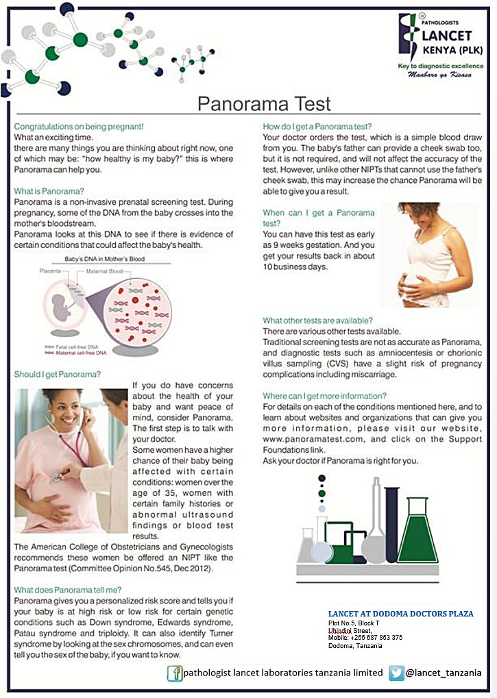
Establishment of paternity . The need for this study arises for various reasons. For example, it may be required if the future father refuses to admit his involvement in the conception. In controversial cases, the results of the examination can serve as evidence in court.
Indications and contraindications for testing
It is natural for any expectant mother to worry about the health of her baby, so the mere desire of a pregnant woman is enough to perform a non-invasive genetic test. This cannot be said about invasive research methods that carry a risk to the fetus and therefore are not carried out without strict indications. But there are situations when genetic analysis is simply necessary for pregnant women. Such a study is highly desirable if:
- Routine screening showed that there is a high risk of giving birth to a child with anomalies. According to the results of ultrasound, it is impossible to draw a conclusion about the presence of Down syndrome and other hereditary pathologies in the fetus, one can only suspect this for a number of reasons.
 To clarify the diagnosis, the pregnant woman is sent for DNA analysis. Previously, it was possible to confirm or refute malformations only through traumatic and risky methods - amniocentesis, cordocentesis and others. Not so long ago, a safe alternative appeared - a non-invasive prenatal genetic test.
 - Expectant mother over 35 years old. With age, the likelihood of having a child with Down syndrome increases. Other chromosomal abnormalities are less dependent on this indicator, but some association can also be traced.
- The woman has a history of miscarriages or miscarriages. A common cause of miscarriage lies precisely in the genetic defects of the fetus. If the last pregnancy ended unsuccessfully, it makes sense to make sure that everything is in order this time.
- In the family of the future mother or father, there were cases of the appearance of children with hereditary pathologies. Until the results of the study are available, a recurrence of such a situation cannot be completely ruled out.
.jpg)
- One of the future parents suffered from alcohol or drug addiction. Even if the problem is in the past, the risk to the child's health still exists.
- In the first weeks of pregnancy, a woman suffered from an acute infectious disease (the most dangerous of them is rubella) or experienced the effects of other teratogenic factors (which can lead to abnormalities in the development of the fetus). The latter include radiation exposure, taking certain medications (for example, tetracycline antibiotics), drinking alcohol, poisoning with salts of heavy metals.
There are cases when it is not possible to carry out genetic testing of pregnant women. The main contraindication to invasive tests is the threat of miscarriage. Also, studies are not performed for acute infections, inflammatory gynecological diseases, myoma. A non-invasive test for genetic abnormalities is not done for multiple pregnancies (triplets or more), since in this case it is difficult to determine the DNA of each fetus and the data may be inaccurate. The analysis is not performed in surrogate mothers and women who conceived by fertilization with a donor egg (with the exception of certain types of testing). It is impossible to obtain reliable test data if a blood transfusion or bone marrow transplantation was performed shortly before pregnancy.
How testing is done
There are many types of genetic tests performed during pregnancy, but all of them can be divided into two groups depending on the method of sampling. From this point of view, invasive and non-invasive methods are distinguished. The former are associated with the penetration of the future mother and fetus into the body, the latter do not imply such an intervention.
Invasive tests
All studies in this group carry a certain risk of spontaneous abortion, so they are resorted to only in extreme cases - if the likelihood of genetic abnormalities in the fetus is high enough.
Chorionic villus biopsy is the earliest invasive test and is performed at 10-14 weeks. The material for analysis is the tissue cells of the chorion - the future placenta. The fence is carried out by puncturing the abdominal wall and uterus with a long needle. The procedure is performed under ultrasound guidance. The accuracy of diagnosing genetic abnormalities by this method is 99% [6] . The same study performed at a later date is called placentocentesis.
Amniocentesis - analysis of fetal DNA contained in amniotic fluid cells. It is used in the second trimester of pregnancy. Using a syringe with a needle, the fetal bladder is punctured through the abdominal wall and a small amount of amniotic fluid (about 30 ml) is taken. The results of the study need to wait two to three weeks. The accuracy of the method also reaches 99%.
Cordocentesis is an analysis of cord blood, which is also taken through the abdominal wall. The procedure is also performed in the II trimester. With the help of cordocentesis, genetic diseases, infections and other intrauterine pathologies can be determined.
Non-invasive genetic test
The technique is relatively new for Russia, and such testing is not yet carried out in all medical centers. This is due to the insufficient equipment of the laboratories of state institutions, and the rather high cost of the study (because it cannot be used everywhere, like screening).
In contrast to all the methods described above, non-invasive testing for fetal genetic abnormalities can be said to be safe. To take a biomaterial, you do not need to resort to a puncture and other traumatic interventions that can lead to complications. For analysis, only the blood of a pregnant woman is needed, which is taken from a vein in the usual way. No special preparation is required for the study.
The test is carried out using high-tech medical equipment. The blood taken from a woman in a centrifuge is divided into erythrocyte mass, a layer of leukocytes and plasma. DNA from the last two fractions is "deciphered" by sequencing, separating the genomes of the mother and fetus. The resulting material is analyzed for the risk of chromosomal and other pathologies. The method of analysis depends on the type of testing. The entire research process takes an average of two weeks.
The informativeness of tests differs depending on their type. Almost all such analyzes are able to determine the presence or absence of Down syndrome, Edwards and Patau, sex chromosome anomalies, the sex of the unborn child. Some methods are suitable for studying the genetic risks of pregnancy after IVF (including with a donor egg). The reliability of the results of non-invasive testing reaches 99%.
In addition to safety and high accuracy, the study has another weight advantage - the ability to perform DNA analysis in early pregnancy. It is done starting at nine to ten weeks, when fetal DNA begins to be detected in the mother's blood. No screening method or invasive test is performed at such times. Early diagnosis of pathologies is crucial in maintaining pregnancy, and the confidence that the unborn child is healthy allows you to expect his birth with joy and without fear.
Thanks to the latest technology in medicine, it has become possible to determine the risk of hereditary diseases in the fetus without resorting to invasive research methods. Genetic analysis of the blood of pregnant women is accurate and safe, and besides, it is the earliest of all possible ways to detect intrauterine pathologies. This testing has two drawbacks: firstly, it is paid, and secondly, it is carried out so far only in a few clinics.
Non-invasive DNA screening (NIPT)
Rus
Petrovsky prospekt 2, bld. ) 77-55555
Clinic phone number
Request a call
+7 (812) 77-55555
Request a call
Fetal chromosome. Non-invasive testing of the fetus from the 9th week of pregnancy
URGENT
ACCURACY
OPERATING
NGC recommends that all pregnant women determine the fetal chromosome set by fetal DNA analysis from the mother's blood without invasive interventions already in the early stages of pregnancy.
Geneticists of the medical center are the first in St. Petersburg and one of the first in Russia for several years to use in prenatal practice the study of fetal chromosomes without invasive interventions - by fetal DNA in the mother's blood.
Non-invasive testing is the most modern method for diagnosing chromosomal pathologies in the fetus (Down syndrome, Edwards, Patau, etc.) on the mother from 9-10 weeks of pregnancy. Benefits of non-invasive diagnostics:
- Possibility: Exclude Down syndrome and other chromosomal abnormalities
- Safety: maternal and fetal - "no puncture"
- Urgency: from 9 weeks pregnant
- Accuracy : DNA test with 99.7% probability
- Efficiency : in one step - only by blood test
- Individuality : the right technology for every application
Consultation with a geneticist before the examination and after the results.
Confidence in the health of the baby from early pregnancy!
Purpose of fetal DNA testing
- DNA screening excludes the most common fetal chromosomal abnormalities - the following syndromes and conditions:
- Dauna
- Edwards
- Patau
- Shereshevsky-Turner
- Klinefelter
- Triploidy
- DNA test reveals the presence of microdeletions - loss of small sections of chromosomes
- According to the results of the study, with a probability of 99.7%, it is possible to exclude a violation of the number 21, 13, 18 and sex chromosomes, the presence of triplodia
- The DNA test is an important test for accurately determining the sex of the fetus in sex-linked gene diseases
What is the research technology based on?
- Unique studies have shown that the blood of pregnant women contains a significant percentage of fetal DNA - up to 25%
- Fetal DNA can be isolated and analyzed
- Molecular genetic study of fetal DNA allows to estimate the number of fetal chromosomes and exclude changes in their number, leading to Down syndrome, Edwards, Patau, Turner and other conditions
- extended study allows to exclude accidental loss of genetic material - microdeletions in other chromosomes
Testing is carried out from the 9th week of pregnancy on the mother's blood, in which there is already a sufficient amount of fetal DNA. The study of fetal DNA has been used in the world for more than a year, but only in 2011 it became possible to conduct a study with high accuracy and strongly recommend it to all pregnant women.
Comparison of the performance of conventional prenatal diagnosis algorithm and non-invasive testing - fetal DNA screening for detection of chromosomal pathology
| Ordinary algorithm
| DNA screening
| |
| CCRINING STATIONS Assess only indirect signs of chromosomal pathology | Direct study of the genetic structure (DNA) of the fetus with an accuracy of 99. | |
| 902 |
Good ultrasound and biochemical screening data cannot rule out chromosomal abnormalities of the fetus, as they do not examine genetic structures (DNA or chromosomes)
What types of non-invasive testing are there?
Various testing technologies are currently used - total DNA quantitation, fetal DNA identification, chromosome count, microdeletion exclusion, etc. Some technologies are used only for singleton pregnancies, others for twins. There are some features during pregnancy after IVF, especially in the case of donor programs and surrogacy. We cooperate with the world's leading laboratories in the field of non-invasive testing - Natera (Panorama), Veracity, Ariosa (Harmony).
Which test is best for your case, a geneticist will help you figure it out.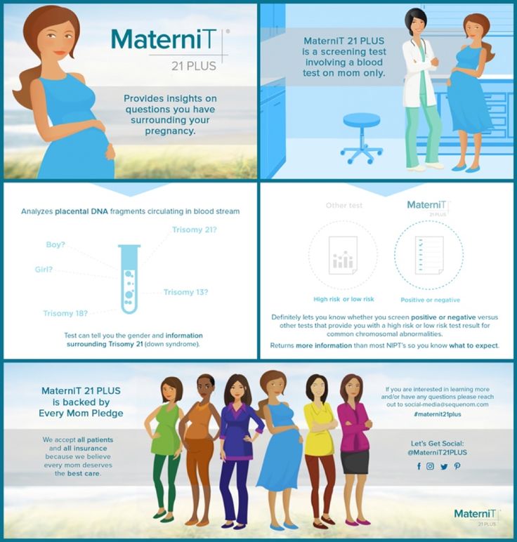 According to the results of medical genetic counseling, you will select the research technology that is optimal for your case.
DNA screening - a test that is prescribed in the early stages of pregnancy, is carried out without intervention and threats. Rules out Down syndrome and other common chromosomal abnormalities.
The result will save you from long-term anxiety, give you confidence in the health of your unborn baby.
To whom and when can non-invasive testing - DNA screening be prescribed?
Fetal DNA testing can be carried out as early as the 9th week of pregnancy. Pregnancy term - "our patient's age" must be confirmed by ultrasound and be at least 2.5 cm. The test is scheduled even before the ultrasound screening in the first trimester. DNA screening eliminates the need for biochemical screening. After receiving the results, only an ultrasound is prescribed.
DNA screening is especially recommended for pregnancy after IVF.
 99%
99% 