How many weeks gestation is full term
Definition of Term Pregnancy | ACOG
Number 579 (Reaffirmed 2022)
The American College of Obstetricians and Gynecologists Committee on Obstetric Practice Society for Maternal-Fetal Medicine
This document reflects emerging clinical and scientific advances as of the date issued and is subject to change. The information should not be construed as dictating an exclusive course of treatment or procedure to be followed.
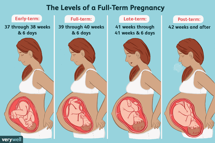
ABSTRACT: In the past, the period from 3 weeks before until 2 weeks after the estimated date of delivery was considered “term,” with the expectation that neonatal outcomes from deliveries in this interval were uniform and good. Increasingly, however, research has shown that neonatal outcomes, especially respiratory morbidity, vary depending on the timing of delivery within this 5-week gestational age range. To address this lack of uniformity, a work group was convened in late 2012, which recommended that the label “term” be replaced with the designations early term (37 0/7 weeks of gestation through 38 6/7 weeks of gestation), full term (39 0/7 weeks of gestation through 40 6/7 weeks of gestation), late term (41 0/7 weeks of gestation through 41 6/7 weeks of gestation), and postterm (42 0/7 weeks of gestation and beyond) to more accurately describe deliveries occurring at or beyond 37 0/7 weeks of gestation. The American College of Obstetricians and Gynecologists and the Society for Maternal-Fetal Medicine endorse and encourage the uniform use of the work group’s recommended new gestational age designations by all clinicians, researchers, and public health officials to facilitate data reporting, delivery of quality health care, and clinical research.
Gestation in singleton pregnancies lasts an average of 40 weeks (280 days) from the first day of the last menstrual period to the estimated date of delivery. In the past, the period from 3 weeks before until 2 weeks after the estimated date of delivery was considered “term” 1, with the expectation that neonatal outcomes from deliveries in this interval were uniform and good. Increasingly, however, research has identified that neonatal outcomes, especially respiratory morbidity, vary depending on the timing of delivery even within this 5-week gestational age range. The frequency of adverse neonatal outcomes is lowest among uncomplicated pregnancies delivered between 39 0/7 weeks of gestation and 40 6/7 weeks of gestation 2, 3. For this reason, quality improvement projects have focused, for example, on eliminating nonmedically indicated deliveries at less than 39 0/7 weeks of gestation 4.
In order to facilitate data reporting, delivery of quality health care, and clinical research, it is important that all clinicians, researchers, and public health officials use both uniform labels when describing deliveries in this period and a uniform approach to determining gestational age. To address the lack of uniformity in neonatal outcomes between 37 0/7 weeks of gestation and 42 0/7 weeks of gestation, a work group was convened in late 2012 to determine whether term pregnancy should be redefined 5. The work group included representatives from the Eunice Kennedy Shriver National Institute of Child Health and Human Development, the American College of Obstetricians and Gynecologists (the College), the Society for Maternal-Fetal Medicine (SMFM), and other professional societies and stakeholder organizations. The work group recommended that the label “term” be replaced by the designations early term, full term, late term, and postterm to more accurately describe deliveries occurring at or beyond 37 0/7 weeks of gestation Box 1. The group recommended that the use of the label “term” to describe all deliveries between 37 0/7 weeks of gestation and 41 6/7 weeks of gestation should be discouraged. Details of the evidence and rationale that are the foundation of these recommendations can be found in published summaries of this conference 5.
The College and SMFM endorse and encourage the uniform use of the work group’s recommended new gestational age designations by all clinicians, researchers, and public health officials to facilitate data reporting, delivery of quality health care, and clinical research.
Early term: 37 0/7 weeks through 38 6/7 weeks
Full term: 39 0/7 weeks through 40 6/7 weeks
Late term: 41 0/7 weeks through 41 6/7 weeks
Postterm: 42 0/7 weeks and beyond
 Defining “term” pregnancy: recommendations from the Defining “Term” Pregnancy Workgroup. JAMA 2013;309:2445–6.
Uniform definitions of term are predicated on a uniform method of determining gestational age. The work group provided a method for determination of gestational age 5 that, like other similar methods 6, focused on a hierarchy of clinical and ultrasonographic criteria. Individual methods may differ in the details of when and how ultrasonographic biometry should be used to change estimated date of delivery based on last menstrual period; however, it is not the purpose of this document to establish the priority of one method over another. The College and SMFM are working with other expert groups to establish evidence-based consensus on criteria for determining gestational age.
Copyright November 2013 by the American College of Obstetricians and Gynecologists, 409 12th Street, SW, PO Box 96920, Washington, DC 20090-6920. All rights reserved.
ISSN 1074-861X
Definition of term pregnancy.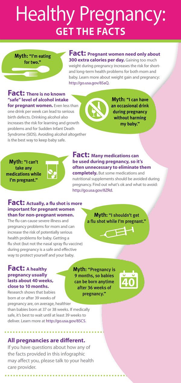 Committee Opinion No. 579. American College of Obstetricians and Gynecologists. Obstet Gynecol 2013;122:1139–40.
Baby due date - Better Health Channel
Summary
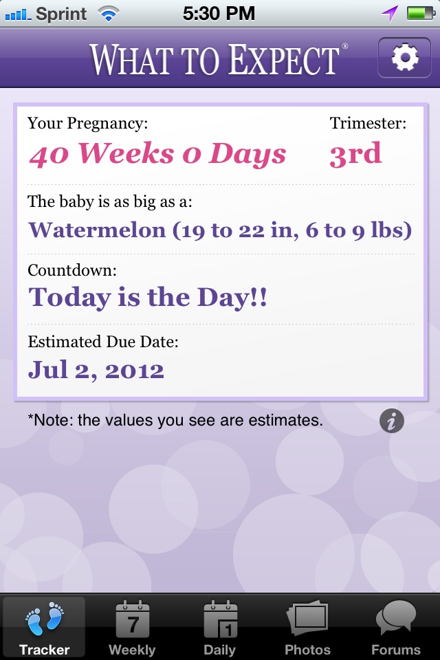
Read the full fact sheet- The unborn baby spends around 38 weeks in the uterus, but the average length of pregnancy, or gestation, is counted at 40 weeks.
- Pregnancy is counted from the first day of the woman’s last period, not the date of conception which generally occurs two weeks later.
- Since some women are unsure of the date of their last menstruation (perhaps due to period irregularities), a baby is considered full term if its birth falls between 37 to 42 weeks of the estimated last menstruation date.
The unborn baby spends around 37 weeks in the uterus (womb), but the average length of pregnancy, or gestation, is calculated as 40 weeks. This is because pregnancy is counted from the first day of the woman’s last period, not the date of conception which generally occurs two weeks later, followed by five to seven days before it settles in the uterus. Since some women are unsure of the date of their last menstruation (perhaps due to period irregularities), a pregnancy is considered full term if birth falls between 37 to 42 weeks of the estimated last menstruation date.
A baby born prior to week 37 is considered premature, while a baby that still hasn’t been born by week 42 is said to be overdue. In many cases, labour will be induced in the case of an overdue baby.
The average length of human gestation is 280 days, or 40 weeks, from the first day of the woman’s last menstrual period. The medical term for the due date is estimated date of confinement (EDC). However, only about four per cent of women actually give birth on their EDC. There are many online pregnancy calculators (see Baby due date calculator that can tell you when your baby is due, if you type in the date of the first day of your last period.
A simple method to calculate the due date is to add seven days to the date of the first day of your last period, then add nine months. For example, if the first day of your last period was 1 February, add seven days (8 February) then add nine months, for a due date of 8 November.
Determining baby due date
Irregular menstrual cycles can mean that some women aren’t sure of when they conceived. Some clues to the length of gestation include:
- Ultrasound examination (especially when performed between six and 12 weeks)
- Size of uterus on vaginal or abdominal examination
- The time fetal movements are first felt (an approximate guide only).
Pregnancy ultrasound
A pregnancy ultrasound is a non-invasive test that scans the unborn baby and the mother’s reproductive organs using high frequency sound waves. The general procedure for a pregnancy ultrasound includes:
- The woman lies on a table.
- A small amount of a clear, conductive jelly is smeared on the woman’s abdomen.
- The operator places the small hand-held instrument called a transducer onto the woman’s abdomen.
- The transducer is moved across the abdomen. The sound waves bounce off internal structures (including the baby) and are transmitted back to the transducer. The sound waves are then translated into a two-dimensional picture on a monitor. The mother doesn’t feel or hear the transmission of the sound waves.
- By measuring the baby’s body parts, such as head circumference and the length of long bones, the operator can estimate its gestational age.
The diagnostic uses of pregnancy ultrasound
Apart from helping to pinpoint the unborn baby’s due date, pregnancy ultrasounds are used to diagnose a number of conditions including:
- Multiple fetuses
- Health problems with the baby
- Ectopic pregnancy (the embryo lodges in the fallopian tube instead of the uterus)
- Abnormalities of the placenta such as placenta praevia, where the placenta is positioned over the neck of the womb (cervix)
- The health of the mother’s reproductive organs.

Premature babies
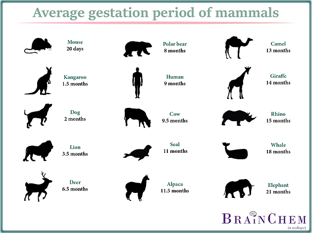
A baby born prior to week 37 is considered premature. The odds of survival depend on the baby’s degree of prematurity. The closer to term (estimated date of confinement, or EDC) the baby is born, the higher its chances of survival - after 34 weeks gestation with good paediatric care almost all babies will survive.
Premature babies are often afflicted by various health problems, caused by immature internal organs. Respiratory difficulties and an increased susceptibility to infection are common.
Often there is no known cause for a premature labour; however, some of the maternal risk factors may include:
- Drinking alcohol or smoking during pregnancy
- Low body weight prior to pregnancy
- Inadequate weight gain during pregnancy
- No prenatal care
- Emotional stress
- Placenta problems such as placenta praevia
- Various diseases such as diabetes and congestive heart failure
- Infections such as syphilis.
Overdue babies
Around five out of every 100 babies will be overdue, or more than 42 weeks gestation. If you have gone one week past your due date without any signs of impending labour, your doctor will want to closely monitor your condition. Tests include:
- Monitoring the fetal heart rate
- Using a cardiotocograph machine
- Performing ultrasound scans.
The placenta starts to deteriorate after 38 weeks or so, which means an overdue baby may not get enough oxygen. An overdue baby could also grow too large for vaginal delivery. Generally, an overdue baby will be induced once it is two weeks past its expected date. Some of the methods of induction include:
- Vaginal prostaglandin gel - to help dilate the cervix
- Amniotomy - breaking the waters, sometimes called an artificial rupture of membranes (ARM)
- Oxytocin - a synthetic form of this hormone is given intravenously to stimulate uterine contractions.
Where to get help
- Your doctor
- Your obstetrician
- Midwife or childbirth educator
Things to remember
- The unborn baby spends around 38 weeks in the uterus, but the average length of pregnancy, or gestation, is counted at 40 weeks.
- Pregnancy is counted from the first day of the woman’s last period, not the date of conception which generally occurs two weeks later.
- Since some women are unsure of the date of their last menstruation (perhaps due to period irregularities), a baby is considered full term if its birth falls between 37 to 42 weeks of its estimated due date.
- Common concerns and discomforts: overdue baby, Mother’s Bliss, UK.
- Going Overdue, 2001, Centre for Reproduction and Minimally Invasive Surgery.
- Premmie-L FAQ and advice sheets, Parents of Premature Babies Inc.(Preemie-L). More information here.
- Pregnancy: what to expect when it’s past your due date, Family Doctor, USA.

- Ultrasound, Women Health Information, Royal Women’s Hospital, Melbourne. More information here.
This page has been produced in consultation with and approved by:
Pregnancy calendar
You are pregnant! Your baby will be born in 40 weeks. What changes will occur in your body, how your baby will grow will tell "Calendar of pregnancy".
1-2 weeks
Pregnancy begins at the moment of fertilization or conception.
Fertilization is a complex biological process of the fusion of female and male germ cells (egg and sperm). The resulting cell (zygote) is a new daughter organism.
A mature egg leaves the ovary approximately on the 12-14th day of the menstrual cycle (ovulation) and enters the fallopian tube, where it remains viable for 24 hours. During an orgasm, a man ejects from 200 to 400 million spermatozoa into the woman's vagina. Some of them penetrate through the cervix into the uterine cavity, and from there into the fallopian tubes.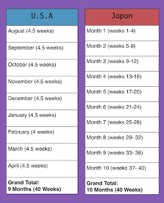 Here, spermatozoa retain the ability to fertilize for 48 hours. Thus, within 6-7 days of a woman's menstrual cycle, conception is possible.
Fertilization of the female egg is performed by a single sperm in the upper part of the fallopian tube. There are two types of sperm: those containing the Y chromosome (“male”) and the X chromosome (“female”). When an egg cell (containing the X chromosome) fuses with a sperm cell, their genetic material is combined and the sex of the child is determined. If there are two X chromosomes in the child's genetic makeup, it's a girl; if an X chromosome and a Y chromosome, it's a boy. It is impossible to change the sex of the child, so you should not follow the "folk beliefs" that guarantee the birth of a child of a given gender.
The fertilized egg begins to divide with the formation of a multicellular organism and move through the fallopian tube into the uterine cavity. During this period, the nutrition of the embryo is carried out at the expense of those substances that have been accumulated in the egg.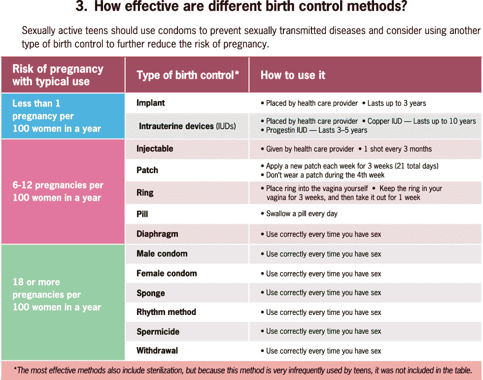 If the peristalsis of the tube is slowed down (due to inflammatory diseases), the embryo penetrates the wall of the fallopian tube with the occurrence of an ectopic pregnancy.
Implantation (introduction) of the embryo into the uterine wall occurs 7-8 days after fertilization.
On the seventh day of pregnancy, the outer layer of the embryo (trophoblast) begins to produce a hormone - chorionic gonadotropin. This hormone gives the mother's body information that pregnancy has occurred, and begins its functional restructuring. Diagnostic test strips detect the chorionic gonadotropin in the urine of a pregnant woman, which makes it possible to diagnose pregnancy at an early stage.
3-4 weeks
You do not have the expected menstruation, nausea in the morning, and frequent urination during the day. You become emotionally labile, irritable, whiny. Basal body temperature is above 37°C.
In appearance, your unborn baby resembles a small auricle measuring 4 mm, surrounded by a small amount of amniotic fluid. On the 21st day after conception, the brain and spinal cord are formed. By the end of the first month, the circulation of embryonic blood is established, the umbilical cord has formed - the connection of the embryo with the future placenta. The eye sockets, the rudiments of arms and legs appeared, the laying and development of other internal organs of the fetus is underway: the liver, kidneys, urinary tract, and digestive organs.
5-6 weeks
You no longer doubt that you are pregnant. Regardless of how you feel, all pregnant women need to visit a antenatal clinic and undergo an examination that will allow you to identify and correct existing health problems in time.
Starting from the 5th week, there may be a threat of termination of pregnancy. This will be evidenced by: periodic pain in the lower abdomen and in the lumbar region, a feeling of pressure on the rectum, an increased amount of mucus. If you experience these symptoms, you should consult a doctor.
By week 6, the face is formed in the embryo: eyes, nose, jaws and limbs.
7-8 weeks
From the 7th week of pregnancy, the yellow body of pregnancy undergoes reverse development, the production of hormones begins to be carried out by the forming placenta.
The baby develops large blood vessels, the heart becomes four-chambered. Bile ducts appear in the liver. There is a development of the endocrine glands, the brain. The auricles are already formed, fingers have appeared on the limbs. The embryo begins to move. At week 8, under the influence of the Y chromosome, the formation of male gonads (testicles) occurs. They begin to produce testosterone - the male sex hormone, which will lead to the formation of the sexual characteristics of the boy.
9-10 weeks
Your metabolism is changing significantly to provide the growing body with all the necessary "building materials" - amino acids, energy. Disadaptation to such a restructuring can result in toxicosis of the 1st half of pregnancy. It is characterized by nausea, vomiting, salivation, weight loss. When the first symptoms appear, consult a doctor.
At the tenth week, the development of the oral cavity, intestines, rectum, and bile ducts ends in the embryo. The formation of the face and hemispheres of the brain was completed. The development of the cerebellum, the main coordinator of movements, begins.
11-12 weeks
The body has adapted to the new conditions. By this time, nausea, vomiting, salivation practically disappear. You become balanced, calm.
After 12 weeks, the growth of the uterus becomes noticeable
13-14 weeks
By this time, the formation of the main organs of the unborn child is completed. In appearance, the fetus resembles a small person.
15-16 weeks
A change in skin pigmentation is possible - the midline of the abdomen, nipples and the skin around them have darkened. These phenomena should pass soon after childbirth.
The formation of the placenta ends. The fetus and placenta represent a single functional system.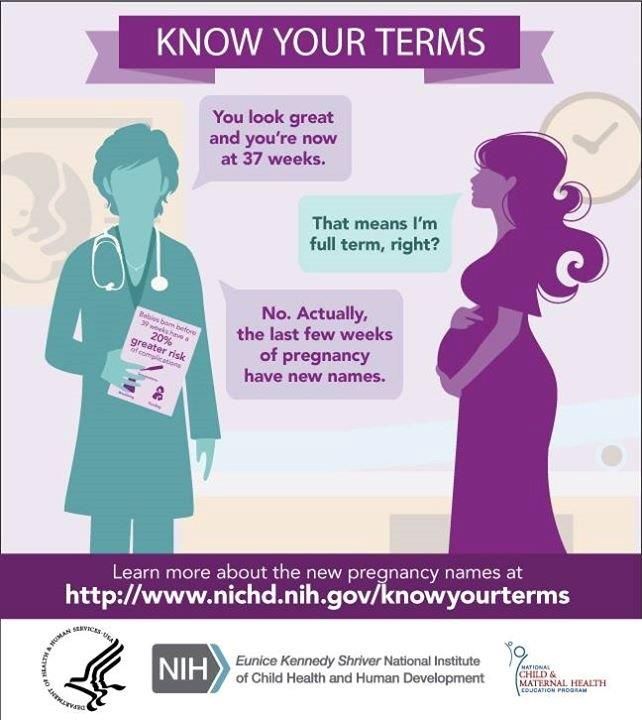 During this period of pregnancy, the fetus floats freely in the amniotic fluid. The composition of the amniotic fluid can determine the condition of the fetus.
17-18 weeks
These days, your unborn child begins to move. His limbs, ligamentous apparatus, cerebellum have already developed enough. By this time, the formation of the immune system is completed.
19-20 weeks
There have been big changes in your body. The pulse quickened, cardiac output increased significantly (40% higher than the initial level) and the volume of circulating blood (almost 500 ml).
Due to the increased volume of plasma compared to the mass of red blood cells, hemoglobin decreases in blood tests.
Some women during this period experience frequent and painful urination, pain in the lumbar region on the right or left, weakness. A large uterus presses down on the bladder, the mouth of the ureters, disrupting the outflow of urine. Stagnation of urine and incomplete emptying of the renal pelvis create conditions for the development of infection.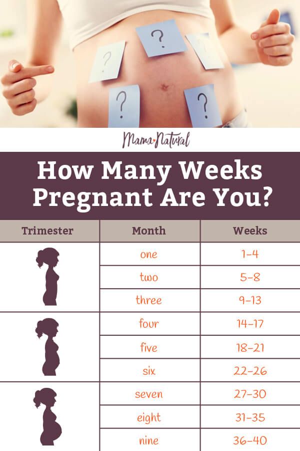 Bacteriuria develops and pyelonephritis of pregnant women may occur. If there is any suspicion of pyelonephritis, you should immediately consult a doctor, because this disease is not only dangerous for your health, but also for the further growth and development of the fetus.
The weight of the baby is 300-350 grams, he often and quite actively moves, swallows amniotic fluid, begins to open his eyes.
21-22 weeks
In these weeks, the fetus already has a mass of 400-500 grams, and it develops very intensively bones and muscles, which require calcium from your body. Therefore, if you do not want to lose your white-toothed smile, then, on the advice of your obstetrician-gynecologist, start taking calcium supplements regularly. This will help save your teeth and get rid of leg cramps. They appear for the same reason of calcium deficiency.
23-24 weeks
At this time, the weight of the fetus is 500-600 g. It already has all the organs and systems fully formed. Until that time, only his lungs remained immature. And now, by 24 weeks, they begin to ripen. And the cells lining the lung alveoli produce surfactant, a substance that, by lubricating the alveoli, prevents them from sticking together during breathing. However, the amount of surfactant is so small that a child born at this time will not be able to breathe on its own. To survive outside the uterus, he needs sophisticated breathing equipment, incubators, a control system, infusors for nutrition, infusion media, artificial surfactant.
There are perinatal centers where children born during these terms of pregnancy are nursed. It is very difficult. And therefore, the longer the pregnancy is prolonged, the more likely the birth of a healthy and viable child. Therefore, try to do everything so that the child is born on time, full-term and healthy.
By this gestational age, the uterus is at a height of about 24 cm above the pubic bone, and now it not only builds up muscles, but is also stretched by the fetus that completely filled its cavity.
25-26 weeks
The fetus already has a mass of 700-750 g. Due to the improvement of the brain structures in his body, a connection is established with the adrenal cortex and they begin to produce corticoids - hormones necessary for adaptation. The pituitary gland of the fetus reaches such a degree of maturity that the production of adrenocorticotropic hormone begins, which also stimulates hormonal production by the adrenal glands. In short, all forces are thrown to the upcoming "publication". But the most obvious changes in these weeks occur in the lungs - there is an increased maturation of cells that produce surfactant. However, a fetus born during this period can only survive in incubators with artificial lung ventilation, artificial feeding with special infusion media. Therefore, try to keep both him and yourself from rash steps.
At this time, it's time to start preparing for the future feeding of the child. Under the influence of placental lactogen, your breasts, that is, the mammary glands, are growing rapidly.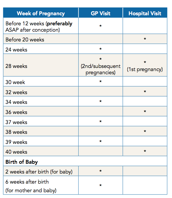 From time to time, droplets of colostrum may appear on the nipples. Daily air baths, washing with cool water, rubbing the nipples with a rough towel will help prepare the nipples for feeding. If the nipples are flat, start to stretch them little by little.
27-28 weeks
This period completes the second trimester of pregnancy. By this time, the fetus weighs up to 1000 g and has a height of up to 35 cm. However, he still cannot live on his own, because. his lungs are not mature enough and special equipment is still needed to nurse him. During these periods of pregnancy, there is an intensive growth of the fetus, the formation of muscles. His movements become more active. Periods of movement alternate with its relatively calm state when the fetus is sleeping. With an ultrasound, you can see that he already knows how to suck his thumb and even smile!
The fundus of the uterus stands on average at a height of 27-28 cm above the womb.
29-30 weeks
The third trimester of pregnancy begins.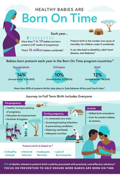 The uterus stands at a height of 29-30 cm, it becomes more difficult for you to breathe. Now one of the most serious complications can develop - toxicosis of the second half of pregnancy, which is characterized by the appearance of edema, increased blood pressure and the appearance of protein in the urine. For early diagnosis of this complication, it is necessary to carefully observe an obstetrician-gynecologist and follow all his recommendations, incl. strict weight control. In the III trimester of pregnancy, the daily weight gain should be no more than 50 g, i.e. no more than 300 g per week. You should also monitor the ratio of drunk and secreted fluid.
31-32 weeks
Have you asked your doctor how the fetus is? Find out now it's very important. Its position can be longitudinal, transverse, oblique. Correct, normal is the longitudinal position of the fetus. Childbirth is safer with cephalic presentation. From this period of pregnancy, it is necessary to wear a prenatal bandage that will support the anterior abdominal wall and help maintain the correct position and presentation of the fetus.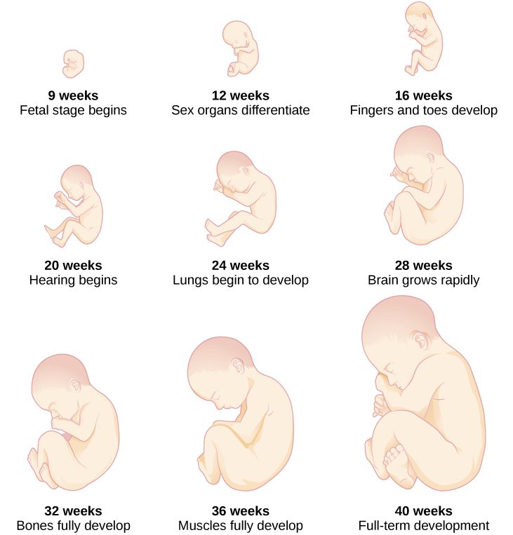 If the presentation of the fetus is breech, i.e. above the entrance to the pelvis is the pelvic end of the fetus, then the bandage should not be worn yet. There is gymnastics to correct the presentation of the fetus.
In the morning and evening for 1 hour, do the following: lie down on the bed on your left side and lie quietly for 15 minutes, then turn over to your right side and lie for the next 15 minutes, and then repeat these turns 2 more times.
Pregnant women with Rh-negative blood and with O (I) blood type need blood tests for Rh - or group immune antibodies. Immunization of pregnant women with Rh-negative blood is carried out from 28 weeks and within 72 hours after childbirth according to the indications, which will be discussed by the doctor observing you in the antenatal clinic.
33-34 weeks
The fetus already has a mass of 1800-2100 g, a height of 40-41 cm. By the end of this period, its lungs will begin to produce surfactant in full and will be able to breathe without special equipment. The fetus is fully developed, its chances of surviving in case of preterm birth are greatly increased. However, there is still extremely little subcutaneous fat, so his skin is thin and has a red color. Such a newborn retains heat very poorly and at birth needs an incubator or a heating pad. His body is still covered with fluff and cheese-like grease, the auricles are still very small, but they are already beginning to straighten out, the boy's testicles descend into the scrotum.
Caring for a premature baby is the hardest work for the whole family, associated with high material costs, physical overload of parents, and this work is not always rewarded, since a child can be born and remain sick. Therefore, up to 37 weeks of pregnancy, a woman should be especially attentive to her condition and, at the slightest suspicion of an increase in the tone of the uterus, starting frequent and regular contractions, immediately consult a doctor.
Doctors know that women, in anticipation of the arrival of a new person in the house, begin to glue walls and paint ceilings during this period. Don't take unnecessary risks. For this, prenatal leave is provided from 30 weeks, so that you can avoid overwork, do not push in transport, and have the opportunity to sleep. So repairs, stuffy shops, queues are no longer for you.
35-36 weeks
The fetus already has a mass of 2100-2700 g and a height of 44-45 cm. It is advisable to see a doctor during this period of pregnancy at least once every 10 days.
37-38 weeks
From this point on, your pregnancy is considered full-term. And if you have a baby in these weeks, he will live. Its development is complete. Now he has a mass of approximately 2700-3000 g. Height is 49-50 cm. The remaining two weeks he will add a little in weight and height.
It becomes easier for you to breathe, as the fetal head is pressed tightly against the entrance to the pelvis, the uterus pulls the anterior abdominal wall more, and therefore its bottom sank lower. Tension of the uterus; small sharp pulling pains in the lumbar region.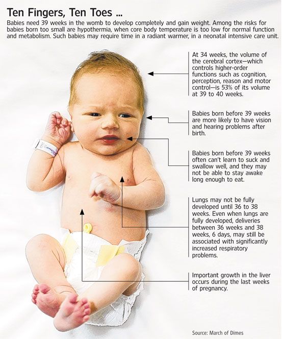
With an exacerbation of extragenital diseases, the appearance of signs of toxicosis in the second half of pregnancy, with an incorrect position of the fetus, with some gynecological diseases, against which pregnancy develops, a scar on the uterus, etc., early prenatal hospitalization is required. Do not forget to take an exchange card, passport, medical insurance policy and birth certificate to the hospital.
39-40 weeks
You can find out the approximate day of delivery by the date of the last normal menstruation - count back three months and add 7 days. The resulting number will be the estimated date of birth. More precisely, according to many parameters, ultrasound data, additional studies, the date of the first fetal movement, the date of the first visit to the obstetrician-gynecologist, especially if the visit was before 11-12 weeks of pregnancy.
The child already has all the signs of maturity. His weight is more than 3000 g, and his height is more than 50 cm, he has fair skin, a sufficient amount of subcutaneous fat, he retains heat and does not need special heating.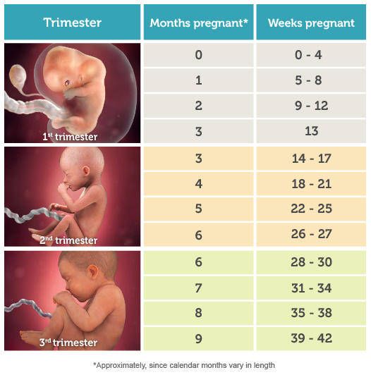 He will scream loudly, breathe, suck. There is a very small amount of lubricant on the skin, which will no longer be able to protect it from the effects of amniotic fluid.
For you, regular contractions (1 contraction every 10 minutes) will become an indicator of the beginning of the birth process, or you will feel the outflow of amniotic fluid, you will see scanty bloody discharge - do not panic, call an ambulance, the telephone number for transportation for pregnant women is written on the margins of your exchange card. While she is driving, change your clothes, prepare your passport, exchange card, medical insurance policy and birth certificate.
3rd trimester of pregnancy: what happens to the fetus
3rd trimester of pregnancy: what happens to the fetus - Private maternity hospital Ekaterininskaya Clinics3rd trimester: 27th week - birth
The baby is almost formed! In the last weeks of pregnancy, when he continues to gain weight and moves in an enclosed space, you may feel more comfortable. Try to get as much rest as possible. Symptoms of the 2nd trimester of pregnancy may include:
- Swelling of hands, feet and face. Excess fluid in the body can cause swelling of the hands, feet and swelling of the face. If possible, try to rest in a horizontal position more often, while raising your legs up, this will improve blood circulation and relieve leg fatigue.
- Discharge from the nipples. Colostrum may begin to come out of the nipples, a liquid that the baby will feed on until milk appears. To avoid stains on clothes, put special pads in your bra.
- Braxton-Hicks contractions. Your baby is getting ready to be born and you may feel light "training" contractions. These contractions that come and go at regular intervals are called Braxton Hicks contractions. If the contractions are strong and prolonged, call your doctor. This could be the start of labor.
3rd trimester milestones
- You will gain 10 to 16 kg of weight during your pregnancy.
 Weight gain is mainly due to the weight of the baby, placenta and amniotic fluid and an increase in the amount of fat and fluid in the woman's body.
 - 37 weeks is considered the full term of pregnancy.
Development of the child in the 3rd trimester of pregnancy
By the end of the 3rd trimester:
- The child takes a head down position in the pelvis, preparing for birth.
- The child's body systems are developed and ready to function independently.
- The soft, fluffy hairs that cover the body have disappeared.
- Baby's body length is about 46-56 cm from head to toes.
Mobile application of the clinic
You can make an appointment with a doctor, get tests
and much more...
Fill out the form to make an appointment or order a call back
I agree with personal data processing policy and user agreement I also give my consent to the processing of personal data.