Abdominal separation test
Diastasis Recti Test: How to Check for Diastasis Recti on Your Own
If you're here, you likely suspect you have diastasis recti. Maybe you've given birth to your first child, recently welcomed a second, or are a seasoned parent with kids long out of the house. Diastasis recti affects over 60% of childbearing women, so no matter what stage of motherhood you are in, you've come to the right place to learn how to check for diastasis recti, and work to resolve it.
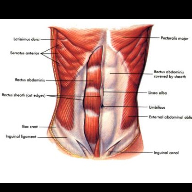
Diastasis recti, also known as abdominal separation, is a separation of the rectus abdominis or "6 pack muscles." Pregnancy is the primary cause of diastasis recti, but anyone can get it from improper exercise or movements that put excessive pressure on the abdominals. DR has historically been treated as a cosmetic issue, but the symptoms far exceed a persistent pooch that lingers long after pregnancy; back pain, urinary stress incontinence, and pelvic floor dysfunction are all hallmarks of the condition.
The good news? You can conduct a diastasis recti test on your own, and can soon be on your way to restoring core strength and function from the comfort of your own home. Here's how to check for diastasis recti, along with frequent questions and answers to ensure you do it correctly.
How to Check for Diastasis Recti on Your Own
Diastasis recti can occur in 3 areas: above the belly button, below the belly button, and at the belly button. To conduct a diastasis recti test on your own, follow these four steps and watch the video below.
How to Check for Diastasis Recti:
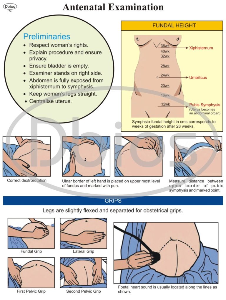
- Lie flat on your back with your knees bent.
- Place your fingers on your belly button, pointing towards your pelvis, and press down.
- Lift your head up about an inch while keeping your shoulders on the ground.
- If you have diastasis recti, you will feel a gap between the muscles that is an inch wide (~ 2 fingers) or greater.
Diastasis Recti Test Gap Chart:
How to Check for Diastasis Recti Video:
Diastasis Recti Test: FAQs
When should you check for diastasis recti?
When it comes to self-checking for DR, there is no strict timeline. In fact, it's never too late to restore core strength and function with exercise! You can self-check as early as 24 hours following a vaginal delivery, one week after a cesarean, or right now if you're suffering from back pain, incontinence, pelvic pain, or a persistent pooch long after childbirth.
How does a c-section delivery impact self-checking for diastasis recti?
A c-section delivery should not hinder your ability to self-check for diastasis recti because the typical incision point is well below the measuring sites. That said, if there is considerable pain or tenderness in any area, wait to check that spot.
Note: Sometimes OB/GYNs will sew together the lower abs when they finish a c-section. This can impact your ability to get an accurate read on your abdominal separation, so it's best to ask their approach before you give birth and state a preference if you have one. If the delivery has already happened, ask to see the surgical report or ask the doctor what she/he did.
How can I tell if I have a diastasis recti induced "gap"?
When checking yourself for diastasis recti, note both the width and the depth of the distance between the abdominal muscles. To accurately measure the gap, lift your head just barely off the floor to trigger a spontaneous activation of the abdominal muscles. If you've followed the steps outlined in the "how to self-check video" and struggle to find the gap, try lifting your head higher a couple of times until you can feel the difference between the engaged muscles (firm tissue) and the connective tissue (softer) that lies between them. Once you are certain you have identified the muscles and that your fingers are aligned vertically between them (pointing downward towards the pelvis), lift your head about an inch off the floor and record the width and depth of the separation.
Note: When checking for diastasis recti, it's important not to lift your head too high because that will result in an erroneously narrow measurement.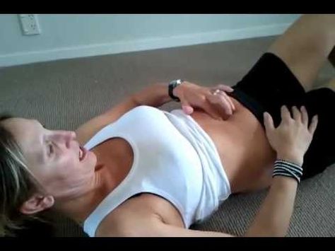
How do I check for diastasis recti if I have a lot of belly fat?
If you have excess belly fat, it's important to press your fingers firmly into your midline when self-checking for diastasis recti. You may need to lift your head and shoulders off the floor to feel the muscles engage. That said, the most accurate measurement will come when the head is lifted about an inch from the ground, and the muscles first begin to grab the sides of the fingers. For this reason, once you are certain you have identified the muscles and that your fingers are pressing down firmly between them (pointing downward towards the pelvis), lift your head about an inch off the floor and record the width and depth of the separation.
How often should I check for diastasis recti?
When participating in the Every Mother program, you will be prompted to perform a self-check and enter your measurements on days 1, 21, 42, 63, and 84 of your EMbody Reclaim Path. It is important not to check more often than every 2-3 weeks because checking is mildly stressful to the tissue you are working to heal.
How do I know if I will need surgery from diastasis recti?
There are times that abdominal separation may require surgical intervention. DR varies from person to person, but if 8 or more fingers sink easily into the gully between your abdominal muscles, follow our EMbody Reclaim stage and consult your doctor to discuss your options. Even if a surgical repair is indicated, surgeons recommend our therapeutic exercises to patients both pre- and post-op to improve outcomes. Our EMbody program holistically supports the health and integrity of the connective tissue, safely strengthens the deep core, coaches patients through safe, functional movements in daily life, and accelerates the healing process.
Every Mother unlocks a scientifically proven method to strengthen the body during pregnancy and rebuild it after birth, regardless of how long it has been since you became a mother. We’re a knowledge circle, a community, and a celebration— of the mother you’ve become, and the woman you’ve been all along. Learn more about the EMbody Program™ here.
Self Testing Diastasis Recti | 4 Simple Ways to Test Yourself
Table of Contents
How to Test for Diastasis Recti?
Believing you may have a diastasis recti can be a worrisome / frustrating experience. However, there is an abundance of information regarding diastasis recti recovery. We at Restore Your Core have designed a program specifically for women who are suffering from diastasis recti or other abdominal/pelvic floor issues. This article will help begin to address what diastasis recti is and instruct you in how to perform a self-test.
What is Diastasis Recti?
Diastasis recti is the stretching or separation of the rectus abdominis (6 pack) muscles caused by the thinning of the linea alba (midline connective tissue). Diastasis recti separation leaves your abdominal organs unsupported, and if severe, can expose your digestive organs creating a stomach bulge.
This separation can range from being isolated above the belly button, within the belly button, and below the belly button sitting above the pubic bone.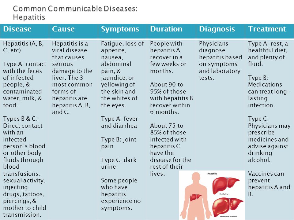 In some cases, the separation emcompasses the entire mid section of the core.
In both men and women, this gap can be created in the midline of your belly anywhere from the pubic bone to the base of your ribcage. During a crunch or sit-up, where one would normally feel tension and closure, there is a space in between.
What Does it Look Like?
Diastasis recti looks different from person to person. Although in some cases the symptoms can be painful and more present, in some people they aren’t noticeable at all. Below I address the most common and present symptoms you should be aware of in determining whether or not you may have a diastasis recti.
Abdominal Bulge
An abdominal bulge is not always an indication of a diastasis recti, yet, it can be a symptom.
This bulge, or stomach “pooch,” occurs when the abdominal organs become unsupported by the rectus abdominis muscles. This can appear as a cone shape or ridge above and within the area located close to the belly button.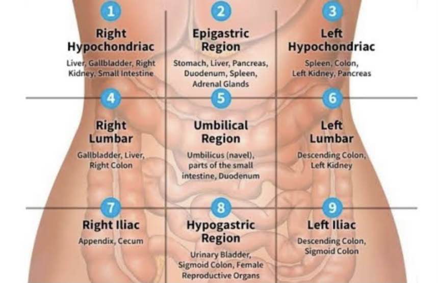 However, depending on where the diastasis recti has become isolated, the bulge can range from above the belly button, on the belly button (causing the belly button to flatten), or below the belly button just above the pubic bone.
Muscle Separation & Linea Alba Stretching
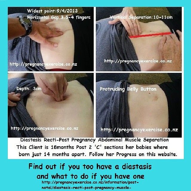
This is the most noticeable and common symptom of diastasis recti (whether you have significant body fat or no body fat at all). A minor separation (one of 1-2 finger widths) is not a significant injury,, but I advised considering rehab or core building exercises to prevent the gap from widening.
In more severe cases, the separation can be that of 5-10 finger widths. This effect is much more noticeable and can be seen as a crevice or significant gap within the abdominal core. Diastasis recti is also measured by shallowness or deepness. Someone could potentially have a 10 finger width separation but it’s shallow. In this case, exercise and safe core strengthening routines can help restore the core to its natural state.
How to Test for Diastasis Recti:
- Lie on your back in a comfortable position. Bend your knees and put your feet flat on the floor.
- Place one hand on the midline of your core with your fingers flat on your midline.
- Place your other hand under your head and neck for support.Lift your head slowly and begin adding pressure through the pads of your fingers.
- With no diastasis recti, there is the sensation of a toned wall as you lift your direct. If you feel a space, or your fingers sink into your core, you likely have diastasis recti.
- Repeat the procedure for the areas directly above your belly button down to the pubis to determine whether the diastasis recti is isolated or in your core as a whole.
How to Tell if You Have a Diastasis
Rectus abdominis separation can lead to a stomach bulge (aka stomach pooch), pelvic floor issues, unnatural posture, and stomach and back pain. The symptoms of diastasis recti include but are not limited to:
- Abdominal Bulge
- Abdominal Gaping
- Lower Back Pain
- Sensation of Bloatedness without Bloat
- Incontinence (leak pee)
- Poor Posture
- Constipation & Bloat
- Doming or invagination of the linea alba when when performing crunches or other traditional ab exercises
- Difficulty with everyday activities due to a lack of core function.
Unless you have a low body fat percentage or have an overly toned core with a visible 6-pack, it is very hard to diagnose a diastasis recti on appearance alone. The linea alba lies beneath the fat layer of your abdomen, so it cannot be seen. Many people have a diastasis recti for years before learning they have it.
Does Coning Always Mean You Have Diastasis Recti
Abdominal coning is most commonly a sign of diastasis recti. Diastasis recti during pregnancy can be a cause of the core doming or coning. This is a result of the expanding uterus as it makes room for your child. Although the separation of the muscles and the stretching of the connective tissues is normal, coning may present its own problems. If your belly is changing its shape from being round and reverting to a coning shape, this could be an indication that diastasis recti is present and will extend into the postpartum period.
What does Mild Diastasis Recti Look Like?
Similarly to the above, mild diastasis recti can show signs of an abdominal bulge or look like the midline of your abdomen is “coning.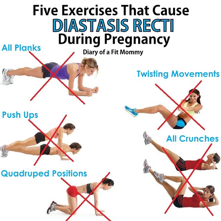 ” During a self-assessment or if you have a physical therapist assess you, diastasis recti looks like a separation in your core. In mild cases, this gap can be 1-2 finger lengths wide, yet present little to no symptoms. It is best to consider core strengthening programs even if the gap is not severe in order to prevent the gap from widening.
Sign up for a delivery of inspiration, exclusive offers, contests and the inside scoop on events.
What to Do If You Think You Have It
Prehab
One of the best things you can do during pregnancy is prehab. I have many long term clients who were able to prevent their diastasis recti from returning with subsequent pregnancies by working their core in a smart, functional way the entirety of their pregnancy. Many report that their core felt stronger than ever with the prehab work that they did. Pregnancy is not an illness, there is no need to halt all exercise. We do, however, want to make good exercise choices. It is very important to exercise your core during pregnancy but not to increase intra ab pressure as you do so.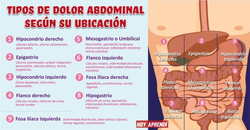
Our online prehab program, One Strong Mama, is designed to prepare your body for the unique demands of pregnancy, birth, and recovery. Everything you need to prepare your body can be found in this program which combines functional exercise, strength training, posture and alignment instruction, and even key educational tools so that you are given a chance to enjoy your pregnancy without stressing over it.
Postpartum
Diastasis recti is often more apparent postpartum. I usually recommend waiting at least 6 to 12 weeks before checking for a diastasis recti. Here are a few tips that can help prevent and/or heal a diastasis recti.
Rest
If I could have a dollar for every mom who wished she had rested more in order to spare herself injury. Rest is so important for healing your body postpartum and ensuring that you do not damage your core and your pelvic floor. We recommend getting back into exercise at least 6 weeks postpartum, but even then, easing into it is key. The “I want to get my body back” sentiment can be very harmful to a recovering body.
Exercise/Rehab
Once you are cleared for exercise by your medical professional, you want to focus on core building exercises which properly strengthen your core without aiding in diastasis recti development. My program Restore Your Core is designed for any woman with weak core issues such as: postpartum issues, diastasis recti, incontinence, and constant back pain.
Restore Your Core is designed so that you can use helpful and intelligent core building strategies not only during exercise, but in everyday motion. I help you approach healing and restoring your body in a personal way. I challenge you to train your whole body to move correctly in order to get you stronger and heal properly.
Join our community and connect with over 30,000 women dedicated to healing their pelvic floor issues. The RYC® Facebook group offers an intimate space for women to openly discuss and learn more about issues such as disastasis recti, leaky pee, prolapses, etc.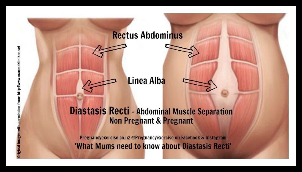 You don’t have to be alone in your journey for a stronger self, join us today!
Page not found
Size:
AAA
Colour: C C C
Pictures On Off
Regular site version
RUENBY
Gomel State
Medical University
- university
- University
- History
- Manual
- Charter and Symbols
- Educational activities
- Organization of the educational process
- International cooperation
- Quality management system
- Tips
- Faculties
- Chairs
- Divisions
- Primary trade union organization of workers
- University publications
- University Pride
- Graduate-2021
- Primary organization "Belarusian Union of Women"
- Single window
- GomGMU in international rankings
- University structure
- Applicants
- Admission Committee
- University Biology Olympiad
- Targeted training
- Conclusion, termination of the "target" contract
- Benefits for young professionals
- Passing score archive
- Map and directions
- Admission procedure for 2023
- Specialties
- Enrollment targets for 2022
- Tuition fees
- Information about the process of receiving documents
- Reception of documents and working hours of the selection committee
- Procedure for admission of citizens of the Russian Federation, Kyrgyzstan, Tajikistan, Kazakhstan
- Admission Campaign Hotline
- Students
- Freshman
- Schedule
- Exam Schedule
- Information for students
- Student Club
- Sports club
- Dormitory
- Regulations
- Practice
- Tuition fees
- Life safety
- BRSM
- Trade union committee of students
- Training Center for Practical Training and Simulation Training
- Multifunctional student card
- Student survey
- Graduates
- Internship and clinical residency
- Doctorate
- Postgraduate
- Master
- Distribution
- Doctors and specialists
- Professorial Advisory Center
- Faculty of advanced training and retraining
- For foreign citizens
- Faculty of Foreign Students
- Tuition fees
- Registration and visas
- Useful information
- Admission rules
- Information on admission opportunities and conditions in 2022
- Official representatives of GomGMU for enrollment of students
- Insurance of foreign citizens
- Admission to the Preparatory Department of foreign citizens
- Reception of foreign citizens for training in English / Training of foreign students in English
- Advanced training and retraining for foreign citizens
- Scientific activity
- Areas of scientific activity
- Scientific and pedagogical schools
- Innovative technologies at GomGMU
- Research unit
- Research laboratory
- Competitions, grants, scholarships
- Scientific events
- Work of the ethics committee
- Help for the researcher
- Young Scientists Council
- Student Scientific Society
- Dissertation Council
- Patents
- Method instructions
- "Horizon Europe"
- State Program (ChNPP)
- Home
Pass a clinical blood test with ESR, leukoformula
I confirm More
- Invitro
- Analyzes
- Hematological ...
- Clinical analysis ...
- Clinical analysis ...
- Covid-19 9002 development of diseases of the cardiovascular system
- Diagnosis of antiphospholipid syndrome (APS)
- Assessment of liver function
- Diagnosis of the condition of the kidneys and genitourinary system
- infections transmitted by sexually (IPP)
- Problems of weight
- VIP examination
- respiratory diseases
- Allergies
- Determination of microelements in the body
- Sports profiles
- Hormonal studies for men
- Differential diagnosis of depression
- Blood coagulation assessment 9Comprehensive immunological studies
- Lymphocytes, subpopulations
- Immunoglobulina
- components of complement
- Regulators and immunity mediators
- Allergological studies - allergen -special tests (allergen -specificestes), venens, ostboats.
- IgG, allergen-specific
- ImmunoCAP technology
- AlcorBio technology
- Systemic connective tissue diseases
- Rheumatoid arthritis, joint damage
- Antiphospholipid syndrome
- Vasculitis and kidney damage
- Autoimmune lesions of the gastrointestinal tract. Celiac disease
- Autoimmune liver diseases
- Neurological autoimmune diseases
- Autoimmune endocrinopathies
- Autoimmune skin diseases
- Lung and heart diseases
- Immune thrombocytopenia0022
- Additional studies (after screening and consultation with a specialist)
- Determination of biological relationship in the family: paternity and motherhood
- Water quality study
- Soil quality testing
- Diagnosis of liver pathology without biopsy: FibroMax, FibroTest, SteatoScreen
- Professional position
- Venous blood for analysis
- Tumor markers.
 View of a practical oncologist. Laboratory justifications.
 - Testosterone: diagnostic threshold, method-dependent reference values
- Laboratory assessment of lipid metabolism parameters in INVITRO
- Lipid profile: fasting or not fasting
1
4 The cost of analyzes is indicated without taking biomaterial
Description
Method of determination See data sheet
Test material Look in the description
Synonyms: Complete blood count, KLA. Full blood count, FBC, Complete blood count (CBC) with differential white blood cell count (CBC with diff), Hemogram.
Brief description of the study CBC: complete analysis, leukocyte formula, ESR
See also: General analysis - see test No. 5, Leukocyte formula - test No. 119, ESR - test No. 139.
Blood is a liquid tissue with various functions. It consists of plasma and formed elements: erythrocytes, leukocytes and platelets.
Complete blood count includes the determination of hemoglobin concentration, the number of erythrocytes, leukocytes and platelets, hematocrit and erythrocyte indices (MCV, RDW, MCH, MCHC).
The leukocyte formula is the percentage of different types of leukocytes (neutrophils, lymphocytes, eosinophils, monocytes, basophils).
CBC is one of the most common laboratory tests used to assess general health.
This analysis plays an important role both in the primary diagnosis of a number of diseases and in the control of their course. This test is used for general assessment of health status, diagnosis, monitoring of the course, evaluation of the effectiveness of therapy for a variety of diseases, including anemia, infections, inflammatory diseases, etc. added anticoagulant into two layers: upper (clear plasma) and lower (settled erythrocytes). With the appearance in the blood plasma of a large number of proteins of the acute phase of inflammation, which include fibrinogen, C-reactive protein, alpha and gamma globulins, etc.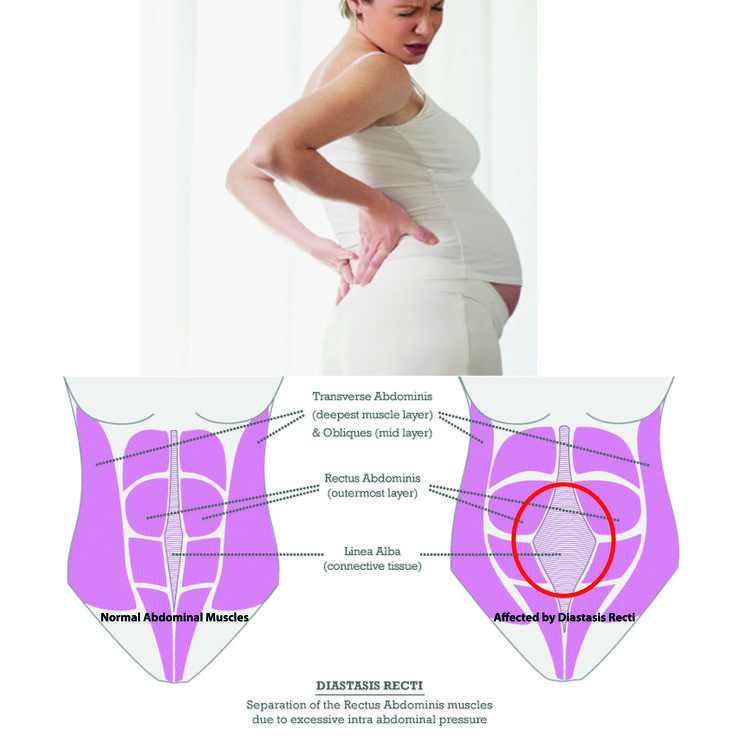 , or paraproteins, the repulsive force between the erythrocytes decreases, and the erythrocytes settle faster (ESR increases). In acute inflammatory diseases, ESR usually rises a day after the onset of the disease, while the normalization of this indicator after recovery is slower, and can take from several days to two or more weeks.
See also test no. 43 CRP (C-reactive protein).
Please note that when performing a clinical blood test (No. 1515) and when calculating the leukocyte formula (No. 119), if significant deviations are found in the samples, and the result requires manual microscopy, INVITRO additionally performs a free manual calculation of the leukocyte formula with counting young forms of neutrophils (including an accurate count of stab neutrophils) and a quantitative assessment of all pathological forms of leukocytes (if any).
What is the purpose of the study "Clinical blood test: general analysis, leukocyte formula, ESR"
Complete blood count together with leukocyte formula is widely used as one of the basic tests of laboratory examination in most diseases, although the changes that occur in peripheral blood are detectable for the most part are non-specific, and are subject to interpretation only in conjunction with an analysis of the clinical situation, anamnesis and the results of other types of examination.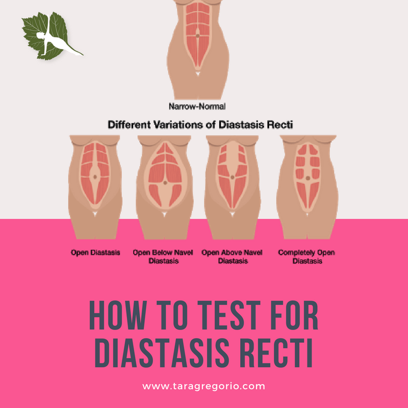 In addition to a general blood test with a leukoformula, in certain clinical situations, it may be useful to evaluate the ESR as a non-specific marker of inflammation activity (see also test No. 43).
What can affect the results of the test "CBC: general analysis, leukoformula, ESR"
Changes in the protein composition of the blood during pregnancy lead to an increase in ESR during this period. The erythrocyte sedimentation rate is influenced by their morphology (poikilocytosis of the erythrocytes of the test sample leads to an underestimation of the ESR, smoothing the shape of the erythrocytes, on the contrary, can accelerate the ESR), as well as the hematocrit value. A decrease in the content of erythrocytes (anemia) in the blood leads to an acceleration of ESR and, conversely, an increase in the content of erythrocytes in the blood slows down the rate of sedimentation.
At different times of the day, at different times after a meal, and also due to excessive dehydration or hyperhydration, fluctuations in the values of clinical blood tests are possible, so it is advisable to carry out routine diagnostic studies under standard conditions (see Preparation for studies).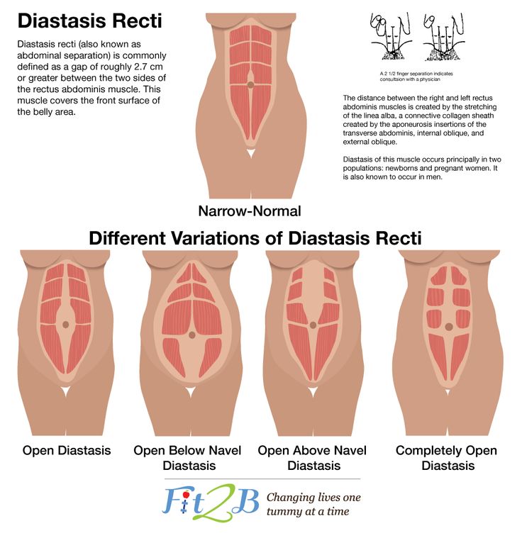 Clinical blood test indicators have age and gender characteristics and are subject to interpretation in comparison with reference values in relation to sex and age.
Preparation
Rules for preparing for the study "Clinical blood test: general analysis, leukoformula, ESR"
It is preferable to take blood in the morning on an empty stomach, after 8-14 hours of an overnight fasting period (you can drink water), it is permissible in the afternoon 4 hours after light meal.
On the eve of the study, it is necessary to exclude increased psycho-emotional and physical activity (sports training), alcohol intake.
Indications for use
In what cases is carried out "Clinical blood test: general analysis, leukoformula, ESR":
- in order to diagnose and control the course of the pathological process;
- hematological diseases;
- inflammatory diseases;
- infections;
- oncological diseases.
Interpretation of results
Interpretation of test results contains information for the attending physician and is not a diagnosis. The information in this section should not be used for self-diagnosis or self-treatment. An accurate diagnosis is made by the doctor, using both the results of this examination and the necessary information from other sources: history, results of other examinations, etc.
Units
See related tests.
Interpretation of the results of the study "Clinical blood test: general analysis, leukoformula, ESR"
See. descriptions of individual tests:
- No. 5 Blood test. Complete blood count (without leukocyte formula and ESR) (Complete Blood Count, CBC)
- №119 Leukocyte formula (differential white blood cell count, leukocytogram, Differential White Blood Cell Count) with blood smear microscopy in the presence of pathological changes.
- №139 ESR (Erythrocyte Sedimentation Rate, ESR)
Questions
and answers
{{{this.