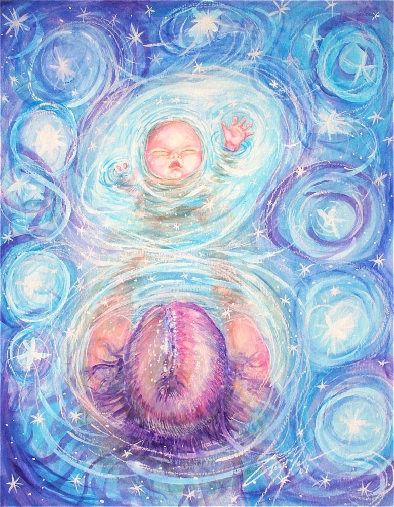
3 month pregnant picture
3 Months Pregnant: Symptoms and Fetal Development
At three months pregnant, you’ve almost made it to the second trimester, and some of the early pregnancy symptoms you may have been experiencing may slowly start to subside. Read on to find out what may be in store for you at three months pregnant, including common symptoms. Plus, find out all the exciting ways your baby is developing this month.
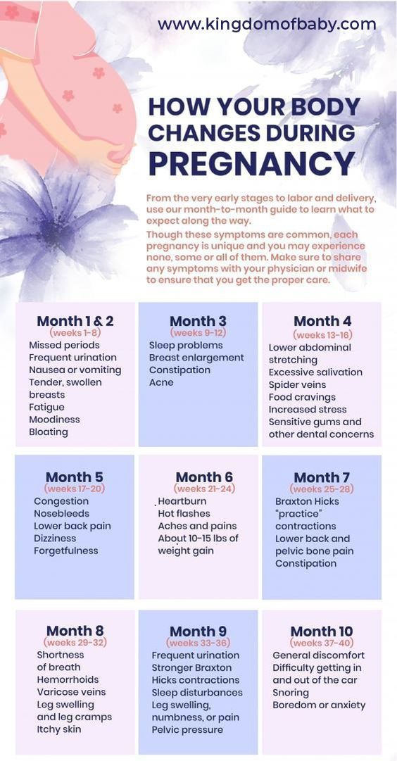
Common Pregnancy Symptoms at 3 Months Pregnant
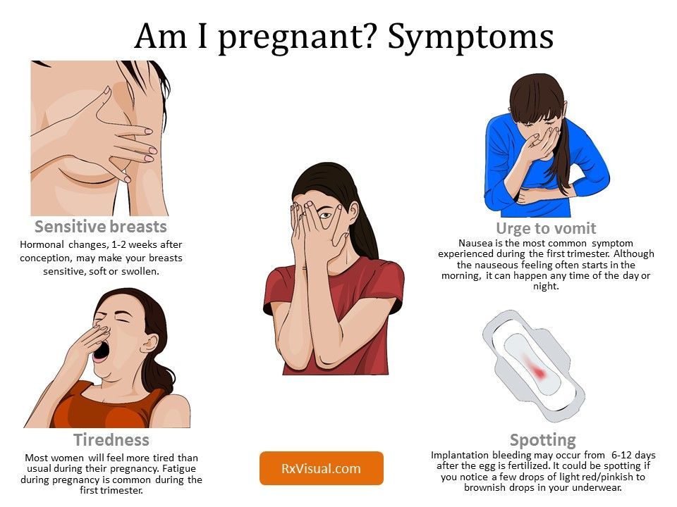
At three months pregnant, you might still be experiencing some of the familiar symptoms of early pregnancy, but some new ones might crop up, too. Some of these symptoms might be quite challenging; keep in mind you may not experience them all.
Increase in vaginal discharge. A combination of pregnancy hormones and the increased blood supply in your body can lead to a bit more vaginal discharge than you might be used to. As long as it’s clear or whitish and doesn’t have a bad smell, it’s probably nothing to worry about.
Try to wear cotton underwear and loose, breathable clothes to help prevent vaginal infections. Chat with your healthcare provider if you’re concerned about what’s happening.
Nausea. You might still be feeling queasy, but perhaps for not much longer. Many moms-to-be say their morning sickness begins to subside during this month, which is great news! If you’re not so lucky, try eating bland foods like toast, rice, or bananas, and sip on ginger ale or ginger tea to soothe your stomach.
Fatigue. The sleepiness may continue this month as your body continues to nourish your little one. Rest when you can, stay hydrated, and do some moderate exercise, as this is shown to improve sleep. Prenatal yoga, walking, and swimming can be good choices, but talk to your healthcare provider before trying any new exercises.
Skin changes. If you’ve noticed that the color of your nipples has started to darken, this is because your body is producing more melanin, a type of pigment.
 This extra melanin can also cause brown patches on your face, which is called chloasma. You might also notice a dark, vertical line that runs from your belly button to the pubic area. This line might start to appear at three months pregnant as your belly size starts to increase. Most of these discolorations will disappear or fade after your little one is born.
 Breast changes. Your breasts may be growing and changing this month, too. Your areolas may grow larger and darker, and your nipples may start to protrude a little more. Under the surface, milk glands are preparing to produce milk, and fat is being added to your breasts. If your bras feel too tight, it’s probably time to go up a size. Go for a professional bra fitting at your local department or specialty lingerie store to get a new bra that is more comfortable.
Constipation. Some pregnancy hormones can cause your digestive system to slow down, leading to constipation. The extra iron in your prenatal vitamins may also be to blame.
 Make sure to stay hydrated and eat more fiber. Fruits, vegetables, and whole grains are great sources of fiber.
How Is Your Baby Developing This Month?
On the inside, your little one’s intestines and musculature system are taking shape. Some bones may start to harden, but the backbone is soft. On the outside, your baby’s hands and feet are growing tiny fingers and toes, which may even have the beginnings of fingernails and toenails at three months pregnant. At some point this month, your little one’s external genitals will start to form, and it won't be long before you'll be able to find out if you're having a girl or a boy!
Keep in mind, you’ll usually find out whether you’re having a boy or girl at your mid-pregnancy ultrasound, but for a little fun now, play around with our Chinese Gender Predictor
How Big Is Your Baby When You’re 3 Months Pregnant?
At the start of this month your baby will be about ½ an inch long, and by the end of this month she’ll be almost 2 inches long and weigh about ½ an ounce.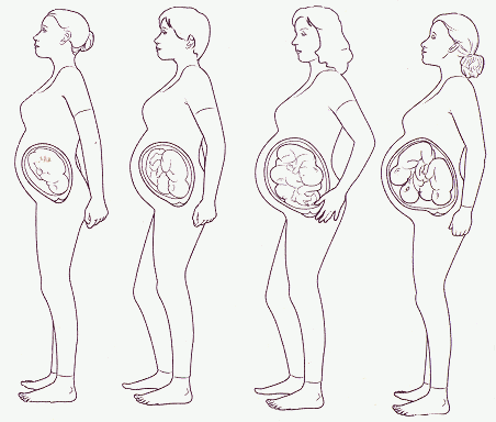
Related pregnancy tool
Fill out your details:
Pre-pregnancy weight (lbs.)
This is a mandatory field.
Height (ft.)
This is a mandatory field.
Height (in.)
Current week of pregnancy (1 to 40)
This is a mandatory field.
Tick the box
I'm expecting twins
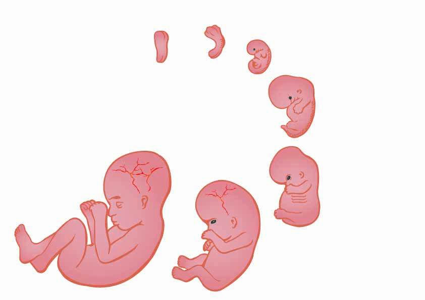
What Does a Fetus Look Like at 3 Months?
Check out these illustrations for a glimpse at what your baby might look like when you’re three months pregnant:
3 Months Pregnant: Your Body’s Changes
It's possible that you might start to project a small baby bump sometime soon, although every mom-to-be starts to show at different times, and you might have to wait another few weeks.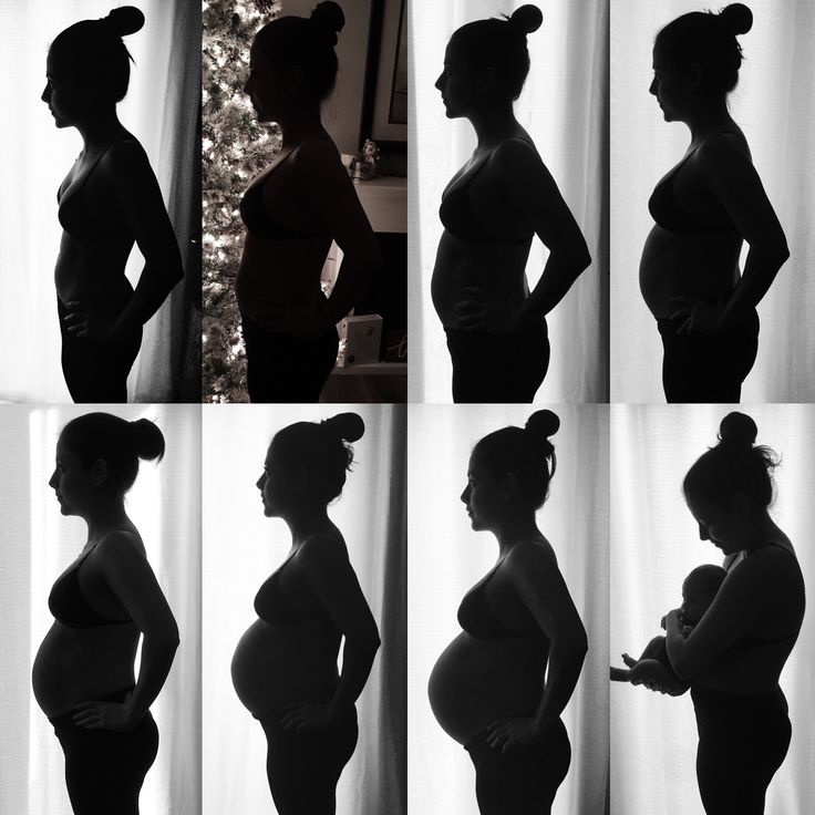 By this month, your uterus has grown to about the size of a large orange. Read more on when you might start to show here.
If your nausea subsides, you may find your appetite is returning. You’ll want to continue eating healthily. Even though there are two of you, you don’t actually need to “eat for two.” Experts recommend adding only about 300 extra calories to your diet each day. This is the equivalent of a light meal or snack. Unless you’ve lost a little weight due to morning sickness, you may end up gaining anywhere from one and a half to four and a half pounds this month. Consult your healthcare provider for advice on how much weight gain is right for you.
Our Weight Gain Calculator can tell you how much weight you may be advised to gain over the course of your pregnancy, based on your pre-pregnancy body mass index (BMI), but your provider is your best resource.
How Far Along Are You at 3 Months Pregnant?
Wondering which weeks are in the third month of pregnancy? Good question! There's no standard answer, but three months pregnant is often defined as covering week nine through week 12 or week 9 through week 13.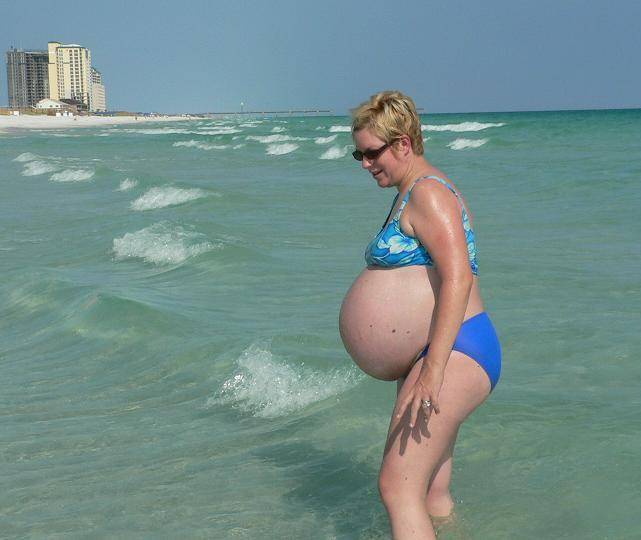 At the end of this month, you’ll be ready to begin the second trimester.
Checklist for When You’re 3 Months Pregnant
Ask your healthcare provider whether any genetic screening tests are recommended for you this month based on your personal situation. Your provider can talk you through the risks and benefits of screening tests like a nuchal translucency test, a cell-free DNA test, or chorionic villus sampling (CVS).
Consider sharing the news with close friends and family soon.
Read up on all the foods and drinks to avoid during pregnancy.
Start thinking about your maternity leave plans, and ask your healthcare provider when is the right time to tell your employer you are pregnant.
Speak to your healthcare provider about safe and gentle exercise options that are suitable for you.
Start to bond with your little one by talking to and singing to your “bump,” or listen to your favorite music together.
 Your little one will soon be able to hear you!
Communicate with your partner about any fears you have or feelings you might want to share. This can help your partner feel more involved, and will take some of the load off of you.
If this is not your first pregnancy, read up on how a second pregnancy can be different to a first.
Sign up for even more weekly pregnancy tips here:
What Happens at 3 Months of Pregnancy?
In This Section
- Month by Month
- What happens in the second month?
- What happens in the third month?
- What happens in the fourth month?
- What happens in the fifth month?
- What happens in the sixth month?
- What happens in the seventh month?
- What happens in the eighth month?
- What happens in the ninth month?
- What happens in the tenth month?
The embryo becomes a fetus when you’re 3 months pregnant. The umbilical cord connects the fetus to your placenta and uterine wall. External sex organs also start to develop.
What happens during week 9 - 10?
The embryo develops into a fetus after 10 weeks. It’s 1–1.5 inches (21–40 mm) long. The tail disappears. Fingers and toes grow longer. The umbilical cord connects the abdomen of the fetus to the placenta. The placenta is attached to the wall of the uterus and it absorbs nutrients from the bloodstream. The cord carries nutrients and oxygen to the fetus and takes waste away from the fetus.
What happens during week 11 - 12?
The fetus is now measured from the top of its head to its buttocks. This is called crown-rump length (CRL).
-
The fetus has a CRL of 2–3 inches (6–7.5 cm).
-
Fingers and toes are no longer webbed.
-
Bones begin hardening.
-
Skin and fingernails begin to grow.
-
Changes triggered by hormones begin to make external sex organs appear — female or male.
 The fetus begins making spontaneous movements.
 -
Kidneys start making urine.
-
Early sweat glands appear.
-
Eyelids are fused together.
What are the symptoms of pregnancy in the third month?
Many of the pregnancy symptoms from the first 2 months continue — and sometimes worsen — during the third month. This is especially true of nausea. Your breasts continue growing and changing. The area around your nipple — the areola — may grow larger and darker. If you’re prone to acne you may have outbreaks.
You probably won’t gain much weight during the first 3 months of pregnancy — usually about 2 pounds. If you’re overweight or underweight you may experience a different rate of weight gain. Talk with your nurse or doctor about maintaining a healthy weight throughout your pregnancy.
Miscarriage
Most early pregnancy loss — miscarriage — happens in the first trimester. About 15 percent of pregnancies end in miscarriage during the first trimester. Learn more about miscarriage.
- Yes
- No
Help us improve - how could this information be more helpful?
How did this information help you?
Please answer below.
Are you human? (Sorry, we have to ask!)
Please don't check this box if you are a human.
You’re the best! Thanks for your feedback.
Thanks for your feedback.
Back to top
We couldn't access your location, please search for a location.
Zip, City, or State
Please enter a valid 5-digit zip code or city or state.
Please fill out this field.
Service All Services Abortion Abortion Referrals Birth Control COVID-19 Vaccine HIV Services Men's Health Care Mental Health Morning-After Pill (Emergency Contraception) Pregnancy Testing & Services Primary Care STD Testing, Treatment & Vaccines Transgender Hormone Therapy Women's Health Care
Filter By All Telehealth In-person
Please enter your age and the first day of your last period for more accurate abortion options. Your information is private and anonymous.
AGE This field is required.
Or call 1-800-230-7526
3D and 4D ultrasound during pregnancy: how to get photos and videos of your baby before birth
11/11/2019
In the life of any woman, pregnancy is an important stage that is filled with vivid emotions and, most importantly, the expectation of the birth of her child. As you know, within 9 months, the expectant mother is recommended to visit doctors of various profiles several times, who will help determine that everything is in order with the baby, or prescribe the necessary treatment. One of these mandatory medical examinations is a fetal ultrasound, which is carried out at least three times during pregnancy. Now, after such a procedure, a woman can get photos and videos with her unborn baby. This opportunity is provided by the service of 3D and 4D ultrasound, which is carried out in the network of clinics "Health", on Sovetsky, 24.
WHY YOU NEED USAGE
Ultrasound examination allows you to assess the general condition of the woman and the fetus, including its weight, size, position and gender. When conducting any type of ultrasound, ultrasound is used. It is tuned to a certain frequency, and the procedure itself does not cause pain or discomfort to both the expectant mother and the baby. A special sensor emits sound waves, they are fully or partially reflected from the tissues of the body and returned in a distorted form. As a result, an image of the unborn child appears on the screen of the device.
The data obtained will help the doctor to monitor the course of pregnancy, the woman's health, intrauterine development of the fetus, identify possible hereditary and congenital pathologies, and prescribe, if necessary, appropriate treatment. The importance of ultrasound is reflected in the orders of the Ministry of Health. So, a pregnant woman must undergo a mandatory three-time screening ultrasound at 11-13 weeks of pregnancy, at 18-21 weeks and at 30-34 weeks.
In the network of clinics "Health", in the office at Sovetsky, 24, expectant mothers can do 3D and 4D ultrasound.
This procedure is safe and informative. At the same time, pregnant women will receive a photo and video of the unborn child on a digital medium as a gift, - explained the network of clinics "Health".
About 3D AND 4D ultrasound
Conventional 2D ultrasound has been used for a long time. It can be done both in the early and late stages of pregnancy - at each stage it will tell you informatively about how the baby is doing in the mother's stomach. Perhaps the most significant disadvantage of this type of study is that its results result in a “flat” picture, which can often only be understood by a doctor.
3D and 4D ultrasound are relatively new and modern procedures. Their main advantage for future parents is obtaining a color three-dimensional image of the unborn child, and for doctors, revealing more accurate and visual data on the development of the fetus.
In the photograph, the result of which will be a 3D ultrasound, you can clearly see the baby's facial features, his fingers on the arms and legs, and then even argue with your spouse about who he already looks like, despite the fact that he is still in the womb. Many parents have already appreciated this type of research and even have a small photo album with it by the time the child is born.
As for 4D ultrasound, it is a real short "movie" with the only main character - a baby. 4D ultrasound shows the fetus in motion in real time. On the recording, you can see how he spends his time, what he does, watch his facial expressions, movements, and even, if you're lucky, enjoy the first smile of the crumbs. Another pleasant and really necessary bonus for future parents is that these invaluable and dear to the heart shots can be recorded on digital media.
Note that such an ultrasound examination is carried out starting from the 23rd week of pregnancy, when all external organs have formed and are visible in the fetus, the source added.
To take a look at your baby before birth, take photos and videos with him - recently, residents of our region are increasingly choosing 3D and 4D ultrasound.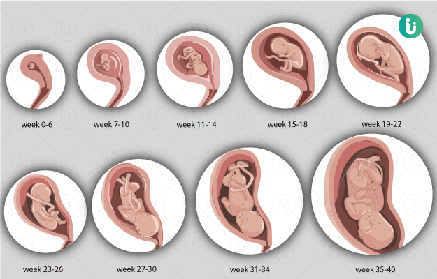 To get a nice bonus from the network of clinics "Health", when registering for this procedure, you just need to name the code word: "A42".
Photo: pixabay.com, pexels.com, Zdorovye clinic network, A42
Source: A42
Weekly development of the child | Regional Perinatal Center
Expectant mothers are always curious about how the fetus develops at a time when it is awaited with such impatience. Let's talk and look at the photos and pictures of how the fetus grows and develops week by week.
What does the puffer do for 9 whole months in mom's tummy? What does he feel, see and hear?
Let's start the story about the development of the fetus by weeks from the very beginning - from the moment of fertilization. A fetus up to 8 weeks old is called embryo , this occurs before the formation of all organ systems.
Embryo development: 1st week
The egg is fertilized and begins to actively split. The ovum travels to the uterus, freeing itself from the membrane along the way.
On the 6th-8th days, implantation of eggs is carried out - implantation into the uterus. The egg settles on the surface of the uterine mucosa and, using the chorionic villi, attaches to the uterine mucosa.
Embryo development: 2-3 weeks
Image of embryo development at 3 weeks.
The embryo is actively developing, starting to separate from the membranes. At this stage, the beginnings of the muscular, skeletal and nervous systems are formed. Therefore, this period of pregnancy is considered important.
Embryo development: 4–7 weeks
Fetal development by week in pictures: week 4
Fetal development by week photo: week 4
Photo of an embryo before the 6th week of pregnancy.
The heart, head, arms, legs and tail are formed in the embryo :) . Gill slit is defined. The length of the embryo at the fifth week reaches 6 mm.
Fetal development by weeks photo: week 5
At the 7th week, the rudiments of the eyes, stomach and chest are determined, and fingers appear on the handles. The baby already has a sense organ - the vestibular apparatus. The length of the embryo is up to 12 mm.
Fetal development: 8th week
Fetal development by weeks photo: weeks 7-8
The face of the fetus can be identified, the mouth, nose, and auricles can be distinguished. The head of the embryo is large and its length corresponds to the length of the body; the fetal body is formed. All significant, but not yet fully formed, elements of the baby's body already exist. The nervous system, muscles, skeleton continue to improve.
Fetal development in the photo already sensitive arms and legs: week 8
The fetus developed skin sensitivity in the mouth area (preparation for the sucking reflex), and later in the face and palms.
At this stage of pregnancy, the genitals are already visible. Gill slits die. The fruit reaches 20 mm in length.
Fetal development: 9–10 weeks
Fetal development by week photo: week 9
Fingers and toes already with nails. The fetus begins to move in the pregnant woman's stomach, but the mother does not feel it yet. With a special stethoscope, you can hear the baby's heartbeat. Muscles continue to develop.
Fetal development by weeks photo: week 10
The entire surface of the fetal body is sensitive and the baby develops tactile sensations with pleasure, touching his own body, the walls of the fetal bladder and the umbilical cord. It is very curious to observe this on ultrasound. By the way, the baby first moves away from the ultrasound sensor (of course, because it is cold and unusual!), And then puts his hands and heels trying to touch the sensor.
It's amazing when a mother puts her hand to her stomach, the baby tries to master the world and tries to touch with his pen "from the back".
Fruit development: 11–14 weeks
The development of the fetus in the photo of the legs: weeks 11
The baby's arms, legs and eyelids are formed, and the genitals become distinguishable (you can find out the gender child). The fetus begins to swallow, and if something is not to its taste, for example, if something bitter got into the amniotic fluid (mother ate something), then the baby will begin to frown and stick out his tongue, making less swallowing movements.
Fruit skin looks translucent.
Development of the fetus: Week 12
Photo of the fetus 12 weeks at 3D ultrasound
The development of a week photo: Week 14
of the kidney is responsible urine. Blood forms inside the bones. And hairs begin to grow on the head. Moves more coordinated.
Fetal development: 15-18 weeks
Fetal development by weeks photo: week 15
The skin turns pink, the ears and other parts of the body, including the face, are already visible. Imagine, a child can already open his mouth and blink, as well as make grasping movements. The fetus begins to actively push in the mother's tummy. The sex of the fetus can be determined by ultrasound.
Fetal development: 19-23 weeks
Fetal development by week photo: week 19
Baby sucks his thumb, becomes more energetic. Pseudo-feces are formed in the intestines of the fetus - meconium , kidneys begin to work. During this period, the brain develops very actively.
Fetal development by weeks photo: week 20
The auditory ossicles become stiff and now they are able to conduct sounds, the baby hears his mother - heartbeat, breathing, voice. The fetus intensively gains weight, fat deposits are formed. The weight of the fetus reaches 650 g, and the length is 300 mm.
The lungs at this stage of fetal development are so developed that the baby can survive in the artificial conditions of the intensive care unit.
Fetal development: 24-27 weeks
Lungs continue to develop. Now the baby is already falling asleep and waking up. Downy hairs appear on the skin, the skin becomes wrinkled and covered with grease. The cartilage of the ears and nose is still soft.
Fetal development by week photo: week 27
Lips and mouth become more sensitive. The eyes develop, open slightly and can perceive light and squint from direct sunlight. In girls, the labia majora do not yet cover the small ones, and in boys, the testicles have not yet descended into the scrotum. Fetal weight reaches 900–1200 g, and the length is 350 mm.
9 out of 10 children born at this term survive.
Fetal development: 28-32 weeks
The lungs are now adapted to breathe normal air. Breathing is rhythmic and body temperature is controlled by the CNS. The baby can cry and responds to external sounds.
Child opens eyes when awake and closes during sleep.
The skin becomes thicker, smoother and pinkish. Starting from this period, the fetus will actively gain weight and grow rapidly. Almost all babies born prematurely at this time are viable. The weight of the fetus reaches 2500 g, and the length is 450 mm.
Fetal development: 33–37 weeks
Fetal development by week photo: week 36
The fetus reacts to a light source. Muscle tone increases and the baby can turn and raise his head. On which, the hairs become silky. The child develops a grasping reflex. The lungs are fully developed.
Fetal development: 38-42 weeks
The fetus is quite developed, prepared for birth and considered mature. The baby has mastered over 70 different reflex movements. Due to the subcutaneous fatty tissue, the baby's skin is pale pink. The head is covered with hairs up to 3 cm.
Fetal development by weeks photo: week 40
The baby perfectly mastered the movements of his mother , knows when she is calm, excited, upset and reacts to this with her movements. During the intrauterine period, the fetus gets used to moving in space, which is why babies love it so much when they are carried in their arms or rolled in a stroller. For a baby, this is a completely natural state, so he will calm down and fall asleep when he is shaken.
Nails protrude beyond the tips of the fingers, the cartilages of the ears and nose are elastic. In boys, the testicles have descended into the scrotum, and in girls, the large labia cover the small ones. The weight of the fetus reaches 3200-3600 g, and the length is 480-520 mm.
After being born, the baby yearns for touching his body, because at first he cannot feel himself - the arms and legs do not obey the child as confidently as it was in the amniotic fluid. Therefore, so that your baby does not feel lonely, it is advisable to carry him in your arms, press him to you while stroking his body.
And one more thing, the baby remembers the rhythm and sound of your heart very well .