What does an ultrasound at 9 weeks look like
What to Expect at a 9 Week Ultrasound
Feb 14, 2022 | 2 minutes Read
PREGNANCY BY WEEK
-
Week
1st Trimester
-
Week
1
-
Week
2
-
Week
3
-
Week
4
-
Week
5
-
Week
6
-
Week
7
-
Week
8
-
Week
9
-
Week
10
-
Week
11
-
Week
12
-
Week
2nd Trimester
-
Week
13
-
Week
14
-
Week
15
-
Week
16
-
Week
17
-
Week
18
-
Week
19
-
Week
20
-
Week
21
-
Week
22
-
Week
23
-
Week
24
-
Week
25
-
Week
26
-
Week
3rd Trimester
-
Week
27
-
Week
28
-
Week
29
-
Week
30
-
Week
31
-
Week
32
-
Week
33
-
Week
34
-
Week
35
-
Week
36
-
Week
37
-
Week
38
-
Week
39
-
Week
40
-
Week
41
-
Week
42
-
Week
4th Trimester
Every pregnant woman is offered ultrasound scans during pregnancy. However, the number of scans and timeline will be different for each woman. Your ultrasound schedule will depend on a few key factors, including:
- The progression and health of your pregnancy
- Your personal preferences
- Your chosen pregnancy provider
- Your medical history
- Your health insurance policy
If you didn’t have an ultrasound at 8 weeks, your 9 week ultrasound will likely be scheduled to assess the gestational age of your baby. If you're not sure when your last menstrual period (LMP) was, a scan at nine weeks will be able to confirm your approximate date of conception.
Your first ultrasound can be a very emotional experience so it's a good idea to take along your partner or close family member for support.
Depending on your unique pregnancy, your chosen provider may schedule an earlier ultrasound at 9 weeks for a few different reasons.
If this is your first ultrasound, it will give you the opportunity to accurately determine your due date. Especially if you haven’t tracked when your LMP was.
Knowing how far along you are in your pregnancy is important. At some point between 11 and 13 weeks your healthcare professional will suggest conducting a nuchal translucency (NT) scan. This scan tests for Down syndrome and for accurate results, you need to know how far along you are.
If you have miscarried a previous pregnancy or experienced some level of vaginal bleeding over the last 9 weeks, you may also be offered an ultrasound. This scan can confirm whether your pregnancy is progressing healthily.
This ultrasound may be conducted vaginally or externally on your abdomen. Know that if your healthcare provider has officially referred you for an early scan your insurance should cover it.
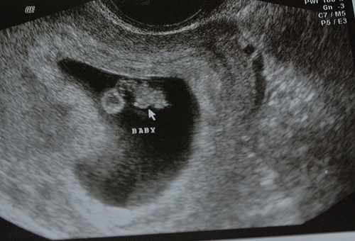
At 9 weeks, you will be able to see your baby's head, body, and limbs. You will also be able to hear your little one's heartbeat for the first time with a Doppler monitor. Bring some tissues with you; this can be a very emotional moment.
It's also important to understand that miscarrying during the first 3 months of pregnancy is quite common.
If your ultrasound shows that your baby is growing slowly or has a lower-than-average heartbeat, your chances of miscarrying are high. If you have been experiencing pain or vaginal bleeding, you might be somewhat prepared for this news.
At nine weeks, your baby will measure approximately 1 inch. The fetus will resemble a green olive and weigh less than 2 grams.
Your little one's eyes will have grown larger and even have some color, but their eyelids will still be fused shut. Your ultrasound may be able to show you the beginnings of what will be your little one's fingers and toes, too.
The information of this article has been reviewed by nursing experts of the Association of Women’s Health, Obstetric, & Neonatal Nurses (AWHONN). The content should not substitute medical advice from your personal healthcare provider. Please consult your healthcare provider for recommendations/diagnosis or treatment. For more advice from AWHONN nurses, visit Healthy Mom&Baby at health5mom.org.
9 Weeks Pregnant Ultrasound: Procedure, Abnormalities and More
Congratulations! You’ve reached the third month of your pregnancy. Two months have passed and now you must be used to the feeling of being pregnant. Well, you can’t feel your baby now, but that tiny one will soon start making small movements (though you may not be able to feel them). No doubt, you must be curious to know everything you can about your baby. In your ninth week of pregnancy, your doctor will recommend an ultrasound to know the size of your growing baby. In the ultrasound scan, you will get to see a tiny blob with a heartbeat in your uterus which will eventually grow to become your baby. Read on to know more about 9-week ultrasound scan.
Why Should You Have an Ultrasound at 9 Weeks?
The first ultrasound scan which will be conducted is called dating and viability scan. This scan is performed for the following purposes.
- To check whether the baby is in the right position inside the uterus or not.
- To rule out ectopic pregnancy and confirm that it is uterine.
- A baby’s heart starts beating around 6 weeks into the pregnancy. And by the 9th week, his heartbeat can be sensed. Therefore, to check the baby’s heartbeat, an ultrasound will be conducted – it also helps a doctor determine whether the pregnancy is viable or not.
- If you experience spotting or bleeding during this time, an ultrasound scan can help determine the cause of the same.
- It can show the number of babies you are carrying. If you are having twins, it can be determined during the 9-week ultrasound scan.
- Couples who have had an IVF treatment need scans to determine successful implantation and viability of the pregnancy.

How to Prepare for Your 9th Week Pregnancy Scan
If you are having a scan around 9 weeks, it will normally be a transvaginal scan (TVS). This is because at this time your baby will be too low in the abdomen or too small for an abdominal scan in early pregnancy. A TVS will allow your doctor to scan the uterus through the vagina. To prepare for the scan, first, you will need to empty your urinary bladder. A full bladder can get in the way of a clear picture during the scan, so a nurse will ask you to use the restroom while she prepares for your scan.
Since the probing is vaginal, you will need to undress from the waist down. It’s suggested that you wear loose clothing such as a long top with slacks or loose pants to avoid being fully undressed.
How Long Does it Take to Perform an Ultrasound Scan?
A typical scan would last for about 20 to 30 minutes. However, if the doctor has a hard time to get clear pictures, it might take a little longer. But, there’s no need to worry about it as women find transvaginal scans more comfortable than abdominal scans.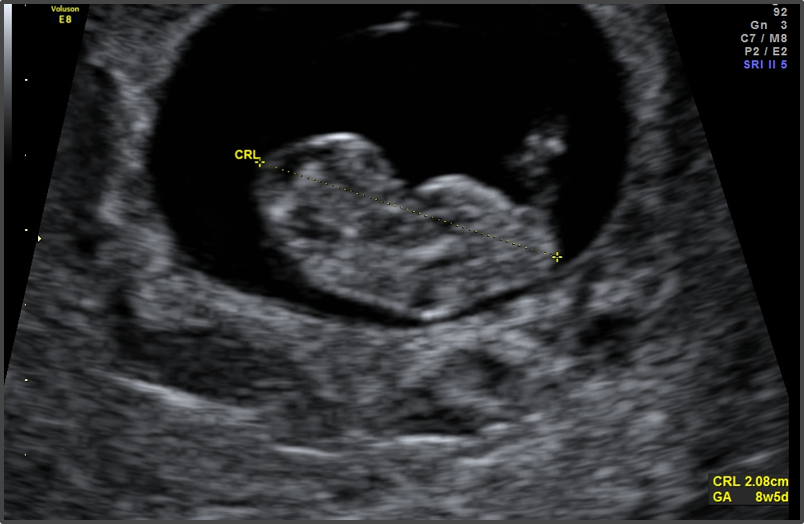
What Happens During an Ultrasound Scan?
During the ultrasound scan, you will be asked to lie down on an examination table or a bed. You will need to raise your knees in a position that the soles of your feet are flat on the bed. Your legs need to be kept apart, so the doctor has space to perform the scan. This is about the same position you’d take during an internal examination.
The probe will be covered with a sterile latex sheath that resembles a condom, and a lubricant gel will be applied on it so that it passes through the vagina with ease and generate clear pictures. The probe will be inserted two to three inches into the vagina to perform the scan. Although the procedure might feel a bit awkward, if you relax, it will be easier for your doctor to insert the probe. If you tense up your muscles, it can become uncomfortable and painful. So remember to take deep breaths and try to relax.
What Will You See on the Scan at the Ninth Week of Your Pregnancy?
Here is what you can expect in your 9-week ultrasound scan:
- Your baby will measure about 2.
 3 cm and weigh close to 2 grams.
 - You will be able to see his head, body, and limbs. By now your baby will no more be an embryo and will be a foetus.
- The heartbeat of a foetus can also be picked up in a 9-week ultrasound scan. It will be usually around 130 to 150 beats per second.
- Your doctor will carefully examine the region around the gestational sac for any bleeding; a condition called subchorionic hematoma.
What Does It Mean to Have an Empty Yolk Sac During the 9th Week of Pregnancy?
The yolk sac envelops the developing foetus and the amniotic fluid. The yolk sac is contained within the gestational sac and is a source of nutrition for the developing foetus in early pregnancy. It is not visible until about 5 to 6 weeks into the gestation; hence it is a marker of the age of the foetus. When no yolk sac is seen around 6 weeks, it may be because there has been an error in remembering the dates of your last period. The doctor will then schedule another scan in a week or two to confirm the presence of a yolk sac. If it is still not seen around 9 weeks of pregnancy, then it is could be a sign of miscarriage. Sometimes there is no need to wait until the follow-up scan, if the 9-week ultrasound pictures show the gestational sac of about 25 mm or more, and there’s no yolk sac or embryo, the doctor will diagnose a miscarriage immediately.
What If Some Abnormalities Are Discovered in the Scan?
Scans can sometimes be inconclusive, hence there are strict guidelines that should be followed. Since doctors adhere to these guidelines, their findings are absolutely sure. Doctors look for minor abnormalities in the scans called “markers” that can indicate a more serious problem or could be just a variation of what is considered normal. So, if they find anything unusual, they may ask you to take tests such as CVS or amniocentesis to check if the chromosomes in the baby are normal.
If the foetus does have a serious problem, you can take time to think through your choices. They may include termination of pregnancy, preparing for a baby who might have special needs, or in extremely rare cases, carrying out a surgery on the foetus.
Ultrasound scans are extremely important to rule out problems and confirm a healthy pregnancy. So do not skip any. Go for ultrasound scans, get everything checked, and have a healthy and safe pregnancy!
Previous Week: 8 Weeks Pregnant Ultrasound
Next Week: 10 Weeks Pregnant Ultrasound
fetal ultrasound at the ninth week
- Fetal development by week
- Our apparatus
- other types...
- Pregnancy management
- Fetometry data at various times
The cost of ultrasound in the first trimester up to 10 weeks is 450 UAH. The price includes biometrics according to protocols, 3D/4D visualization.
The cost of ultrasound in the first trimester for a period of 10 weeks 6 days is 550 hryvnia. The price includes prenatal screening, biometric protocols, 3D/4D visualization. nine0015
The cost of a comprehensive PRISCA prenatal screening (ultrasound + beta hCG + PAPP with the calculation of the individual risk of chromosomal pathologies (for example, Down syndrome or Edwards syndrome) is UAH 1,005.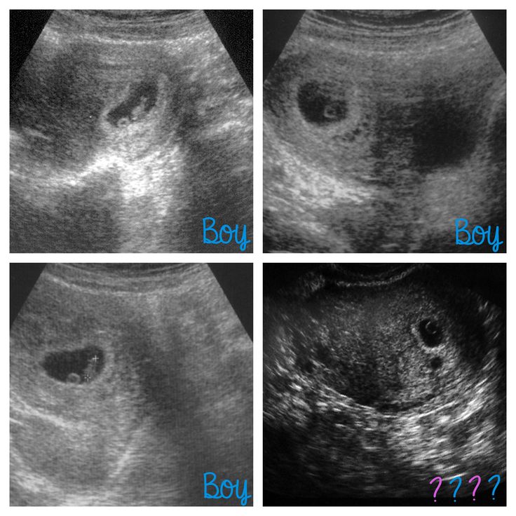
The most important event of the ninth week of pregnancy is the new status of your unborn child, based on
Your unborn child will now officially be called a fetus, not an embryo, until birth.0023 Ultrasound of the fetus at 9 weeks of pregnancy the dimensions correspond to the average olive, it is 14-15 mm, and the fetus already has tiny hands and feet, the fingers on which are still webbed. Bone and tendon formation begins. The umbilical cord and placenta form and grow, on the ultrasound of the fetus at 9 weeks of gestation, the thickness of the chorion (placenta) is measured, the umbilical cord is determined. The fetus is able to respond with movements to touching the anterior abdominal wall in the projection of the uterus, for example, during ultrasound during pregnancy. The fetal heart becomes quite large and practically formed, the heart rate reaches 180 during this period, which can be noted with fetal ultrasound at 9weeks of pregnancy using Doppler. The face of the fetus becomes more formed, the ears more protruding, the liver, spleen and gallbladder continue to improve. Of course, it is still impossible to see all these organs on an ultrasound scan of the fetus at 9 weeks of gestation, this will be possible a little later. The internal organs of the fetus at nine weeks of gestation can still protrude in the form of an umbilical hernia. This can be seen on an ultrasound of the fetus at 9 weeks of gestation and this is normal for this stage of development. The complete return of the internal organs within the anterior abdominal wall of the fetus normally occurs after 10 weeks of pregnancy. With a fetal ultrasound at nine weeks of pregnancy, the ovaries are necessarily evaluated. The corpus luteum with ultrasound of the ovaries begins its reverse development, as a full-fledged placenta is formed, capable of taking on the full hormonal support of a developing pregnancy. The yolk sac is still visualized, it has not yet fulfilled its main hematopoietic function. Sometimes it can even be seen with a fetal ultrasound at 12 weeks of gestation. nine0015
From the ninth week of pregnancy, you begin to put on weight. If you lose weight due to nausea and vomiting after nine weeks of pregnancy, tell your doctor about your pregnancy. Under the influence of progesterone and estrogen, swelling of the mucous membranes occurs. Difficulty breathing, a feeling of congestion in the ears, swelling of the vaginal mucosa may be noted. There may be slight bleeding from the nose. The gums soften, you should brush your teeth regularly to avoid the development of gum disease. You still feel the early symptoms of pregnancy, which may be present all at once or selectively. All pregnancies are different, even for the same woman. nine0015
read more: 10th week of pregnancy
- Hydrotubation (echohydrotubation): study of the patency of the fallopian tubes (ultrasonic hysterosalpingoscopy)
- Transvaginal
- Ovaries
- Uterus
- Mammary glands
Female ultrasound
- Duplex Scan
- Cerebral vessels
- Neck vessels (duplex angioscanning of the main arteries of the head)
- Veins of the lower extremities
Vascular ultrasound
- Transrectal (trusion): prostate
- Scrotum (testicles) nine0003 Vessels of the penis
Male ultrasound
- Appendicitis
- Abdomen
- Gallbladder
- Stomach
- Intestines
- Bladder
- Soft tissue
- Pancreas
- Liver
- Kidney
- Joints
- Thyroid
- Echocardiography (ultrasound of the heart)
Organ ultrasound
- Varicose veins: ultrasound diagnosis of varicose veins
- Hypertension: Ultrasound diagnosis of hypertension
- Thrombosis: ultrasound diagnosis of vein thrombosis
- Ultrasound diagnosis of chronic pancreatitis
- for kidney stones
- for cholecystitis
Ultrasound diagnostics of diseases
- Hip joints in newborns (if hip dysplasia is suspected)
Pediatric ultrasound
9-12 weeks of pregnancy
Ninth week for the baby
During the ninth week, the weight of the fetus changes from 1 gram to 10 grams, the length is 30-45 mm. The back of the fetus straightens and the embryonic tail disappears. The future child becomes completely similar to a small person. The head at this stage is pressed to the chest, the neck is bent, the arms are also brought to the chest.
The development of the brain, which is quite intensive, is one of the main processes of this period. The cerebellum begins to function, the hemispheres acquire a clear outline. Since the cerebellum is responsible for the coordination of movements, in the fetus they cease to be spontaneous and become clear and active, the fetus begins to feel the movement of its own body. nine0015
The heart rate at this stage is 120-150 beats per minute, the heart has two ventricles and two atria. There is a circulatory system of vessels, blood begins to flow through them. While blood circulation in the upper part of the fetus is characterized by greater intensity than in the lower. Therefore, the arms are more developed in comparison with the legs. There is an elongation of the fingers, and the membranes between them slowly disappear. By the end of the ninth week, the formation of the eye ends, they are tightly covered with eyelids. There is a closing of the facial bones, on the head it is already possible to distinguish the nose and nostrils, auricles and lobes, and the upper lip. At this stage, the fetus becomes more and more like a human face. nine0015
Intensive development of internal organs leads to rounding of the tummy of the fetus. The digestive organs and the liver develop, which is important because it is responsible for hematopoiesis (the formation of new blood cells). Thus, "fetal" blood appears.
At this stage in the life of the fetus, such an important event occurs as the beginning of the synthesis of hormones, which include adrenaline. Intensive growth of the adrenal glands provides this process. Also, the beginning of the synthesis of hormones is associated with the complication of the structure of the adrenal glands. All this helps the fetus to comfortably adapt to a variety of changes and extreme conditions.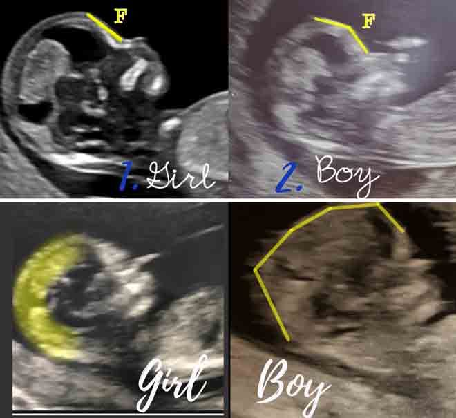 It is the presence of adrenaline in the body that allows it to withstand various stresses. The fetus acquires the ability to endure stress, since adrenaline provides the regulation of a special mode of "survival". nine0015
Ninth week for the expectant mother
At this stage, the woman may still have drowsiness, fatigue, frequent mood swings and dizziness. Manifestations of toxicosis can reach their maximum. It is this period that is optimal in order to visit a gynecologist and register.
The tenth week for the baby
This week is important and significant, it is from it that the fetal stage of development begins, and the unborn child is now officially called a fetus, not an embryo. All the internal organs have already been laid, which in the future will only have to grow and develop. Experts rightly consider the tenth week to be the final one in the first critical period: from that time on, the likelihood of developing defects that may arise due to chemical factors of various nature is no longer so high.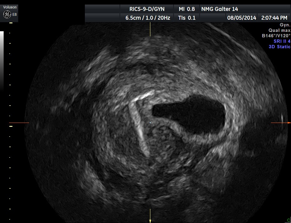 nine0015
The fetus at this time is freely located in the uterine cavity, practically not touching its walls. The future baby has an intensive formation of the nervous system, the transmission of impulses by neuromuscular pathways is being established. This process leads to the emergence of intense movements. Such movements are reflex, they are active and are caused by contact with the walls of the uterus. The fetus already makes fairly clear movements with its legs, head and handles. The woman is not yet able to feel the movements of the fetus, but they are clearly visible during an ultrasound examination. nine0015
At this time, the diaphragm is finally formed - a flat muscle designed to separate the abdominal and chest cavities. There is a further development of internal organs.
The tenth week for the expectant mother
The woman feels increased anxiety, she retains emotional lability. All this is a consequence of ongoing hormonal changes. The only thing to be understood is that the balance will soon be restored, which means the return of a stable good mood. nine0015
The situation changes with the manifestations of toxicosis. Nausea begins to disturb less and less, and vomiting, as a rule, stops altogether. Nausea mostly in the morning. If toxicosis completely disappears, increased appetite may occur. At this time, it is important to monitor the diet and prevent a sharp increase in weight. It is necessary to exclude overeating and the use of high-calorie foods. For a woman in position, this is harmful and can cause shortness of breath, swelling, deterioration of well-being. Excess weight is the reason for increasing the load on all body systems, which already spends a lot of energy on the development of the fetus. nine0015
Women in the tenth week may notice a change in the abdomen. The reason may be overeating, as well as the redistribution of subcutaneous fat and muscle relaxation due to the influence of the pregnancy hormone - progesterone.
The uterus enlarges during this period, but not so much as to influence the shape of the abdomen, it reaches the size of a large apple or grapefruit. Accordingly, for others, pregnancy is still completely invisible.
Eleventh week for baby
The fetus at the eleventh week continues intensive growth. Outwardly, it looks like this: a fairly large head, small legs pressed to the tummy, a small torso and well-developed long arms. Such an uneven development is due to the fact that it was the upper part of the body that received the bulk of the nutrients and the proportion of oxygen throughout the entire previous period, in which such vital organs as the heart and brain are located.
The fetus continues to form joints and bones, muscle growth. Not only large joints develop, but also small ones. In the jaws, the rudiments of teeth are formed, on the fingers - nails. nine0015
The movements of the unborn child become more and more purposeful. Loud noises and sudden movements begin to cause a response in him. Grasping and sucking reflexes develop - this can be seen in the movement of the fingers and lips. The formation of olfactory and taste buds begins.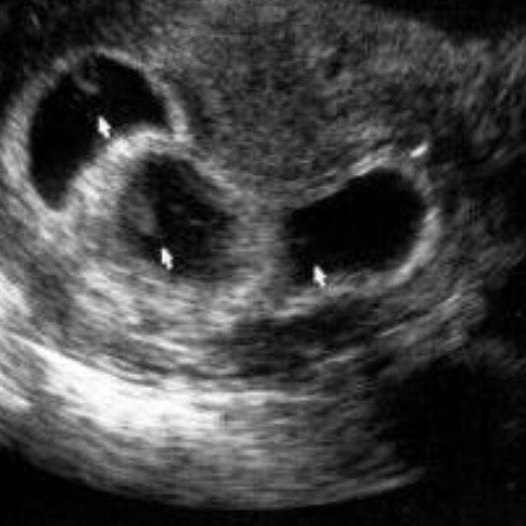 If amniotic fluid enters the nose or mouth, the fetus is able to taste it.
At this stage, the formation of the iris of the eyes also takes place, which, after birth, determines their color. In newborns, in most cases, the eyes are blue or blue, brown are quite rare. The final color of the iris is formed by five months. It depends on the accumulated melanin pigment located in the iris. Genetic inheritance determines the amount of this pigment. nine0015
Eleventh week for the expectant mother
At this time, in most cases, vomiting and nausea, as well as intolerance to certain odors, disappear. Thus, a woman gets the opportunity to form a complete diet and start eating varied, giving preference to various healthy foods. It is advisable to eat freshly prepared food. If you follow a certain diet, you can avoid any problems at this time, the main of which is a problem with digestion. The relaxing hormone progesterone causes the bowel muscles to become lazy, leading to bloating and constipation. If even strict adherence to the diet does not help to cope with problems, you need to contact a specialist who can prescribe safe medications. nine0015
As the fetus grows, blood volume also increases. As a result of such changes, a woman may experience increased sweating. Increased kidney function leads to more frequent urination. If there is no discomfort and pain with frequent urination, there is no reason to worry. Otherwise, you will also need to pay a visit to a specialist. Discomfort and pain can be symptoms of inflammation of the bladder, i.e. cystitis syndromes.
At eleven weeks, the first prenatal screening is performed, which is aimed at identifying malformations. An ultrasound and biochemical study is performed. The first screening is not only aimed at identifying malformations. This examination allows you to find out the state of the chorion, the growth and degree of development of the fetus, the exact gestational age and other details. nine0015
Twelfth week for the baby
Twelfth week ends the first trimester of pregnancy. By the end of this period, the length of the fetus is 90 mm, and its weight is approximately 20 g. At this time, many significant events take place in the life of the fetus.
He has an intensive development of the brain, the formation of connections between the spinal cord and the cerebral hemispheres. If we consider the structure of the brain, it resembles a smaller version of the brain of an adult. During all the first months, only erythrocytes were in the blood of the fetus, but at the twelfth week, leukocytes, which are the body's defenders and belong to the immune system, are added to them. nine0015
The digestive tract also develops. The liver, which at this stage is the most developed organ and occupies most of the abdominal cavity, begins to produce bile, and not only provide hematopoiesis, as it was before. It is from the twelfth week that the intestine actively grows and begins to fit into loops, which can later be seen in an adult. The first peristaltic movements occur, i. e., the contraction of the muscles of the intestine, which should in the future ensure the movement of food through it. The fetus, starting from this week, swallows amniotic fluid, and they pass through the intestines. This happens before birth. Peristaltic waves are a training of the intestinal muscles. nine0015
In addition, rhythmic muscle movements occur in the fetus, which are also training and imitate breathing. The glottis is tightly closed, so amniotic fluid is not able to penetrate to the respiratory organs.
The kidneys begin to function in the fetus, urine in them is collected in small portions and exits through the urethra, entering the amniotic fluid.
At the twelfth week, the formation of the placenta is completed, which becomes able to function independently. The placenta is the most important organ for the fetus; through it, not only is the exchange of nutrients between the woman and the fetus. The placenta is an effective protector against internal and external toxins. nine0015
Twelfth week for the expectant mother
Twelve weeks is rightly considered the best in pregnancy. The woman's well-being returns to normal due to the transfer of control of the process from the corpus luteum, which produced progesterone, which is the culprit of many troubles, to the placenta. All manifestations of toxicosis disappear, women become relaxed and calm.
At this stage, the uterus has already enlarged enough and reached the edge of the pubis, but this is not able to affect the shape of the abdomen. nine0015
As a rule, the first trimester is not accompanied by weight gain. If toxicosis manifested itself to a large extent, even weight loss is possible. If a woman's appetite has not changed as a result of pregnancy, she should gain no more than 10% of her total weight during the entire pregnancy. Such an increase is usually 1-2 kg.
From the twelfth week, if the woman is doing well and there are no contraindications, it is recommended to start playing sports.
