What can an ultrasound detect in pregnancy
What To Expect, Purpose & Results
Overview
What is an ultrasound in pregnancy?
A prenatal ultrasound (or sonogram) is a test during pregnancy that checks on the health and development of your baby. An obstetrician, nurse midwife or ultrasound technician (sonographer) performs ultrasounds during pregnancy for many reasons. Sometimes ultrasounds occur to check on your baby and make sure they’re growing properly. Other times your pregnancy care provider orders an ultrasound after they detect a problem.
During an ultrasound, sound waves are sent through your abdomen or vagina by a device called a transducer. The sound waves bounce off structures inside your body, including your baby and your reproductive organs. Then, the sound waves transform into images that your provider can see on a screen. It doesn’t use radiation, like X-rays, to see your baby.
Even though prenatal ultrasounds are safe, you should only have them when it’s medically necessary. If there’s no reason for an ultrasound (for example, if you just want to see your baby), your insurance company might not pay for it.
Prenatal ultrasounds may be called fetal ultrasounds or pregnancy ultrasounds. Your provider will talk to you about when you can expect ultrasounds during pregnancy based on your health history.
Why is a fetal ultrasound important during pregnancy?
An ultrasound is one of the few ways your pregnancy care provider can see and hear your baby. It can help them determine how far along you are in pregnancy, if your baby is growing properly or if there are any potential problems with the pregnancy. Ultrasounds may occur at any time in pregnancy depending on what your provider is looking for.
What can be detected in a pregnancy ultrasound?
A prenatal ultrasound does two things:
- Evaluates the overall health, growth and development of the fetus.
- Detects certain complications and medical conditions related to pregnancy.
In most pregnancies, ultrasounds are positive experiences and pregnancy care providers don’t find any problems.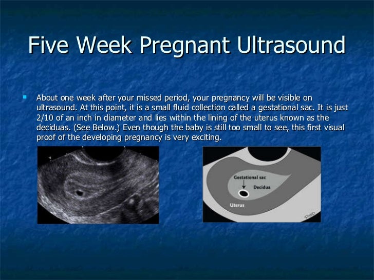 However, there are times this isn’t the case and your provider detects birth disorders or other problems with the pregnancy.
Reasons why your provider performs a prenatal ultrasound are to:
- Confirm you’re pregnant.
- Check for ectopic pregnancy, molar pregnancy, miscarriage or other early pregnancy complications.
- Determine your baby’s gestational age and due date.
- Check your baby’s growth, movement and heart rate.
- Look for multiple babies (twins, triplets or more).
- Examine your pelvic organs like your uterus, ovaries and cervix.
- Examine how much amniotic fluid you have.
- Check the location of the placenta.
- Check your baby’s position in your uterus.
- Detect problems with your baby’s organs, muscles or bones.
Ultrasound is also an important tool to help providers screen for congenital conditions (conditions your baby is born with). A screening is a type of test that determines if your baby is more likely to have a specific health condition. Your provider also uses ultrasound to guide the needle during certain diagnostic procedures in pregnancy like amniocentesis or CVS (chorionic villus sampling).
An ultrasound is also part of a biophysical profile (BPP), a test that combines ultrasound with a nonstress test to evaluate if your baby is getting enough oxygen.
How many ultrasounds do you have during your pregnancy?
Most pregnant people have one or two ultrasounds during pregnancy. However, the number and timing vary depending on your pregnancy care provider and if you have any health conditions. If your pregnancy is high risk or if your provider suspects you or your baby has a health condition, they may suggest more frequent ultrasounds.
When do you have your first prenatal ultrasound?
The timing of your first ultrasound varies depending on your provider. Some people have an early ultrasound (also called a first-trimester ultrasound or dating ultrasound). This can happen as early as seven to eight weeks of pregnancy. Providers do an early ultrasound through your vagina (transvaginal ultrasound). Early ultrasounds do the following:
- Confirm pregnancy (by detecting a heartbeat).
- Check for multiple fetuses.
- Measure the size of the fetus.
- Help confirm gestational age and due date.
Some providers perform your first ultrasound closer to 12 weeks of pregnancy.
20-week ultrasound (anatomy scan)
You can expect an ultrasound around 18 to 20 weeks in pregnancy. This is known as the anatomy ultrasound or 20-week ultrasound. During this ultrasound, your pregnancy care provider can see your baby’s sex (if your baby is in a good position for viewing their genitals), detect birth disorders like cleft palate or find serious conditions related to your baby’s brain, heart, bones or kidneys. If your pregnancy is progressing well and with no complications, your 20-week ultrasound may be your last ultrasound during pregnancy. However, if your provider detects a problem during your 20-week ultrasound, they may order additional ultrasounds.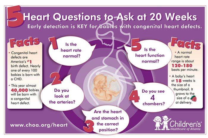
How soon can you see a baby on an ultrasound?
Pregnancy care providers can detect an embryo on an ultrasound as early as six weeks into the pregnancy. An embryo develops into a fetus around the eighth week of pregnancy.
If your last menstrual period isn’t accurate, it’s possible that it may be too early to detect a fetal heart rate.
Which ultrasound is most important during pregnancy?
All ultrasounds during pregnancy are important. Your pregnancy care provider uses ultrasound to tell them important information about your pregnancy.
Test Details
What are the two main types of pregnancy ultrasounds?
The two main types of pregnancy ultrasound are transvaginal ultrasound and abdominal ultrasound. Both use the same technology to produce images of your baby. Your pregnancy care provider performs a transvaginal ultrasound by placing a wand-like device inside your vagina. They perform an abdominal ultrasound by placing a device on the skin of your belly.
Transvaginal ultrasound
During a transvaginal ultrasound, your pregnancy care provider places a device inside your vaginal canal (similar to how you place a tampon). In early pregnancy, this ultrasound helps to detect a fetal heartbeat or determine how far along you are in your pregnancy (gestational age). Images from a transvaginal ultrasound are clearer in early pregnancy as compared to abdominal ultrasound.
Abdominal ultrasound
Your pregnancy care provider performs an abdominal ultrasound by placing a transducer directly on your skin. Then, they move the transducer around your belly (abdomen) to capture images of your baby. Sometimes slight pressure has to be applied to get the best views. Providers use abdominal ultrasounds after about 12 weeks of pregnancy.
Traditional ultrasounds are 2D. More advanced technologies like 3D or 4D ultrasound can create better images. This is helpful when your provider needs to see your baby’s face or organs in greater detail. Not all providers have 3D or 4D ultrasound equipment or specialized training to conduct this type of ultrasound.
Your provider may recommend other types of ultrasounds. Examples of additional ultrasounds are:
- Doppler ultrasound: This type of ultrasound checks how your baby’s blood flows through its blood vessels. Most Doppler ultrasounds occur later in pregnancy.
- Fetal echocardiogram: This type of ultrasound looks at your baby’s heart size, shape, function and structure. Your provider may use it if they suspect your baby has a congenital heart condition, if you had another child that had a heart condition or if you have certain health conditions that warrant taking a closer look at the heart.
How do I prepare for the test?
There’s no special preparation for an ultrasound. Some pregnancy care providers ask that you come with a full bladder and don’t use the restroom before the test. This helps them view your baby better on the ultrasound. You can bring a support person, but bringing children is discouraged as this is an important test that requires complete focus.
You may be asked to change into a hospital gown, but this isn’t usually required for abdominal ultrasounds. If your provider is performing a transvaginal ultrasound in your first trimester, you’ll put on a hospital gown or undress from the waist down.
What should I expect during a prenatal ultrasound?
You’ll lie on a padded examining table during the test. Most ultrasounds occur in a dimly lit room, which helps your ultrasound technician (or sonographer) see the screen. Your sonographer applies a small amount of water-soluble gel to the skin of your belly. The gel doesn’t harm your skin or stain your clothes, but it may feel cold. This gel helps transmit sound waves more efficiently.
Next, the sonographer places a transducer on the skin of your abdomen. The transducer sends sound waves into your body, which reflect off internal structures, including your baby.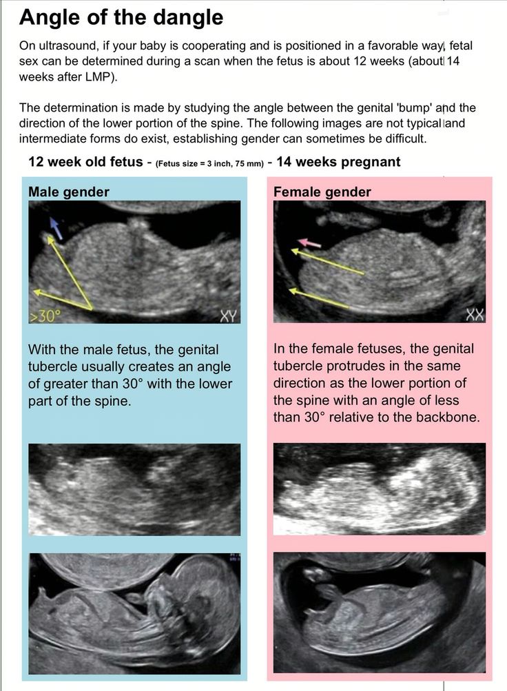 The sound waves that reflect back create pictures on a screen. Your sonographer uses these images to take important measurements such as your baby’s head circumference and length. You may see them making lines on the screen or clicking a button to “freeze” certain angles.
There’s virtually no discomfort during a prenatal ultrasound. You may feel mild discomfort if you have to pee. The ultrasound test takes about 30 minutes to complete.
If you have a transvaginal ultrasound, the process is only different in that the transducer is inside your vagina and not on your belly.
What should I expect after a pregnancy ultrasound?
If you had an abdominal ultrasound, your sonographer wipes the gel off your belly. They may print off some ultrasound pictures for you to take home with you.
In most cases, your sonographer won’t discuss the results of your test with you. If your obstetrician performs your ultrasound, they may discuss what they see as they go along.
If a sonographer performs your ultrasound, an obstetrician will look at the images, then discuss their findings with you at your next appointment. Most practices schedule your appointment right after your ultrasound so you get your results the same day.
What are the risks of prenatal ultrasounds?
Studies have shown ultrasounds are safe during pregnancy. There are no harmful side effects to you or your baby.
Is it safe to do an ultrasound every month during pregnancy?
While ultrasounds are safe for you and your baby, most major medical associations recommend that pregnancy care providers should only do ultrasounds when the tests are medically necessary. If your ultrasounds are normal and your pregnancy is uncomplicated or low risk, repeat ultrasounds aren’t necessary.
Results and Follow-Up
What results do you get on a pregnancy ultrasound?
Your ultrasound results will be normal or abnormal. A normal result means your pregnancy care provider didn’t find any problems and that your baby is growing and developing normally. An abnormal result means your provider noticed something irregular. If they do, your provider will order additional ultrasounds or diagnostic tests to determine if something is wrong.
Occasionally, the ultrasound is incomplete if there’s difficulty seeing all the structures needed for that particular ultrasound. Your baby’s position or movement sometimes makes it difficult to see everything your provider needs to see. If this is the case, you’ll need a repeat ultrasound and they’ll try again.
There are some limitations to ultrasounds, so your provider may not find certain abnormalities until after birth.
What are reasons you need more ultrasounds during pregnancy?
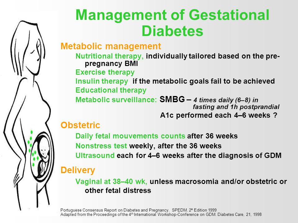
There are several reasons your pregnancy care provider may order additional ultrasounds during your pregnancy. Some of these reasons include:
- Problems with your ovaries, uterus, cervix or other pelvic organs.
- Your baby is measuring small for their gestational age or your provider suspects IUGR (intrauterine growth restriction).
- Problems with the placenta like placenta previa or placental abruption.
- You’re pregnant with twins, triplets or more.
- Your baby is breech.
- You have too much amniotic fluid (polyhydramnios).
- You have too little amniotic fluid (oligohydramnios).
- You have a condition like gestational diabetes or preeclampsia.
- Your baby has a congenital disorder.
Normal results on pregnancy ultrasounds can vary. Generally, a normal result means your baby appears healthy and your provider didn’t find any issues.
Why do some pregnancy providers schedule ultrasounds differently?
The number of ultrasounds you’ll have and when you have them can vary between providers. Every practice operates differently and some providers do things differently based on your health history or symptoms.
When does a pregnancy ultrasound determine sex?
Your baby’s sex isn’t visible on an ultrasound until about 18 to 20 weeks. Be sure to tell your pregnancy care provider whether or not you want to know the sex of your baby before your ultrasound.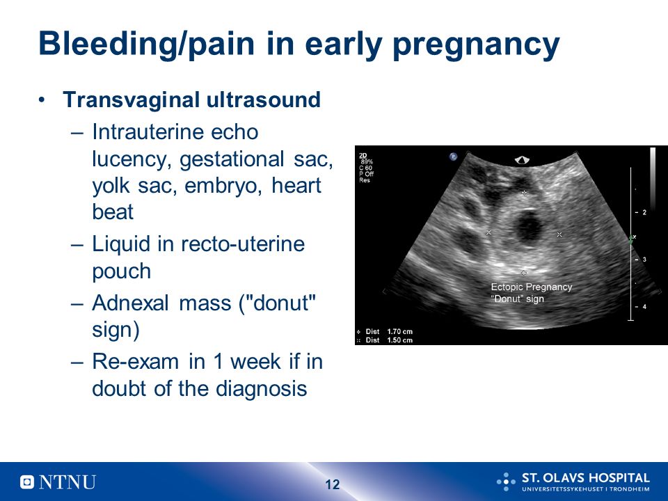
A note from Cleveland Clinic
An ultrasound during pregnancy can be both exciting and terrifying. Your pregnancy care provider uses ultrasound to get a better idea of how your baby is growing and developing. There are different types of ultrasounds, and the exact timing may vary depending on your provider. Most pregnant people have two ultrasounds — one in the first trimester and one in the second trimester. However, if there’s a potential complication or medical reason for more ultrasounds, your provider will order more as a precaution. Talk to your provider about the ultrasound schedule during pregnancy and what you can expect.
Ultrasound during pregnancy | March of Dimes
A prenatal ultrasound uses sound waves and a computer screen to show a picture of your baby inside the womb.
Ultrasounds can help your health care provider see how your baby is growing and developing.
Your provider also may use ultrasounds to see if other tests need to be done to check on your baby’s health.

There are several types of ultrasounds and they are safe for you and your baby when done by a trained health care provider.
What is an ultrasound?
Ultrasound (also called sonogram) is a prenatal test offered to most pregnant women. It uses sound waves to show a picture of your baby in the uterus (womb). Ultrasound helps your health care provider check on your baby’s health and development.
Ultrasound can be a special part of pregnancy—it’s the first time you get to “see” your baby! Depending on when it’s done and your baby’s position, you may be able to see his hands, legs and other body parts. You may be able to tell if your baby’s a boy or a girl, so be sure to tell your provider if you don’t want to know!
Most women get an ultrasound in their second trimester at 18 to 20 weeks of pregnancy. Some also get a first-trimester ultrasound (also called an early ultrasound) before 14 weeks of pregnancy. The number of ultrasounds and timing may be different for women with certain health conditions like as asthma and obesity.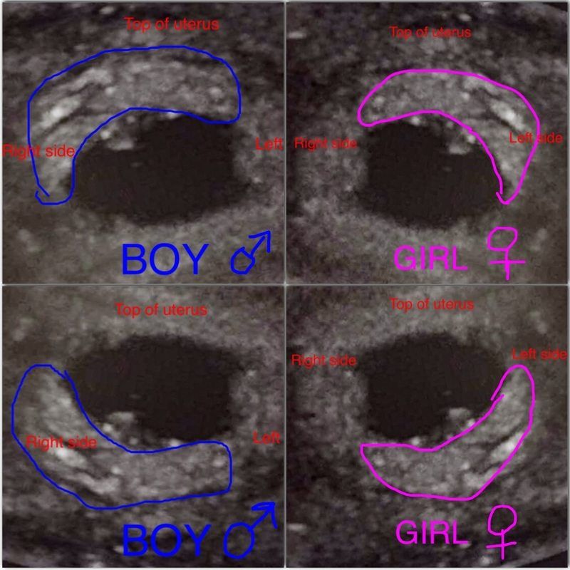
Talk to your provider about when an ultrasound is right for you.
What are some reasons for having an ultrasound?
Your provider uses ultrasound to do several things, including:
- To confirm (make sure) you’re pregnant
- To check your baby’s age and growth. This helps your provider figure out your due date.
- To check your baby’s heartbeat, muscle tone, movement and overall development
- To check to see if you’re pregnant with twins, triplets or more (also called multiples)
- To check if your baby is in the heads-first position before birth
- To examine your ovaries and uterus (womb). Ovaries are where eggs are stored in your body.
Your provider also uses ultrasound for screening and other testing. Screening means seeing if your baby is more likely than others to have a health condition; it doesn’t mean finding out for sure if your baby has the condition. Your provider may use ultrasound:
- To screen for birth defects, like spina bifida or heart defects.
 After an ultrasound, your provider may want to do more tests, called diagnostic tests, to see for sure if your baby has a birth defect. Birth defects are health conditions that a baby has at birth. Birth defects change the shape or function of one or more parts of the body. They can cause problems in overall health, in how the body develops, or in how the body works.
 - To help with other prenatal tests, like chorionic villus sampling (also called CVS) or amniocentesis (also called amnio). CVS is when cells from the placenta are taken for testing. The placenta is tissue that provides nutrients for your baby. Amnio is a test where amniotic fluid and cells are taken from the sac around your baby.
- To check for pregnancy complications, including ectopic pregnancy, molar pregnancy and miscarriage.
Are there different kinds of ultrasound?
Yes. The kind you get depends on what your provider is checking for and how far along you are in pregnancy. All ultrasounds use a tool called a transducer that uses sound waves to create pictures of your baby on a computer. The most common kinds of ultrasound are:
- Transabdominal ultrasound. When you hear about ultrasound during pregnancy, it’s most likely this kind. You lay on your back on an exam table, and your provider covers your belly with a thin layer of gel. The gel helps the sound waves move more easily so you get a better picture. Then he moves the transducer across your belly. You may need to drink several glasses of water about 2 hours before the exam to have a full bladder during the test. A full bladder helps sound waves move more easily to get a better picture. Ultrasound is painless, but having a full bladder may be uncomfortable. The ultrasound takes about 20 minutes.
- Transvaginal ultrasound. This kind of ultrasound is done through the vagina (birth canal). You lay on your back on an exam table with your feet in stirrups.
 Your provider moves a thin transducer shaped like a wand into your vagina. You may feel some pressure from the transducer, but it shouldn’t cause pain. Your bladder needs to be empty or just partly full. This kind of ultrasound also takes about 20 minutes.
In special cases, your provider may use these kinds of ultrasound to get more information about your baby:
- Doppler ultrasound. This kind of ultrasound is used to check your baby’s blood flow if he’s not growing normally. Your provider uses a transducer to listen to your baby’s heartbeat and to measure the blood flow in the umbilical cord and in some of your baby’s blood vessels. You also may get a Doppler ultrasound if you have Rh disease. This is a blood condition that can cause serious problems for your baby if it’s not treated. Doppler ultrasound usually is used in the last trimester, but it may be done earlier.
- 3-D ultrasound. A 3-D ultrasound takes thousands of pictures at once.
 It makes a 3-D image that’s almost as clear as a photograph. Some providers use this kind of ultrasound to make sure your baby’s organs are growing and developing normally. It can also check for abnormalities in a baby’s face. You also may get a 3-D ultrasound to check for problems in the uterus.
 - 4-D ultrasound. This is like a 3-D ultrasound, but it also shows your baby’s movements in a video.
Does ultrasound have any risks?
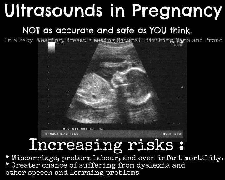
Ultrasound is safe for you and your baby when done by your health care provider. Because ultrasound uses sound waves instead of radiation, it’s safer than X-rays. Providers have used ultrasound for more than 30 years, and they have not found any dangerous risks.
If your pregnancy is healthy, ultrasound is good at ruling out problems, but it can’t find every problem. It may miss some birth defects. Sometimes, a routine ultrasound may suggest that there is a birth defect when there really isn’t one.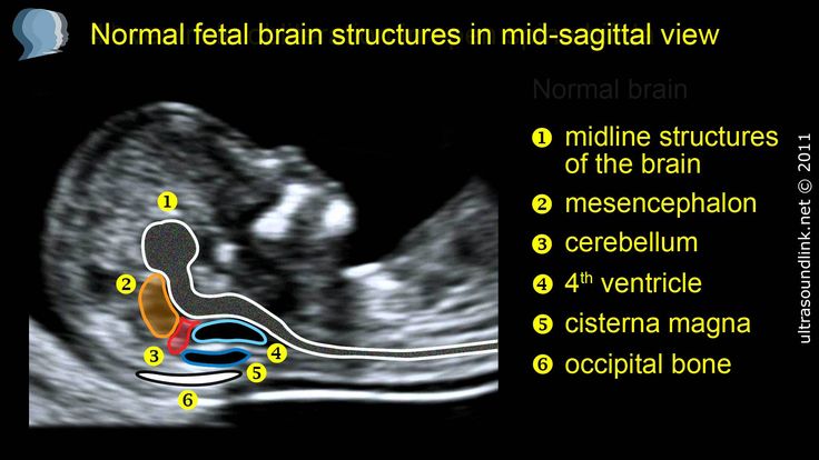 While follow-up tests often show that the baby is healthy, false alarms can cause worry for parents.
You may know of some places, like stores in a mall, that aren’t run by doctors or other medical professionals that offer “keepsake” 3-D or 4-D ultrasound pictures or videos for parents. The American College of Obstetricians and Gynecologists (ACOG), the Food and Drug Administration (FDA) and the American Institute of Ultrasound in Medicine (AIUM) do not recommend these non-medical ultrasounds. The people doing them may not have medical training and may give you wrong or even harmful information.
What happens after an ultrasound?
For most women, ultrasound shows that the baby is growing normally. If your ultrasound is normal, just be sure to keep going to your prenatal checkups.
Sometimes, ultrasound may show that you and your baby need special care. For example, if the ultrasound shows your baby has spina bifida, he may be treated in the womb before birth. If the ultrasound shows that your baby is breech (feet-down instead of head-down), your provider may try to flip your baby’s position to head-down, or you may need to have a cesarean section (also called c-section). A c-section is surgery in which your baby is born through a cut that your doctor makes in your belly and uterus.
No matter what an ultrasound shows, talk to your provider about the best care for you and your baby.
Last reviewed: October, 2019
Ultrasound in the first month of pregnancy at the Medical Center "Medicenter"
Ultrasound examination (ultrasound) is a modern examination method that allows you to "see" the internal organs of the human body. The basis of this method is the different frequency of ultrasonic waves that pass through tissues with different densities. The receiving device of the ultrasound machine converts the waves into an image on the screen. This study is completely safe, so it can be used during pregnancy at the earliest possible time. nine0005
You can confirm the fact of pregnancy by different methods, but not all of them give a 100% result. The first suspicions a woman usually begins to experience when menstruation does not start on time. However, delay is not always an accurate symptom of pregnancy.
The next step is usually methods based on determining the level of hCG (human chorionic hormone) in urine or blood. The level of this hormone begins to rise when the embryo implants in the uterine wall. Household tests that determine the level of hCG in the urine are very popular: they are affordable, easy to use and have sufficient accuracy. However, sometimes they give false negative results. A blood test for hCG is even more accurate. However, pregnancy can be accurately confirmed only with the help of ultrasound - in some diseases, the level of hCG increases in non-pregnant women, and even in men. nine0005
Ultrasound in the first week of pregnancy
The first obstetric week of pregnancy is counted from the date of the start of the last menstruation. In fact, there is no pregnancy at this moment, and even ovulation has not yet occurred. It would seem, why is this the first week? It turns out that gynecologists use this method of calculation, because it is difficult to determine the exact date of conception. Due to the fact that in the first week there is not even a fertilized egg, ultrasound will not show pregnancy.
Ultrasound in the second week of pregnancy
At the end of the second obstetric week of pregnancy (or at the beginning of the third) of the standard cycle, ovulation usually occurs and fertilization can occur. However, this is too early for ultrasound - no visible changes in the reproductive organs have yet been observed.
Ultrasound in the third week of pregnancy
In the third obstetric week of pregnancy, during the standard cycle, the embryo usually implants, that is, it attaches to the wall of the uterus. After about a week, it will be possible to see the fetal egg and thickening of the mucosa at the site of its implantation.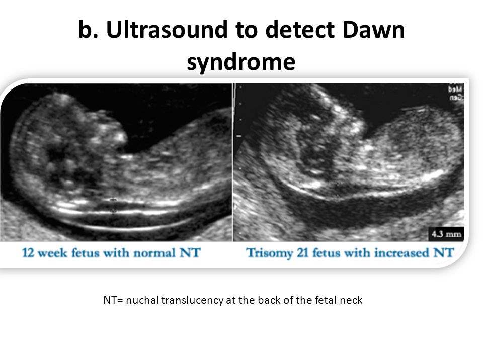 In the third week, ultrasound is uninformative. nine0005
Ultrasound at the fourth week of pregnancy
To confirm pregnancy, ultrasound can be done at the end of the first month - at 4 weeks - usually this period corresponds to a slight delay in menstruation. At this time, the doctor can usually see a small fetal egg attached to the wall of the uterus, and determine the gestational age. The embryo is usually not yet visible.
In addition to confirming the fact of pregnancy, ultrasound examination allows early detection of pathologies and other features. So, with the help of ultrasound in the first month, it is possible to exclude ectopic pregnancy, cystic drift, the threat of miscarriage. nine0005
Early ultrasound can also show multiple pregnancies. Carrying twins or even triplets is a special challenge for both the doctor and the expectant mother.
How is ultrasound done in the first month of pregnancy?
Ultrasounds for pregnant women are done in two ways: transvaginally (using a vaginal probe) and transabdominally (through the abdominal wall). The first method is more accurate and allows you to determine earlier if a woman is pregnant. Therefore, with ultrasound in the first month of pregnancy, it is used. nine0005
For early ultrasound examinations, a special probe is used: it is a plastic rod 3 cm in diameter and about 12 cm long. A research condom is put on the probe, and gel is applied to its surface. The transducer is then inserted into the vagina a few centimeters. This procedure does not cause discomfort, is carried out quickly and accurately. Within a few minutes, the doctor will record all the necessary data on the screen, and the study will end. nine0005
Special preparation for transvaginal ultrasound in early pregnancy is not necessary, only in case of increased gas production, the doctor may prescribe drugs for flatulence.
Thus, an ultrasound scan in the first month will help determine whether you are expecting a baby (or maybe more than one), accurately determine the period, and also exclude ectopic pregnancy and other pathological conditions.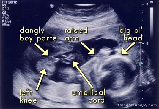 This safe and modern research method, which is highly informative, can be used starting from 4-5 weeks (or a few days after a missed period). nine0005
Our clinics in St. Petersburg
You can get detailed information and make an appointment by calling +7 (812) 640-55-25
Make an appointment
Early pregnancy ultrasound
Two stripes on the test is always a complete surprise for a woman, even if she already knows about her position in her soul. After the first confirmation, the girl most often wants to see the baby with her own eyes and make sure that he is developing normally. This can be done using an instrumental method such as ultrasound. Usually, expectant mothers, especially expecting their first child, have a lot of questions. nine0005
How to avoid missing ectopic and non-developing pregnancies
Often, pregnant women at the very beginning are afraid of two conditions - an ectopic or missed pregnancy. Especially experienced by those who have already experienced it once.
In an ectopic pregnancy, the fertilized egg does not reach the uterus and is implanted in other places - most often these are the fallopian tubes. If nothing is done, the embryo will grow and burst the tube. In this case, the woman can not be saved. Therefore, an ectopic pregnancy should be terminated without hesitation. Since the embryo is not removed separately, the entire tube is cut out. This can be done twice in a lifetime, after which pregnancy occurs only after artificial replanting using IVF. An ectopic pregnancy can be suspected indirectly by the level of β-hCG, but only the presence of a fetal egg in the uterus on ultrasound clearly refutes it. nine0005
But the experiences of the expectant mother do not end there. Frozen pregnancy is impossible to predict. In some cases, doctors shrug their shoulders, and why this happened remains unknown. It is dangerous to wait for a non-developing egg to come out by itself for a long time, since it can begin to decompose, and the uterus can become inflamed. Up to 6 weeks, the gynecologist can offer a less traumatic medical interruption, and after - classic abortive methods. It is possible to unambiguously identify a frozen one only by ultrasound - it shows that the fetal egg is smaller than it should be for its term or a heartbeat is not heard. At random, such a diagnosis is never made, because the life of the unborn child depends on it, and most often an ultrasound is prescribed again after a few days. If it turns out that the dimensions have not changed, and there is still no heartbeat, then - alas! nine0005
Why early ultrasound is needed
Ultrasound diagnostics is a safe method that has no contraindications. But it is pointless to do it before 4-5 weeks, the fetal egg cannot be seen so early. In this case, a transvaginal probe is used for ultrasound. In public clinics, the first ultrasound diagnosis is carried out as part of the first screening at 12 weeks, and earlier it is prescribed only if the woman herself or her doctor is worried about something:
- a woman complains of pain in the lower part of the abdominal region and bloody “daub”;
- expectant mother has serious chronic diseases;
- pregnancy occurred, although the couple was protected by a spiral;
- the patient took dangerous and heavy drugs, had an infection, was irradiated.


