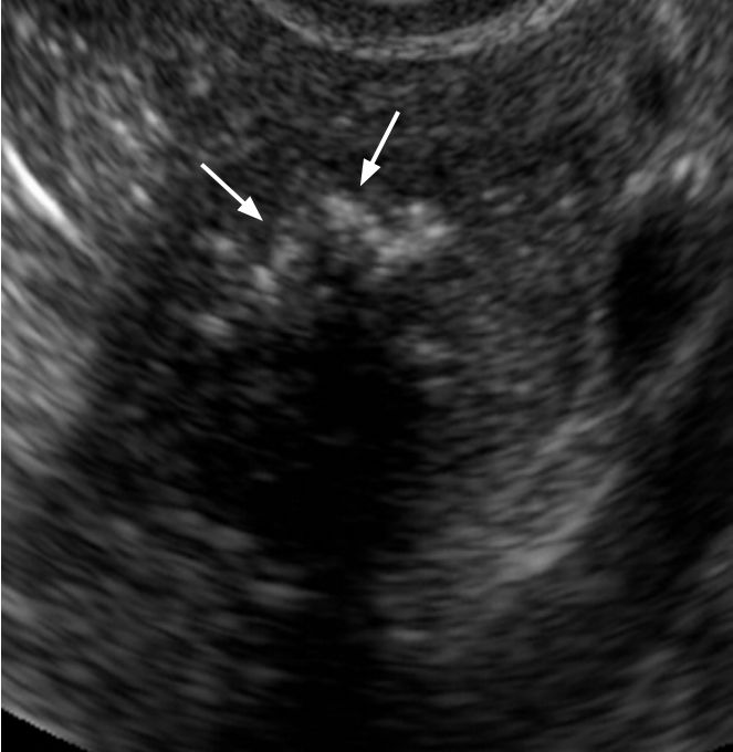
Vaginal ultrasound results
Transvaginal Ultrasound: Purpose, Procedure, and Results
What is a transvaginal ultrasound?
An ultrasound test uses high-frequency sound waves to create images of your internal organs. Imaging tests can identify abnormalities and help doctors diagnose conditions.
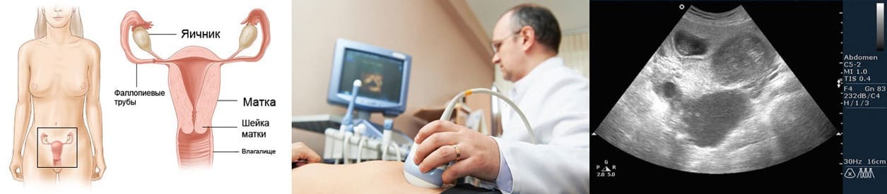
A transvaginal ultrasound, also called an endovaginal ultrasound, is a type of pelvic ultrasound used by doctors to examine female reproductive organs. This includes the uterus, fallopian tubes, ovaries, cervix, and vagina.
“Transvaginal” means “through the vagina.” This is an internal examination.
Unlike a regular abdominal or pelvic ultrasound, where the ultrasound wand (transducer) rests on the outside of the pelvis, this procedure involves your doctor or a technician inserting an ultrasound probe about 2 or 3 inches into your vaginal canal.
There are many reasons why a transvaginal ultrasound might be necessary, including:
- an abnormal pelvic or abdominal exam
- unexplained vaginal bleeding
- pelvic pain
- an ectopic pregnancy (which occurs when the fetus implants outside of the uterus, usually in the fallopian tubes)
- infertility
- a check for cysts or uterine fibroids
- verification that an IUD is placed properly
Your doctor might also recommend a transvaginal ultrasound during pregnancy to:
- monitor the heartbeat of the fetus
- look at the cervix for any changes that could lead to complications such as miscarriage or premature delivery
- examine the placenta for abnormalities
- identify the source of any abnormal bleeding
- diagnose a possible miscarriage
- confirm an early pregnancy
In most cases, a transvaginal ultrasound requires little preparation on your part.
Once you’ve arrived at your doctor’s office or the hospital and you’re in the examination room, you have to remove your clothes from the waist down and put on a gown.
Depending on your doctor’s instructions and the reasons for the ultrasound, your bladder might need to be empty or partially full. A full bladder helps lift the intestines and allows for a clearer picture of your pelvic organs.
If your bladder needs to be full, you have to drink about 32 ounces of water or any other liquid about one hour before the procedure begins.
If you’re on your menstrual cycle or if you’re spotting, you have to remove any tampon you’re using before the ultrasound.
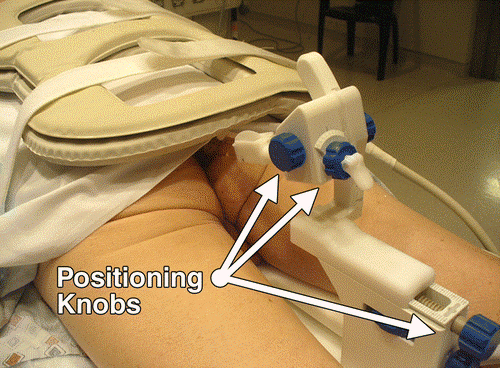
When it’s time to begin the procedure, you lie down on your back on the examination table and bend your knees. There may or may not be stirrups.
Your doctor covers the ultrasound wand with a condom and lubricating gel, and then inserts it into your vagina. Make sure your provider is aware of any latex allergies you have so that a latex-free probe cover is used if necessary.
You might feel some pressure as your doctor inserts the transducer. This feeling is similar to the pressure felt during a Pap smear when your doctor inserts a speculum into your vagina.
Once the transducer is inside of you, sound waves bounce off your internal organs and transmit pictures of the inside of your pelvis onto a monitor.
The technician or doctor then slowly turns the transducer while it’s still inside of your body. This provides a comprehensive picture of your organs.
Your doctor may order a saline infusion sonography (SIS). This is a special kind of transvaginal ultrasound that involves inserting sterile salt water into the uterus before the ultrasound to help identify any possible abnormalities inside the uterus.
The saline solution stretches the uterus slightly, providing a more detailed picture of the inside of the uterus than a conventional ultrasound.
Although a transvaginal ultrasound can be done on a pregnant woman or a woman with an infection, SIS cannot.
There are no known risk factors associated with transvaginal ultrasound.
Performing transvaginal ultrasounds on pregnant women is also safe, for both mother and fetus. This is because no radiation is used in this imaging technique.
When the transducer is inserted into your vagina, you’ll feel pressure and in some cases discomfort. The discomfort should be minimal and should go away once the procedure is complete.
If something is extremely uncomfortable during the exam, be sure to let the doctor or technician know.
You might get your results immediately if your doctor performs the ultrasound. If a technician performs the procedure, the images are saved and then analyzed by a radiologist. The radiologist will send the results to your doctor.
A transvaginal ultrasound helps diagnose multiple conditions, including:
- cancer of the reproductive organs
- routine pregnancy
- cysts
- fibroids
- pelvic infection
- ectopic pregnancy
- miscarriage
- placenta previa (a low-lying placenta during pregnancy that may warrant medical intervention)
Talk with your doctor about your results and what type of treatment, if any, is necessary.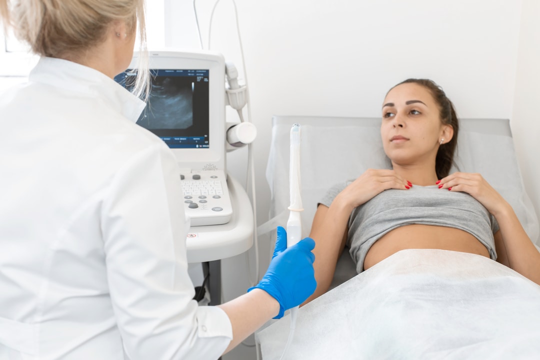
There are virtually no risks associated with a transvaginal ultrasound, although you might experience some discomfort. The entire test takes about 30 to 60 minutes, and the results are typically ready in about 24 hours.
If your doctor is unable to get a clear picture, you might be called back to repeat the test. A pelvic or abdominal ultrasound is sometimes done before a transvaginal ultrasound depending on your symptoms.
If you experience too much discomfort from a transvaginal ultrasound and can’t tolerate the procedure, your doctor may perform a transabdominal ultrasound. This involves your doctor applying gel to your stomach and then using a handheld device to view your pelvic organs.
This approach is also an option for children when pelvic images are needed.
Transvaginal Ultrasound: Purpose, Procedure, and Results
What is a transvaginal ultrasound?
An ultrasound test uses high-frequency sound waves to create images of your internal organs.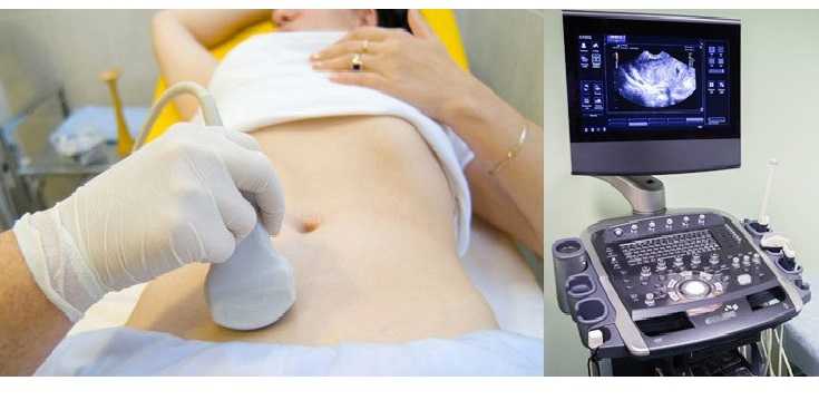 Imaging tests can identify abnormalities and help doctors diagnose conditions.
A transvaginal ultrasound, also called an endovaginal ultrasound, is a type of pelvic ultrasound used by doctors to examine female reproductive organs. This includes the uterus, fallopian tubes, ovaries, cervix, and vagina.
“Transvaginal” means “through the vagina.” This is an internal examination.
Unlike a regular abdominal or pelvic ultrasound, where the ultrasound wand (transducer) rests on the outside of the pelvis, this procedure involves your doctor or a technician inserting an ultrasound probe about 2 or 3 inches into your vaginal canal.
There are many reasons why a transvaginal ultrasound might be necessary, including:
- an abnormal pelvic or abdominal exam
- unexplained vaginal bleeding
- pelvic pain
- an ectopic pregnancy (which occurs when the fetus implants outside of the uterus, usually in the fallopian tubes)
- infertility
- a check for cysts or uterine fibroids
- verification that an IUD is placed properly
Your doctor might also recommend a transvaginal ultrasound during pregnancy to:
- monitor the heartbeat of the fetus
- look at the cervix for any changes that could lead to complications such as miscarriage or premature delivery
- examine the placenta for abnormalities
- identify the source of any abnormal bleeding
- diagnose a possible miscarriage
- confirm an early pregnancy
In most cases, a transvaginal ultrasound requires little preparation on your part.
Once you’ve arrived at your doctor’s office or the hospital and you’re in the examination room, you have to remove your clothes from the waist down and put on a gown.
Depending on your doctor’s instructions and the reasons for the ultrasound, your bladder might need to be empty or partially full. A full bladder helps lift the intestines and allows for a clearer picture of your pelvic organs.
If your bladder needs to be full, you have to drink about 32 ounces of water or any other liquid about one hour before the procedure begins.
If you’re on your menstrual cycle or if you’re spotting, you have to remove any tampon you’re using before the ultrasound.
When it’s time to begin the procedure, you lie down on your back on the examination table and bend your knees. There may or may not be stirrups.
Your doctor covers the ultrasound wand with a condom and lubricating gel, and then inserts it into your vagina. Make sure your provider is aware of any latex allergies you have so that a latex-free probe cover is used if necessary.
You might feel some pressure as your doctor inserts the transducer. This feeling is similar to the pressure felt during a Pap smear when your doctor inserts a speculum into your vagina.
Once the transducer is inside of you, sound waves bounce off your internal organs and transmit pictures of the inside of your pelvis onto a monitor.
The technician or doctor then slowly turns the transducer while it’s still inside of your body. This provides a comprehensive picture of your organs.
Your doctor may order a saline infusion sonography (SIS). This is a special kind of transvaginal ultrasound that involves inserting sterile salt water into the uterus before the ultrasound to help identify any possible abnormalities inside the uterus.
The saline solution stretches the uterus slightly, providing a more detailed picture of the inside of the uterus than a conventional ultrasound.
Although a transvaginal ultrasound can be done on a pregnant woman or a woman with an infection, SIS cannot.
There are no known risk factors associated with transvaginal ultrasound.
Performing transvaginal ultrasounds on pregnant women is also safe, for both mother and fetus. This is because no radiation is used in this imaging technique.
When the transducer is inserted into your vagina, you’ll feel pressure and in some cases discomfort. The discomfort should be minimal and should go away once the procedure is complete.
If something is extremely uncomfortable during the exam, be sure to let the doctor or technician know.
You might get your results immediately if your doctor performs the ultrasound. If a technician performs the procedure, the images are saved and then analyzed by a radiologist. The radiologist will send the results to your doctor.
A transvaginal ultrasound helps diagnose multiple conditions, including:
- cancer of the reproductive organs
- routine pregnancy
- cysts
- fibroids
- pelvic infection
- ectopic pregnancy
- miscarriage
- placenta previa (a low-lying placenta during pregnancy that may warrant medical intervention)
Talk with your doctor about your results and what type of treatment, if any, is necessary.
There are virtually no risks associated with a transvaginal ultrasound, although you might experience some discomfort. The entire test takes about 30 to 60 minutes, and the results are typically ready in about 24 hours.
If your doctor is unable to get a clear picture, you might be called back to repeat the test. A pelvic or abdominal ultrasound is sometimes done before a transvaginal ultrasound depending on your symptoms.
If you experience too much discomfort from a transvaginal ultrasound and can’t tolerate the procedure, your doctor may perform a transabdominal ultrasound. This involves your doctor applying gel to your stomach and then using a handheld device to view your pelvic organs.
This approach is also an option for children when pelvic images are needed.
Ultrasound of the small pelvis, interpretation of results, preparation for ultrasound examination
Preparation for pelvic ultrasound, interpretation of the results of ultrasound.
Ultrasound is an informative and painless way to study the organs located in the small pelvis. Using this method, various disorders in the work of the genital organs of men and women are revealed. This study is suitable for almost all people.
When is pelvic ultrasound indicated
During the ultrasound in women, you can see various pathologies in the reproductive organs. This study may be prescribed for:
- pain in the groin and lumbar region;
- pain when urinating;
- mucus and blood in urine;
- changes in the length of the menstrual cycle;
- inflammation of the organs of the reproductive system;
- inability to conceive;
- to confirm pregnancy;
- after difficult delivery or termination of pregnancy; nine0012
- installation of the Navy;
- possible ectopic pregnancy.
If pregnancy is confirmed, then ultrasound should be done 3 times - 1 time in each trimester. If necessary, the doctor will prescribe additional ultrasound to monitor the identified pathologies.
Ultrasound of the pelvic organs is prescribed for men with:
- inability to conceive a child;
- pain during urination;
- violation in the work of the prostate and bladder.
 nine0012
Adolescents may be prescribed an ultrasound if the genital organs are not properly developed, too early or, on the contrary, late puberty.
Ultrasound can be done by almost everyone, however, the vaginal method cannot be performed with bleeding, as well as girls who have not had sexual intercourse.
Rectal ultrasound can not be performed in cases where there is available:
- anal fissures;
- hemorrhoids;
- after surgery on the rectum. nine0012
Do not perform an ultrasound after an X-ray. This may skew the results. It is better to postpone the study for a few days.
How to prepare for an ultrasound
Preparation depends on which method the ultrasound is performed - transvaginally (performed through the vagina), transabdominally (through the abdominal wall) or transrectal (through the rectum). The uzist should tell you in advance how the procedure will be carried out, because only the vaginal method does not require preparation.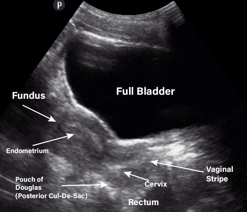 nine0003
Preparation for transabdominal ultrasound:
- do not eat gas-producing foods It is better to eat cereals, steamed vegetables, low-fat fish and meat;
- If the diet did not bring results, then to get rid of gases, you can take activated charcoal a couple of days before the ultrasound;
- In the morning, on the day of the examination, breakfast is prohibited. It is allowed to have dinner the night before and also do an enema;
- an hour before the start of the ultrasound, the patient drinks 1.5-2 liters of water so that the bladder is full during the study. nine0012
Preparation for transrectal examination
Before a rectal examination (approximately 2-3 hours before) you need to do an enema. If a study of the work of the prostate is carried out, the search for the causes of erectile dysfunction and infertility of the bladder should be complete. To do this, one hour before the start of the ultrasound, you should drink 800-1000 ml of water.
Transvaginal examination method
With the vaginal method, the bladder does not need to be filled. The study can be performed on any day of the cycle (with the exception of the days of menstruation), but it will be more effective immediately after the end of the discharge. To assess the correct functioning of the ovaries, as well as to make sure that the follicles are maturing, ultrasound can be scheduled on different days of the cycle. nine0003
Features of different ultrasound methods
Each research method has its own characteristics:
- The transvaginal method is performed using a special transducer. The patient takes off his clothes below the waist, lies on his back, spreading his bent legs. The doctor must put a condom on the sensor, then lubricate it with a gel, thanks to which the organ under study will be better visible. When the probe is inserted into the vagina, the patient does not feel any pain. This method will most informatively tell about the state of the female genital organs.
 nine0012
 - Transabdominal method. The patient lies on the couch, the specialist applies a special gel to the lower abdomen. With the help of this study, all organs located in the small pelvis are evaluated.
- Transrectal method. It is often used to study the condition of the male genital organs, as well as the bladder. The patient needs to undress below the waist, lie down in the fetal position. The ultrasound specialist applies a gel to the transducer for painless insertion into the rectum.
During the study, you can take material for a biopsy.
Ultrasound interpretation for women
The walls of the bladder in the normal state are of the same thickness, there are no stones in the cavity. In the presence of stones on ultrasound, dark areas with even and clear boundaries are visible.
The thickened walls of the bladder can indicate tumors, polyps, injuries with the formation of hematomas, cystitis.
Normal indicators of the reproductive organs in women: nine0003
- The length of the uterus varies on average from 4 to 7.
 5 cm, and the width is 4.5 - 6 cm. The contours are clear and even, echogenicity is uniform.
 - Endometrium can vary in thickness depending on cycle day
- The cervix is 2-3 cm in length and width in a healthy state. At the same time, the anterior-posterior size is about 1.5-2 cm. The ovaries have the same value.
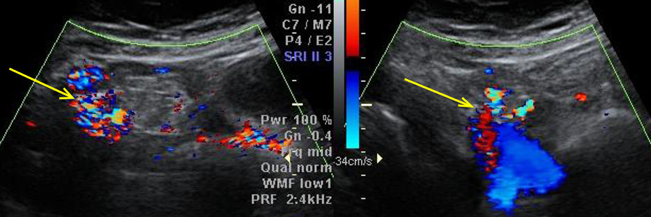
In the presence of an accumulation of benign tumors, echogenicity is reduced, and the size is increased. With endometriosis, the echogenicity of the muscular layer of the uterus is increased, retroversion of the uterus is often present. nine0003
If the ovaries are enlarged, and there are many small follicles in them, then the question of polycystic disease may arise.
Interpretation of ultrasound in men
In the study, the ultrasound doctor determines the shape, size, structure and genital organs, as well as the bladder.
The normal prostate is about 2.5 to 3.5 cm long, 2.5 to 4 cm wide, and about 0.2 cm thick.
Seminal vesicles no larger than 1 cm, without seals.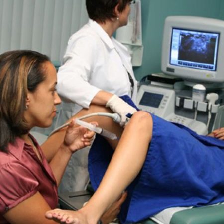 nine0003
Dmitrieva Olga Nikolaevna
Chief physician of "Polyclinika.ru" on Frunzenskaya, neurologist, ENMG specialist
reviews
Clinic
m. Frunzenskaya
Stolbova Tatyana Sergeevna 1905, internist, gastroenterologist
reviews
Clinic
m. Street 1905 Goda
Yuliy Sergeevich Gromov
Head physician of "Polyclinic.ru" on Sukharevskaya, surgeon
reviews
Clinic
m. Sukharevskaya
Agarkova Elena Valentinovna
Head physician "Polyclinika.![]() ru" in Zelenograd, gastroenterologist
ru" in Zelenograd, gastroenterologist
reviews
Clinic
Zelenograd
Aydinov Gennady Ivanovich Academician Yangelya, therapist
reviews nine0003
Clinic
m. st. Academician Yangel
Godulyan Aleksey Viktorovich
chief physician of "Polyclinika.ru" at Krasnye Vorota, endocrinologist, KMN
reviews
Clinic
m. Red Gate
Kupriyanova Olga Sergeevna
Head doctor "Polyclinika.ru" on Avtozavodskaya, general practitioner
reviews
Clinic
m.