Small soft spot on baby's head
About the fontanelle | Pregnancy Birth and Baby
About the fontanelle | Pregnancy Birth and Baby beginning of content5-minute read
Listen
What is a fontanelle?
A fontanelle is a ‘soft spot’ of a newborn baby’s skull. It is a unique feature that is important for the normal growth and development of your baby’s brain and skull. Your health team will check your baby’s fontanelles during routine visits.
If you touch the top of your baby’s head you can feel a soft spot in between the bones — this is a fontanelle.
A newborn baby’s skull is made up of sections of bone known as plates that are joined together by fibrous joints called sutures. The sutures provide some flexibility and allow your baby’s head to narrow slightly as it travels through the birth canal. The sutures also enable your baby’s head to grow in the first years of life.
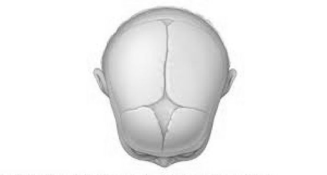
There are 2 fontanelles on your baby’s skull. These are the skin-covered gaps where the skull plates meet. The anterior fontanelle is at the top of your baby’s head, and the posterior fontanelle is located at the back of your baby’s head.
Illustration showing the anterior and posterior (front and back) fontanelles of a baby's skull.When will my baby’s fontanelles close?
The posterior fontanelle usually closes by the time your baby is 2 months old. The anterior fontanelle can close any time between 4 and 26 months of age. Around 1 in every 2 babies will have a closed fontanelle by the time they are 14 months old.
Can I touch my baby’s fontanelles?
Yes, you can gently touch your baby’s fontanelles. If you run your fingers softly along your baby’s head you are can probably feel them. Your doctor will touch your baby’s fontanelles as part of their routine medical examination. There is no need to be concerned or worried about touching your baby’s fontanelles as long as you are gentle.
What does a normal fontanelle look like?
Your baby’s fontanelle should feel soft and flat. If you softly touch a fontanelle, you may at times feel a slight pulsation — this is normal. If a fontanelle changes, or feels different to how it usually does, show your doctor or midwife as it may be a sign that your baby’s health may need to be checked.
Sunken fontanelle
If you notice that your baby’s fontanelles are low or sunken, your baby may be dehydrated.
However, you may notice other signs of dehydration in your baby before their fontanelles becomes sunken.
Other signs of dehydration include:
- having fewer wet nappies
- not feeding well
- loosing fluids from vomiting or diarrhoea
- perspiration (or sweating) in very hot weather
- being less alert or floppy
Bulging fontanelle
A bulging or swollen fontanelle may be a sign of a number of serious but rare conditions including meningitis or encephalitis (infections in the brain), cerebral haemorrhage (bleeding in the brain), hydrocephalus, an abscess or another cause of increased pressure in the brain.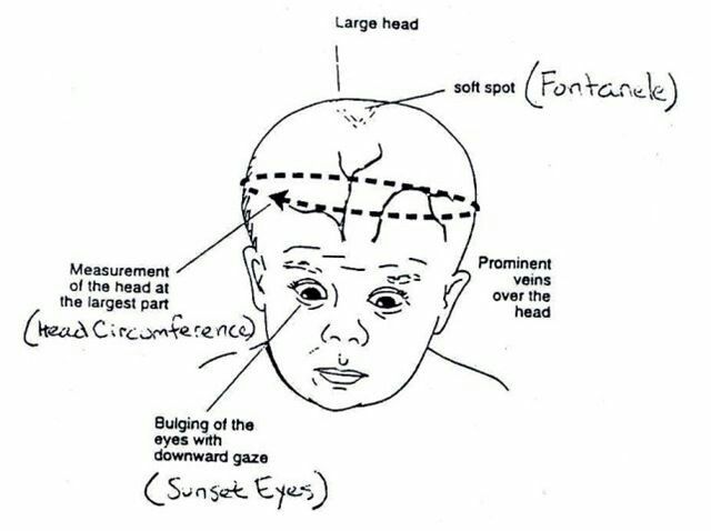
If you think that your baby’s fontanelles are bulging or sunken, seek medical advice immediately.
What if a fontanelle closes too soon?
Your baby’s fontanelles may close early. This can happen for several reasons. Your baby may have hyperthyroidism (high levels of the thyroid hormone) or hyperparathyroidism (high levels of parathyroid hormone). Another cause of early fontanelle closure is a condition known as craniosynostosis. Craniosynostosis occurs when one or more of the fibrous joints (sutures) between the bone plates in a baby’s skull fuse too early, before the brain has finished growing. As the brain continues to grow, it pushes on the skull from the inside but cannot expand into the closed over area. This causes the skull to have an unusual shape.
If you notice that your baby’s fontanelles seem to have closed early, if you can feel a ridge along your baby’s skull, or if you think that your baby’s head has an unusual shape, take your baby to see their GP or paediatrician.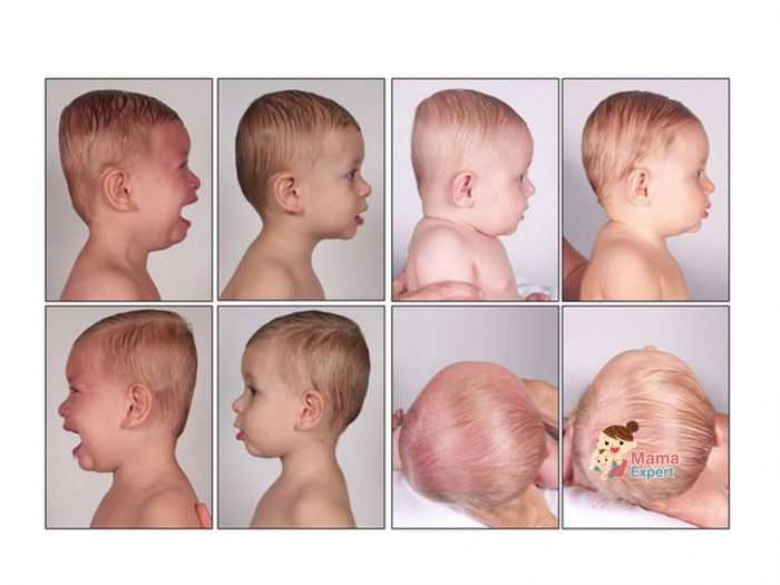
What if a fontanelle doesn’t close?
Your baby’s fontanelles may not close on time for several reasons. Common reasons for delayed fontanelle closure include congenital hypothyroidism (low thyroid hormones from birth), Down syndrome, increased pressure inside the brain, rickets and familial macrocephaly (a genetic tendency to have a large head).
If one or both of your baby’s fontanelles haves not closed by the time they are 2 years old, speak to your GP or paediatrician.
If you have any concerns about your baby’s fontanelles you should make an appointment to see your child health nurse, GP or paediatrician.
Speak to a maternal child health nurse
Call Pregnancy, Birth and Baby to speak to a maternal child health nurse on 1800 882 436 or video call. Available 7am to midnight (AET), 7 days a week.
Sources:
Children’s Health Queensland Hospital and Health Service (Craniosynostosis), American Family Physician (The Abnormal Fontanel), Australian Family Physician (The 6 week check - An opportunity for continuity of care), WA Health (Bulging Anterior Fontanelle), The Royal Children's Hospital Melbourne (Clinical Practice Guidelines: Dehydration)Learn more here about the development and quality assurance of healthdirect content.
Last reviewed: February 2022
Back To Top
Related pages
- How to know when your baby is well - video
- Knowing your baby is well - podcast
- Regular health checks for babies
- How your baby’s brain develops
Need more information?
Newborn baby essentials
Find out some of the essentials for looking after your newborn. Find out when your baby will need to have health checkups and immunisations. There is also lots of information on nappies, giving your baby a bath and teeth development.
Read more on Pregnancy, Birth & Baby website
Colic in infants - MyDr.com.au
Colic is a pattern of unexplained, excessive crying in an otherwise healthy and well-fed baby and happens to 1 in 5 Australian babies.
Read more on myDr website
Knowing your baby is well - podcast
Listen to Dianne Zalitis, midwife and Clinical Lead at Pregnancy, Birth and Baby, talk to Feed Play Love with Shevonne Hunt about signs your baby is well.
Read more on Pregnancy, Birth & Baby website
Common worries and fears for parents
New parents often worry that they don't know what to do. However, there are practical ways to deal with the challenges so you can enjoy your baby more.
Read more on Pregnancy, Birth & Baby website
Flattened head
Plagiocephaly (flattened or misshapen head) means an uneven or asymmetrical head shape. Plagiocephaly won't affect your baby's brain development but it should be treated.
Read more on Pregnancy, Birth & Baby website
Glossary of pregnancy and labour
Glossary of common terms and abbreviations used in pregnancy and labour.
Read more on Pregnancy, Birth & Baby website
Disclaimer
Pregnancy, Birth and Baby is not responsible for the content and advertising on the external website you are now entering.
OKNeed further advice or guidance from our maternal child health nurses?
1800 882 436
Video call
- Contact us
- About us
- A-Z topics
- Symptom Checker
- Service Finder
- Subscribe to newsletters
- Sign in
- Linking to us
- Information partners
- Terms of use
- Privacy
Pregnancy, Birth and Baby is funded by the Australian Government and operated by Healthdirect Australia.
Pregnancy, Birth and Baby’s information and advice are developed and managed within a rigorous clinical governance framework.
This site is protected by reCAPTCHA and the Google Privacy Policy and Terms of Service apply.
Healthdirect Australia acknowledges the Traditional Owners of Country throughout Australia and their continuing connection to land, sea and community. We pay our respects to the Traditional Owners and to Elders both past and present.
This information is for your general information and use only and is not intended to be used as medical advice and should not be used to diagnose, treat, cure or prevent any medical condition, nor should it be used for therapeutic purposes.
The information is not a substitute for independent professional advice and should not be used as an alternative to professional health care. If you have a particular medical problem, please consult a healthcare professional.
Except as permitted under the Copyright Act 1968, this publication or any part of it may not be reproduced, altered, adapted, stored and/or distributed in any form or by any means without the prior written permission of Healthdirect Australia.
Support this browser is being discontinued for Pregnancy, Birth and Baby
Support for this browser is being discontinued for this site
- Internet Explorer 11 and lower
We currently support Microsoft Edge, Chrome, Firefox and Safari. For more information, please visit the links below:
- Chrome by Google
- Firefox by Mozilla
- Microsoft Edge
- Safari by Apple
You are welcome to continue browsing this site with this browser. Some features, tools or interaction may not work correctly.
5 Warning Signs From Your Baby’s Soft Spot – Cleveland Clinic
When you’re a new parent, you learn that you need to protect that soft spot, or fontanelle, on your baby’s head.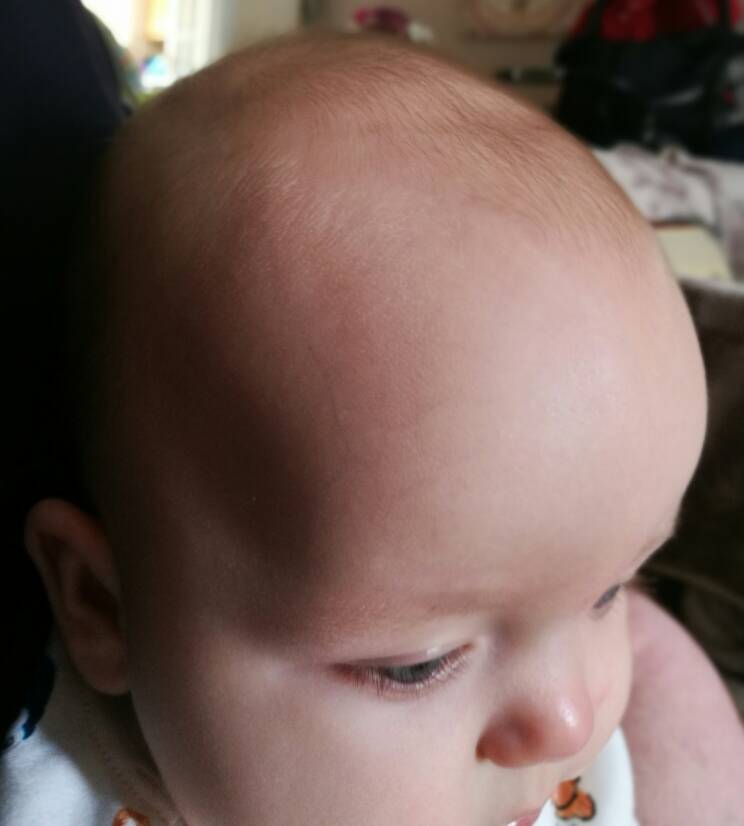 It rarely requires much attention, but it reminds you just how fragile your infant is.
That’s why, if something changes — is the soft spot sunken in a little? — you may worry there’s something wrong.
A change in the fontanelle isn’t always a major problem, but it can sometimes reveal internal issues, says Violette Recinos, MD, Section Head of Pediatric Neurosurgery at Cleveland Clinic.
“It is a good indicator of the baby’s potential hydration status and brain status,” she says. “It’s like an automatic pressure sensor.”
What is the fontanelle?
The fontanelle is the space between different plates of a baby’s skull that will eventually come together. This aspect of an infant’s skull structure typically allows for easy delivery through the birth canal and for rapid head growth during the first year of life, Dr. Recinos says.
There are actually two soft spots — one at the back of the head and another on top. The posterior one closes within a few months of birth, while the top fontanelle typically remains until just past a child’s first birthday.
Dr. Recinos explains what changes in the fontanelle can tell you about your infant’s health.
Sunken in soft spot
This is often a sign of dehydration, she says. It may occur if your child is sick and not getting enough fluids.
What you should do: See your pediatrician if the sunken appearance persists and you can’t get your baby to take in more fluids.
Advertising Policy
Swollen soft spot
After a fall, a swollen soft spot (particularly if it’s accompanied by vomiting) is sometimes a sign of head trauma.
What you should do: Seek medical treatment right away.
Bulging soft spot
Fluid buildup (hydrocephalus) can cause rapid head growth and can make the soft spot look “full,” Dr. Recinos says.
A bulging fontanelle also might signal internal bleeding or a tumor or mass causing pressure in the head.
What you should do: If their soft spot is bulging, that’s a reason to seek care from your pediatrician, she says.
Seek emergency care if your infant exhibits fatigue, vomiting or unusual mental status along with the fontanelle’s fullness. Treatment for these conditions may include surgery to insert a shunt that relieves fluid buildup or to remove any underlying mass, Dr. Recinos says.
Disappearing soft spot
Most of the time the soft spot is obvious, particularly on a newborn. But at times it can seem to disappear quickly.
This may scare parents, but it typically means it’s just a “quiet fontanelle,” not that it has fused together prematurely, Dr. Recinos says.
Advertising Policy
As long as your child’s head is growing normally, all is probably well, she says. But your pediatrician may suggest an imaging test to make sure the fontanelle is still open.
Occasionally, though, the skull bones do close earlier than normal on one side, causing craniosynostosis. Depending on which bones fused, the baby may develop an abnormal head shape. For example, sagittal craniosynostosis, the most common form, results in a longer head that is shaped somewhat like a football.
What you should do: Children with craniosynostosis may need surgery to open the fused bones and reshape the skull. In some cases, the child will wear a helmet afterward until the site heals and the head shape normalizes.
Soft spot that doesn’t close
If the soft spot stays big or doesn’t close after about a year, it is sometimes a sign of a genetic condition such as congenital hypothyroidism.
What you should do: Talk to your doctor about treatment options.
The bottom line? If you have any questions or concerns about your baby’s soft spot, it’s a good idea to check in with your pediatrician to make sure all is well.
Congenital nevus pigmentosa in children, removal of the nevus of the child's skin on the head, neck or trunk, treatment of vascular nevus in newborns
Congenital malformations of the skin and subcutaneous fat
It can occur during pregnancy or appear soon after birth.
To one degree or another, the following factors can lead to the development of pathology:
- gene breakdown;
- the presence of genitourinary infection in the mother during pregnancy; nine0010
- heredity;
- the impact of negative external factors on the mother's body during pregnancy;
- incorrect mother's diet during pregnancy, the predominance of artificial colors, preservatives, flavors;
- long-term use of hormonal contraceptives before pregnancy.

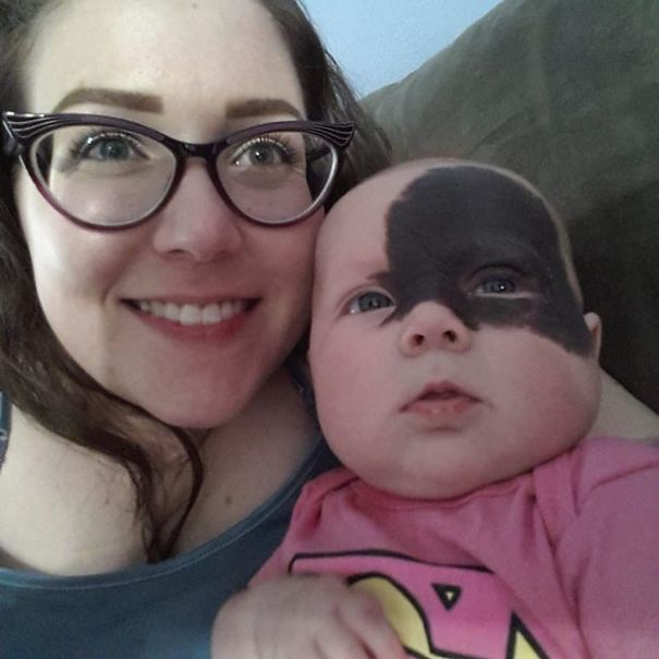
Initially, a pigmented nevus in children is defined as a benign formation. However, it may have melanocytic activity, which means it can grow. With the age of the child, not only the growth of the spot is possible, but also the appearance of new ones, as well as their degeneration into malignant tumors. A skin tumor - melanoma - is aggressive, as it rapidly progresses and metastasizes to the nearest organs and tissues. nine0005
Pigmentary nevus is determined in approximately 1% of children.
Boys and girls are equally susceptible to the appearance of spots on the skin. A nevus can look different: a medium-sized or large-sized knot, a stain that is different from the main skin tone, a wart. Localization is also diverse: limbs, torso, head. In most cases, a nevus occurs on a child's head, neck, chest, or upper back.
The sizes of formations vary from a few mm to several cm. In about 5%, the number of nevi is so large that they occupy a significant proportion of the skin surface. Normally, when palpated, they can be both soft and nodular, but without pain. nine0005
Varieties of nevi
The spots have clear borders and are pale brown to black in color.
Small and medium-sized formations, due to their light tone, can be practically indistinguishable in the first 2-3 months after the birth of a child. However, as they grow, they darken and proportionally grow in size. Nevi are also non-pigmented, then they can be identified by touch by the different structure of the skin.
Spots are divided by size into: nine0005
- small - up to 1.6 mm;
- medium - from 1.6 to 10 mm;
- large - from 10 to 20 cm;
- giant - from 20 cm.
Another criterion for classification is the clinical type. According to him, there are nevi:
- Adenomatous. They are located on the face and head, have a convex shape, clear boundaries, the color varies from yellowish to dark brown.
- Warty.
 Located on the torso or limbs. They are characterized by uneven outlines and surface, dark color. nine0010
 - Giant pigment. Often they develop in utero, they can be located on one or both sides of the body. The surface is covered with hairs, often accompanied by a scattering of small neoplasms around them.
- Blue. They are located in the upper body, on the face and hands. The color is bluish or gray-blue, the surface is dense and glossy. Nevus slightly protrudes above the skin surface and does not exceed 2 cm in diameter.
- Papillomatous. They are located on the head, do not have clear boundaries and outlines. The surface is heterogeneous, the color is from light to almost black. When injured, it becomes inflamed. nine0010
- epidermal. They are usually located on the limbs one at a time. They have an uneven surface and color from light to dark.
A characteristic sign of a non-malignant neoplasm is the presence of hairs on the surface.
Nevus treatments
Treatment is allowed only in patients over the age of 2 years. Therapy has 4 main areas:
- surgical removal; nine0009 laser excision;
- cryodestruction;
- electrocoagulation.
Surgical removal, as a rule, of nevi that have grown deep into the tissue. Giant neoplasms are removed in several steps, but the disadvantage of this method is the formation of scars at the site of nevi.
Laser photocoagulation is used to remove dysplastic pigmented nevi and other clinical types of macules. The method allows you to remove the formation without pain, without a subsequent scar. nine0005
Cryodestruction involves exposure to the affected area of the skin at very low temperatures. As a result, the altered cells die and are replaced by healthy ones. After cryodestruction, a small crust appears, which quickly disappears.
Electrocoagulation affects the nevus with high temperatures. Usually this method is used to remove small and medium-sized stains.
What do the growths on the head say?
Neoplasms can appear anywhere on the body. The scalp is no exception. However, this is a rather difficult place to independently determine the type of neoplasm that has appeared. Therefore, we have prepared a list of the most common growths on the head, dividing them into three groups: malignant, borderline and benign. nine0005
In one of our articles "Skin growths: benign, malignant and borderline", we have already talked about skin growths and classified them according to the danger to human health. Today we will analyze those that are most often found on the head and, sometimes, are invisible under the hair. Experts recommend regularly probing and, if possible, examining the head for the presence of neoplasms, since the proximity of a malignant neoplasm to the brain can lead to irreparable consequences. If you notice a strange growth on your head, contact your dermatologist or oncologist immediately. nine0005
The scalp is also the most traumatized and exposed area. Many people use combs with hard teeth, which can easily rip off the build-up.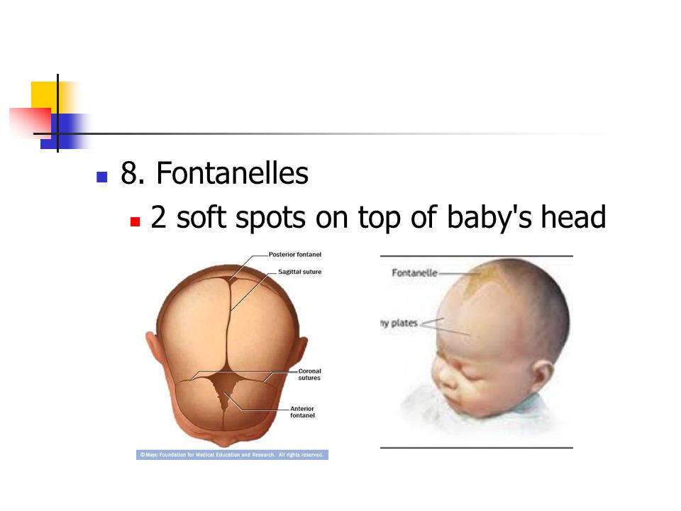 In addition, we wash our hair, dye, apply masks that can corrode the neoplasm and provoke its degeneration into a malignant tumor. Another risk factor is long exposure to the sun without a hat. As you know, many moles tend to become malignant with prolonged and strong exposure to ultraviolet radiation. nine0005
That is why, if you find or feel a growth in your hair that:
- hurts;
- bleeds;
- itches;
- peeling off;
- festering
Need to see a specialist urgently.
Types of neoplasms on the head
First, let's consider malignant neoplasms on the scalp.
1. Melanoma.
First of all, malignant neoplasms include melanoma - skin cancer. You can learn more about it in our special article "Skin Cancer: Melanoma.". The neoplasm looks like a small light brown or black plaque with a rough surface. Melanoma is dangerous, it metastasizes and can lead to irreparable consequences.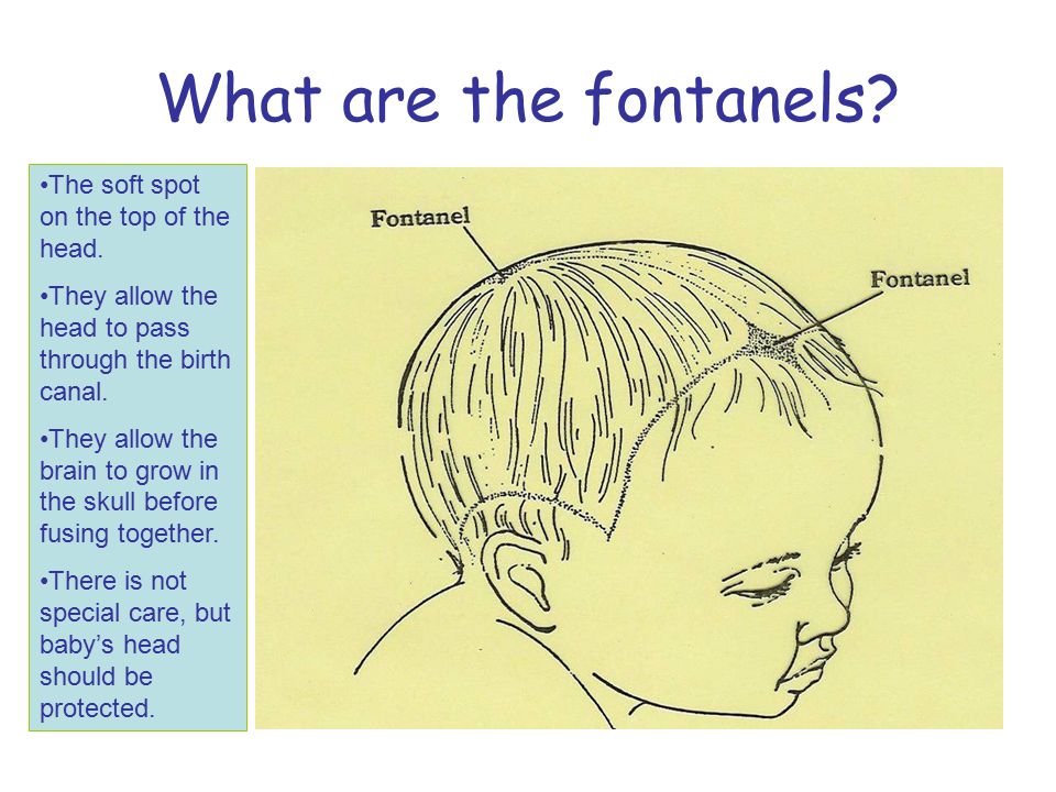 Therefore, if it is found on the scalp, you should immediately consult a doctor. One of the most effective treatments for melanoma is laser therapy, which helps skin cells regenerate faster while killing all harmful cells. nine0005
2. Basalioma.
The second reason not to waste time and go for an examination to an oncologist may be a neoplasm similar to a nodule with a crust, light pink or red - this is a basalioma. It develops from the cells of the basal layer of the skin and is often accompanied by the formation of ulcers and erosions. You can learn more about this neoplasm in our special article "Basalioma. Skin cancer: basal cell carcinoma.". The advanced method of treating basalioma is photodynamic therapy (for more details, see PDT), this is a sparing method of exposure, which already after several sessions gives visible results. nine0005
3. Epithelioma.
Skin epithelioma is a tumor that develops on the surface layer of the epidermis.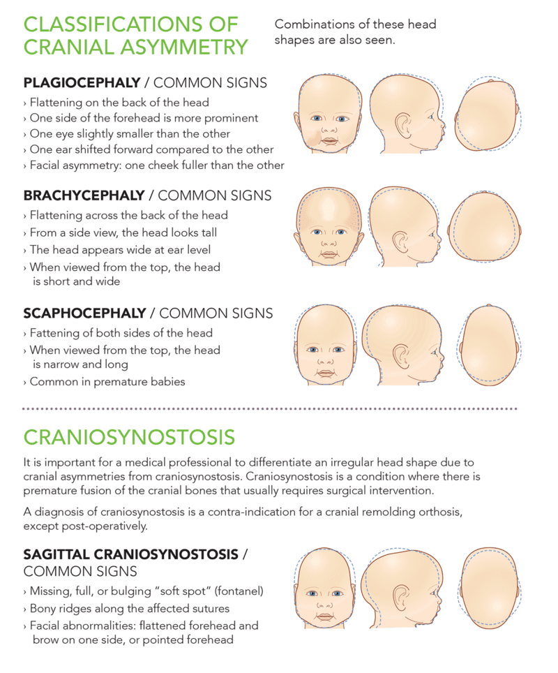 Also, epithelioma of the sebaceous gland is distinguished - a neoplasm that occurs on the scalp, with inflammation of the sebaceous glands. Epithelioma looks like a growth of pink or light brown color, can reach 5 cm in diameter. This neoplasm is dangerous because it metastasizes to the lymph nodes very quickly. It can occur against the background of previous dermatological diseases, as well as strong UV radiation. nine0005
Borderline neoplasms on the scalp
1. Keratosis.
Keratosis of the scalp is the keratinization of the upper layer of the skin. Most often found on the face and scalp, it can be located both on a small area of \u200b\u200bthe skin and affect the entire surface of the head. The neoplasm looks like multiple warts, from light to dark brown. Specialists also detect seborrheic keratosis of the scalp, its appearance indicates pathologies occurring in the body. With such a diagnosis, a thorough examination is necessary, since this may be evidence of cancer of the internal organs.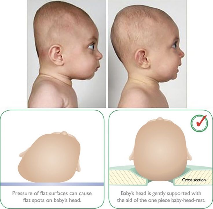 Treatment of keratosis of the scalp is prescribed by an oncologist or dermatologist, after receiving all the tests. Treatment options may include medication and peeling, massage, and laser therapy. nine0005
2. Keratoacanthoma.
Keratoacanthoma of the scalp is a benign tumor of the hair follicles, most often seen in the elderly. It is a spherical dense neoplasm affecting the scalp. Keratoacanthoma and its multiple form, has a flesh color, and can grow rapidly, reaching 2-3 cm. In some cases, the neoplasm can degenerate into a malignant one, especially if it is often injured. Treatment of keratoacanthoma is possible only with its complete excision with a scalpel, electric current or laser. nine0005
Benign tumors on the scalp
1. Moles.
A mole is a small pigmented formation on the skin that can appear at any age and on any part of the skin, even on the head in the hair. In large numbers, moles can grow during puberty, with hormonal failure or pregnancy, which you can read more about in our special article. According to statistics, a mole on the scalp is not dangerous, the likelihood of degeneration into skin cancer is extremely small. However, it is necessary to check moles, RTM-diagnostics is the best way to cope with this. Based on the results of the check, you may be shown the removal of a mole. nine0005
2. Warts.
Perhaps the most common neoplasms on the scalp are warts and papillomas. They appear due to the human papillomavirus, which is most often activated with reduced immunity, with severe stress, infectious diseases, and a lack of vitamins in the body. Learn more about what HPV is and how it manifests on the body in our article "Human papillomavirus". Warts are classified as benign neoplasms, however, if the wart on the head in the hair is often injured, then it must be removed. The most effective method in such cases is laser removal. nine0005
3. Hemangioma.
Scalp hemangioma is a vascular tumor that appears due to abnormal development of blood vessels.