Scan of down's syndrome baby
Prenatal Testing for Down Syndrome | Patient Education
Screening tests
All pregnant individuals in California have access to California prenatal screening (CA PNS), also called sequential integrated screening. This noninvasive process is carried out in two steps.
In the first step, which is performed when the pregnancy is between 10 and 14 weeks, a blood sample is taken from the pregnant person and a nuchal translucency ultrasound is performed to measure the fluid at the back of the baby's neck. If the blood test is scheduled prior to the ultrasound, we can provide those results at the end of your ultrasound appointment. The blood test results, nuchal translucency measurement and pregnant person's age are together used to estimate the risk for Down syndrome and trisomy 18 (a genetic condition, also called Edwards syndrome, that affects fetal development).
The second step is a test performed with a blood sample from the pregnant person when the pregnancy is between 15 and 20 weeks. When the results of this blood test are combined with the results from the first trimester blood test and nuchal translucency ultrasound, the detection rate for Down syndrome increases. This test also provides a personal risk assessment for having a fetus with trisomy 18, Smith-Lemli-Opitz syndrome (a genetic condition that can slow growth and cause intellectual disability), an open neural tube defect (a problem with the formation of the embryo's nervous system, such as spina bifida) or an abdominal wall defect (an abnormal opening in the abdomen).
Diagnostic tests
Amniocentesis, chorionic villus sampling (CVS) and ultrasound are the three primary procedures for diagnostic testing.
Amniocentesis is the test we most commonly use to identify chromosomal problems, such as Down syndrome. (In at-risk fetuses, it can be used to detect other genetic diseases, such as cystic fibrosis, Tay-Sachs disease and sickle cell disease.)
An amniocentesis procedure for genetic testing is typically performed when the pregnancy is between 15 and 20 weeks. Under ultrasound guidance, a needle is inserted through the abdomen to remove a small sample of amniotic fluid. Cells from the fluid are cultured and a karyotype test – an analysis of the cells' chromosomal makeup – is performed. It takes about two weeks to receive the results. Amniocentesis detects most chromosomal disorders with a high degree of accuracy.
There is a low risk of miscarriage as a result of amniocentesis – about 1 in 900. Miscarriage rates for amniocentesis performed at UCSF are extremely low.
Like amniocentesis, chorionic villus sampling is most commonly used to identify chromosomal problems, such as Down syndrome. (It can also be used to detect other genetic diseases – including cystic fibrosis, Tay-Sachs disease and sickle cell disease – in at-risk fetuses.) The main advantage over amniocentesis is that CVS is done much earlier in pregnancy, at 10 to 12 weeks rather than 15 to 20 weeks.
CVS involves removing a tiny piece of tissue from the placenta for analysis.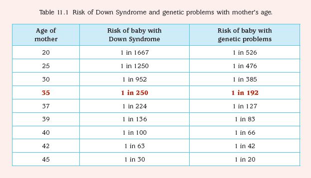 Under ultrasound guidance, this sample is obtained either with a needle inserted through the abdomen or a catheter inserted through the vagina and into the cervix (outer end of the uterus). The tissue is cultured and a karyotype test of the cells' chromosomal makeup is performed. It takes about two weeks to receive the results.
While it can provide information earlier in pregnancy than amniocentesis, CVS does not detect spinal cord defects. However, we can screen for spinal cord defects later in the pregnancy using expanded alpha-fetoprotein (AFP) blood testing or ultrasound.
There is a low risk of miscarriage as a result of CVS – about 1 in 450. Miscarriage rates for CVS procedures performed at UCSF are very low.
While the primary purpose of ultrasound is to determine the pregnancy's status – due date, size of the fetus and whether there's more than one baby – ultrasound can also provide some information about possible birth defects. All pregnant UCSF patients undergo a comprehensive ultrasound exam before any invasive tests are performed. We will explain your ultrasound results at the time of your visit.
In some patients, an ultrasound raises concern for a fetal abnormality. This possibility makes ultrasound expertise crucial, so you may find it reassuring that we have extensive experience in performing and interpreting ultrasound exams in pregnancy.
The meaning of a positive result
If you receive positive results on a screening test, we recommend that you discuss its implications and your options with your doctor and a genetic counselor. They will explain what types of diagnostic testing are available. Whether to have invasive genetic testing is your decision.
If a diagnostic test finds a genetic abnormality, we recommend discussing the significance of the result with experts on the condition, including a medical geneticist and a genetic counselor, as well as your doctor.
Screening for Down syndrome | Pregnancy Birth and Baby
Screening for Down syndrome | Pregnancy Birth and Baby beginning of content5-minute read
Listen
If you or someone you care about has a child with Down syndrome, or is pregnant with a Down syndrome baby, you may have many questions or feel overwhelmed by what this means.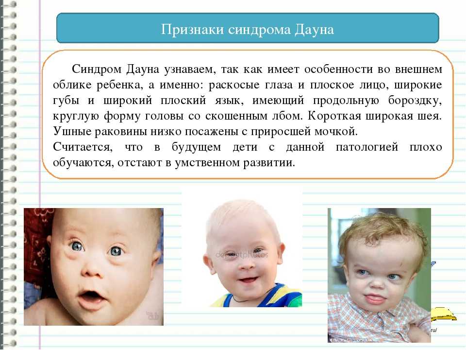 The most important thing that babies with Down syndrome need — just like any baby — is a loving, secure environment in which they feel nurtured and supported.
What is Down syndrome?
Down syndrome is the most common chromosomal condition that a baby can be born with. Around 1 in every 1,000 babies born in Australia has Down syndrome.
Down syndrome is also known as Trisomy 21. People with Down syndrome usually look different from other people and may have some difficulty learning new things.
What causes Down syndrome?
Normally, every cell in the human body — except egg and sperm cells — has 46 chromosomes. The egg and sperm cells normally have 23 chromosomes each.
The most common cause of Down syndrome is when an extra copy of chromosome 21 randomly appears in either the egg or sperm. At conception, when the egg and sperm meet, the extra copy of chromosome 21 grows throughout all of the embryo’s cells, giving them each 47 chromosomes instead of the usual 46.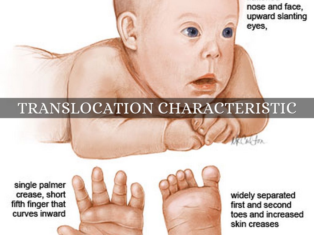
Less commonly, the extra copy of the chromosome can appear in some of the embryo’s cells after conception. This is called mosaicism and results in some cells having 46 chromosomes and some having 47.
Another rare form of the syndrome is caused by ‘Robertsonian translocation’. This occurs when a piece of chromosome 21 breaks off and becomes joined to another chromosome before or after conception. In this case, the embryo’s cells will all have 46 chromosomes, but each cell still contains an extra copy of chromosome 21.
What are the most common features of Down syndrome?
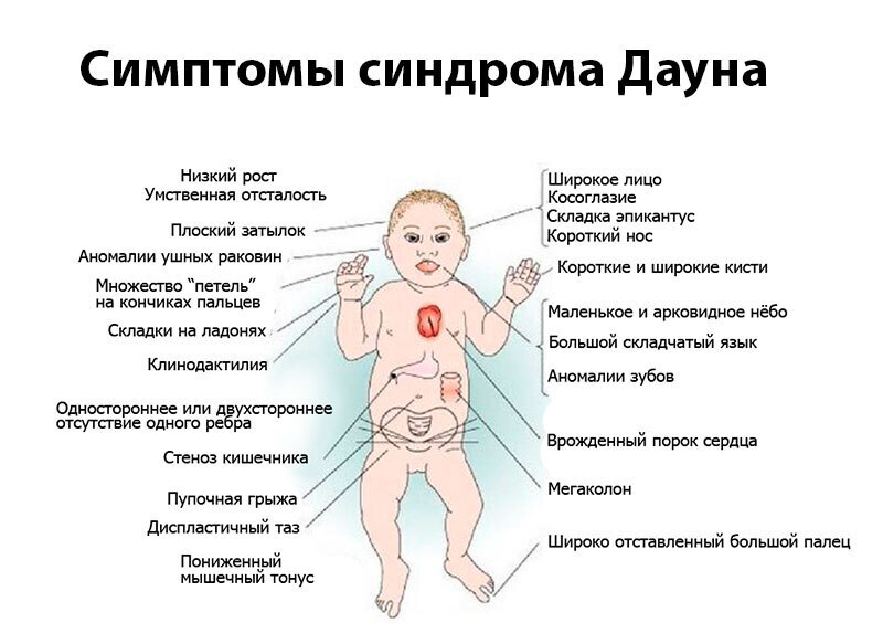
Many babies with Down syndrome have one or more of the following features at birth:
- low muscle tone ('floppy' muscles)
- a flatter face with eyes slanting upward
- small ears and a wider neck than usual
- a single crease across the palm of the hand
- a gap between the first and second toes
Down syndrome is also associated with intellectual disability.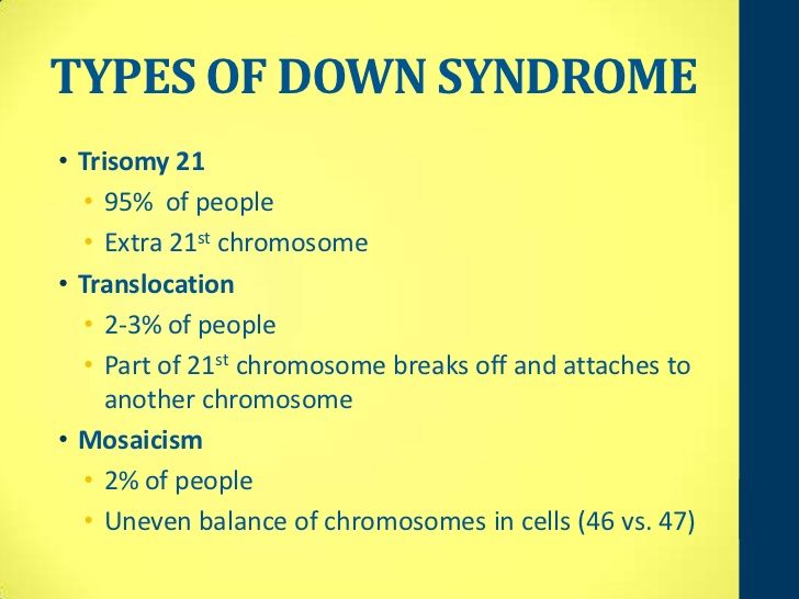
However, not everyone with Down syndrome will have all of these features.
What medical complications might come with Down syndrome?
About 1 in every 2 babies born with Down syndrome will have heart problems and approximately 1 in 10 will have gastrointestinal (gut) problems. Hearing and vision problems are also more common in people with Down syndrome.
How is Down syndrome diagnosed?
Pregnant women are offered routine antenatal tests at different times during pregnancy and for different reasons. These include screening tests, such as ultrasounds and blood tests, that can help estimate your baby’s risk of being born with a range of conditions, including Down syndrome.
Non-invasive prenatal testing (NIPT) is a new blood test that can be done as an alternative screening test.
Diagnostic tests (such as chorionic villus sampling or amniocentesis) will show whether a baby actually has Down syndrome.
After the baby is born, chromosome testing from a blood sample, may be done to confirm Down syndrome.
What is the treatment for Down syndrome?
There is currently no way to prevent or cure Down syndrome. Prenatal testing allows you and your family to make informed decisions, including ending the pregnancy. For this reason, before you have the test it’s a good idea to think about why you are choosing to do it, and how you will feel once you get the results.
Consider also who you want to discuss any important decisions with — your partner, a friend or family member, or a health professional such as your doctor or midwife are all good options.
Early diagnosis can also help you and your doctor continue to check your baby for complications and to act early if needed. Some people with Down syndrome may need surgery to repair heart defects or gut blockages. Medicines to treat thyroid disease may also be needed in some cases.
Children all learn and develop at their own pace. However, there are effective early intervention programs for children with Down syndrome that can help them reach their learning potential.
Resources and support
Down Syndrome Australia provides support, information and resources for people with Down syndrome and their families across Australia.
Support services include:
- information and advice about Down syndrome
- information about local support groups
- home visits
- new parent packs
- workshops and webinars
- online support groups for you and your family
A national phone number (1300 881 935) can connect you with your local state or territory Down syndrome organisation for further information.
Speak to a maternal child health nurse
Call Pregnancy, Birth and Baby to speak to a maternal child health nurse on 1800 882 436 or video call. Available 7am to midnight (AET), 7 days a week.
Sources:
Centre for Genetics education (Trisomy 32 - Down Syndrome), Lab Tests Online (Down syndrome), Down Syndrome Australia (Prenatal and New Parent)Learn more here about the development and quality assurance of healthdirect content.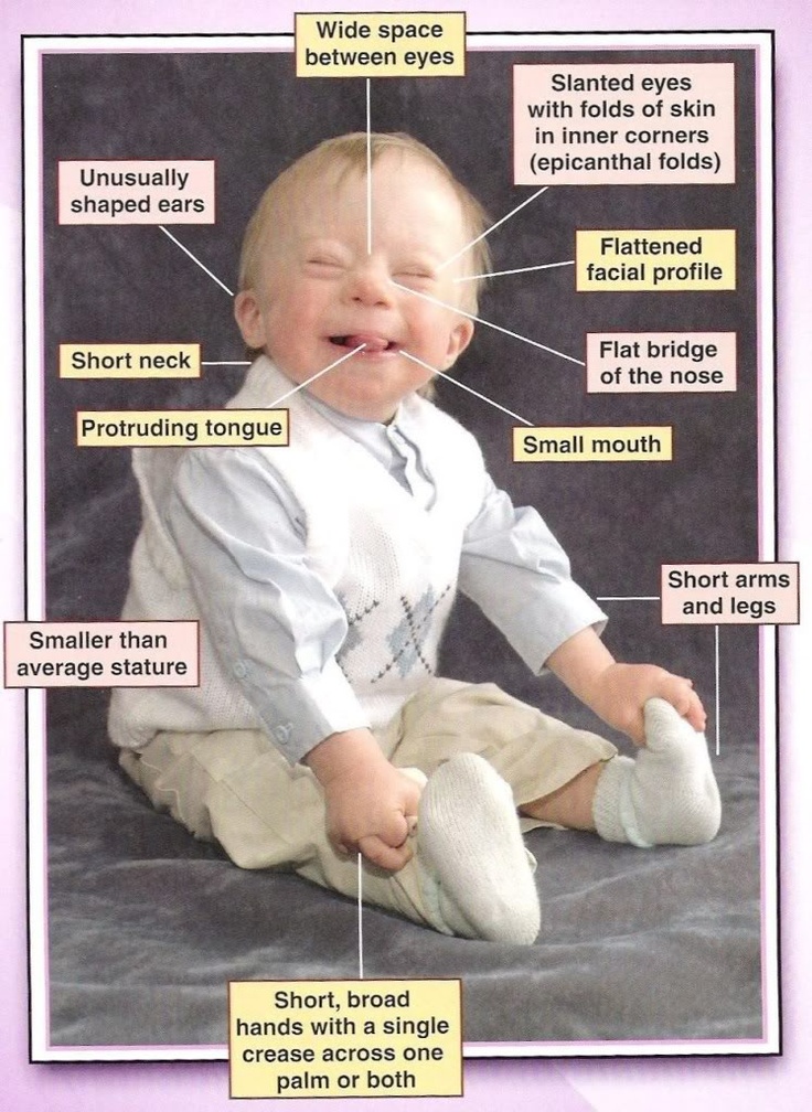
Last reviewed: March 2022
Back To Top
Related pages
- Caring for a child with Down syndrome
- What is Down syndrome?
- Questions to ask your doctor about tests and scans
- Routine antenatal tests
- Pregnancy checkups, screenings and scans
Need more information?
Support - Prenatal Screening
People often contact our service to better understand their screening results
Read more on Prenatal Screening - Down Syndrome Queensland website
Maternal screening - Pathology Tests Explained
Why and when to get tested for maternal screening
Read more on Pathology Tests Explained website
DSA Prenatal Testing Factsheet - Prenatal Screening
Support through the Down Syndrome Queensland support service is available for any prospective parent, health care professional, community service, carer or family members supporting someone who has received unexpected news about their pregnancy
Read more on Prenatal Screening - Down Syndrome Queensland website
Prenatal Screening for Chromosomal and Genetic Conditions
Read more on RANZCOG - Royal Australian and New Zealand College of Obstetricians and Gynaecologists website
Non-invasive prenatal testing (NIPT)
A non-invasive prenatal test (NIPT) is a sensitive test to screen for Down syndrome and some other chromosomal disorders early in pregnancy.
Read more on Pregnancy, Birth & Baby website
What it's like to Receive an Unexpected Result - Prenatal Screening
Support through the Down Syndrome Queensland support service is available for any prospective parent, health care professional, community service, carer or family members supporting someone who has received unexpected news about their pregnancy
Read more on Prenatal Screening - Down Syndrome Queensland website
Dear Doctor, we want you to know... - Prenatal Screening
Support through the Down Syndrome Queensland support service is available for any prospective parent, health care professional, community service, carer or family members supporting someone who has received unexpected news about their pregnancy
Read more on Prenatal Screening - Down Syndrome Queensland website
Maternity Consumer Network Patient Information Brochure - Prenatal Screening
Support through the Down Syndrome Queensland support service is available for any prospective parent, health care professional, community service, carer or family members supporting someone who has received unexpected news about their pregnancy
Read more on Prenatal Screening - Down Syndrome Queensland website
What is Down syndrome?
Down syndrome is a genetic disorder characterised by mental and developmental impairments.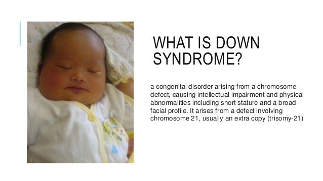
Read more on Pregnancy, Birth & Baby website
What is Down syndrome? – Down Syndrome Australia
Down syndrome is a genetic condition and is also sometimes known as trisomy 21. You can find out more about Down syndrome below. You can also turn on the Easy Read for this page.
Read more on Down Syndrome Australia website
Disclaimer
Pregnancy, Birth and Baby is not responsible for the content and advertising on the external website you are now entering.
OKNeed further advice or guidance from our maternal child health nurses?
1800 882 436
Video call
- Contact us
- About us
- A-Z topics
- Symptom Checker
- Service Finder
- Linking to us
- Information partners
- Terms of use
- Privacy
Pregnancy, Birth and Baby is funded by the Australian Government and operated by Healthdirect Australia.![]()
Pregnancy, Birth and Baby is provided on behalf of the Department of Health
Pregnancy, Birth and Baby’s information and advice are developed and managed within a rigorous clinical governance framework. This website is certified by the Health On The Net (HON) foundation, the standard for trustworthy health information.
This site is protected by reCAPTCHA and the Google Privacy Policy and Terms of Service apply.
This information is for your general information and use only and is not intended to be used as medical advice and should not be used to diagnose, treat, cure or prevent any medical condition, nor should it be used for therapeutic purposes.
The information is not a substitute for independent professional advice and should not be used as an alternative to professional health care. If you have a particular medical problem, please consult a healthcare professional.
Except as permitted under the Copyright Act 1968, this publication or any part of it may not be reproduced, altered, adapted, stored and/or distributed in any form or by any means without the prior written permission of Healthdirect Australia.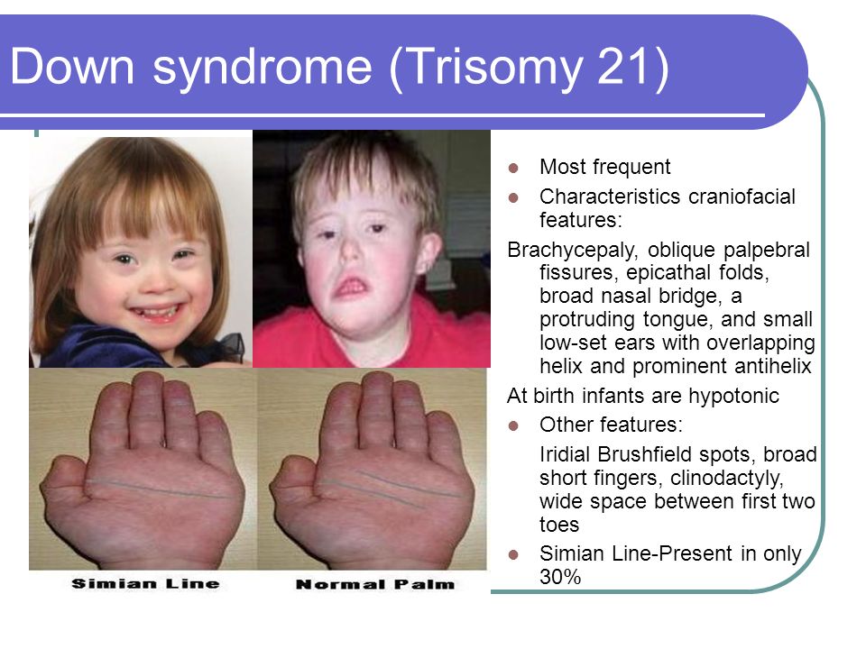
Support this browser is being discontinued for Pregnancy, Birth and Baby
Support for this browser is being discontinued for this site
- Internet Explorer 11 and lower
We currently support Microsoft Edge, Chrome, Firefox and Safari. For more information, please visit the links below:
- Chrome by Google
- Firefox by Mozilla
- Microsoft Edge
- Safari by Apple
You are welcome to continue browsing this site with this browser. Some features, tools or interaction may not work correctly.
Diagnosis of Down's syndrome according to ultrasound, timing of the examination, signs of fetal chromosomal pathology
It is possible to diagnose Down's syndrome already in the early stages of pregnancy according to ultrasound examination (ultrasound) of the fetus . The method is based on the use of high-frequency sound, which is reflected from various surfaces with different intensities. Deciphering the signals allows the doctor to create a two-dimensional image of the internal structure of the area under study. nine0003
Is it possible to detect Down syndrome by ultrasound
Down syndrome in the fetus is diagnosed by ultrasound in 60-90% of cases. High accuracy of the examination is achieved by comparing the development of the fetus with the norm. In addition to Down syndrome, ultrasound can detect signs of a number of other genetic malformations. Pathologies in the structure of the heart, incomplete rotation of the intestine, duodenal atresia can also serve as signs of Down syndrome in the fetus, all these malformations are recorded by ultrasound. nine0003
Tests before marriage
Down syndrome is observed in one case in 700-800 pregnancies and is characterized by the presence of an extra 21st chromosome. The syndrome affects the future life of the child.
The possibility of having a child with Down syndrome increases with the age of the parents. In addition, the risk of a chromosomal abnormality increases significantly if one of the parents already has Down syndrome. In some cases, the pathology is mosaic in nature and may be invisible even to very close people. nine0003
Down's syndrome is reliably diagnosed in the perinatal period using ultrasound and a number of other methods.
How does Down syndrome manifest itself after birth?
Children with Down syndrome have a number of external differences. Their faces are flatter, with a slightly pronounced bridge of the nose and an epicanthal fold at the inner corners of the eyes. The skull of the child is shortened, with a flat occiput.
After birth, children with Down syndrome are somewhat behind in physical and mental development. Over time, the mental retardation, in comparison with other children, will be more and more noticeable. nine0003
In addition, people with Down syndrome are more likely to suffer from congenital heart defects (occur in almost 40% of cases). The presence of an extra chromosome significantly increases the risk of developing some other diseases: cataracts, Alzheimer's, myeloid leukemia, and often there are disorders in the digestive system. Weakened immunity makes people with the syndrome more vulnerable to viruses and colds.
Methods for diagnosing chromosomal abnormalities
Chromosomal abnormalities can be diagnosed by non-invasive and invasive methods. The first allow you to establish a number of pathologies characteristic of chromosomal abnormalities. Invasive methods are used to make an accurate diagnosis. nine0003
ultrasound
Establishing the risk of Down syndrome in a fetus by ultrasound is called ultrasound screening. Often the method is used in combination with a biochemical analysis of the mother's blood. The accuracy of diagnosing Down syndrome on ultrasound in the second trimester of pregnancy can reach 91%. The examination is carried out at 11-13 and 16-18 weeks of pregnancy.
Invasive techniques
Invasive prenatal diagnosis can lead to a number of complications. The risk to the fetus and mother is small. However, the procedure is prescribed only if necessary, if Down's syndrome in the fetus is confirmed by ultrasound data and maternal blood tests. nine0003
Biochemical screening
The main task of screening is to identify risk groups in terms of having a child with serious illnesses, including those with chromosomal disorders. Biochemical screening is recommended for all women in the first and second trimesters of pregnancy.
Diagnostics in the first trimester of pregnancy
In the first trimester of pregnancy, Down syndrome can be diagnosed by ultrasound, due to the presence of a number of pathologies. These include an increased thickness of the nuchal space in the fetus, reduced, compared with the norm, the size of the cerebellum and frontal lobe, impaired bone formation, heart defects, absence of the nasal bone, etc.
It is important to remember that no single marker will be considered sufficient for a reliable diagnosis of Down syndrome - there is not enough ultrasound data for this. Therefore, having received the results of the examination, you do not need to be upset ahead of time - you need to consult a specialist.
What to do if the diagnosis is confirmed?
Examination can show pathology in the fetus. It is up to the parents to decide what to do next. If they are ready for responsibility, then, even if Down syndrome is confirmed by ultrasound and invasive research methods, parents can leave the child. Despite the peculiarities of development, he can live a long and happy life. In cases where there are too many pathologies, doctors may recommend terminating the pregnancy. However, even in this case, the decision will be made by the parents. nine0003
pregnancy bronchi abdomen vagina genitals pituitary eyes eye orbits shin head brain throat larynx rib cage thoracic region diaphragm for kids glands stomach gallbladder stomach retroperitoneum back of the head teeth brush intestines collarbone knee limbs contrast agent coccyx bone sacrum lungs lymph node facial skeleton elbow scapula small pelvis uterus period breast bladder scrotum soft tissues adrenal glands leg nose nasopharynx finger groin liver esophagus pancreas spine penis kidneys small of the back lumbosacral region forearms appendages prostate calcaneus ribs arm sciatic nerve spleen heart vessels joints back foot joints tendon pelvis hip joint trachea Turkish saddle an ear jaw scull cervical cervical region neck thyroid ovaries
Sign up for a consultation or diagnostic today!
You can make an appointment by phone: +7 (812) 901-03-03
Or leave a request
full name
Phone number
By clicking the "Make an appointment" button, I accept the terms of the Personal Data Processing and Security Policy and consent to the processing of my personal data.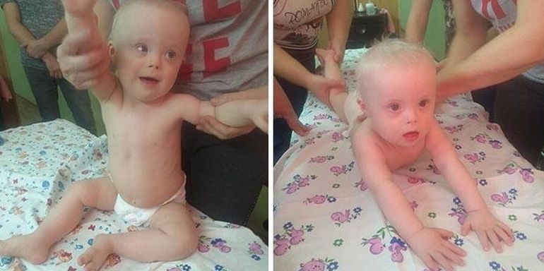 nine0003
Our medical centers
Making an appointment
Patient's last name *
Incorrect first name
nine0002 Name *Middle name
nine0002 Contact phone *E-mail *
nine0002 By clicking the "Make an appointment" button, I accept the terms of the Personal Data Processing and Security Policy and consent to the processing of my personal data.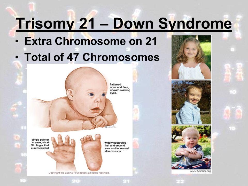
Registration and payment for repeated online appointment
Patient's last name *
Incorrect first name
nine0002 Name *Middle name *
nine0002 Contact phone *E-mail *
nine0002 By clicking the "Submit request" button, I accept the terms of the Personal Data Processing and Security Policy and consent to the processing of my personal data.
About cookies on this website
We use cookies, IP addresses and device data for analytics to make your visit to the site convenient and personalized. You can disable cookies in your browser settings. By continuing to use our site, you consent to the processing of the listed data and accept the terms of the Privacy Policy. nine0003
Ultrasound diagnostics of Down syndrome and other chromosomal abnormalities
- Ultrasound of the fetus is the main method for diagnosing the likelihood of Down syndrome
- Basic ultrasound markers of Down syndrome
- Signs of Down syndrome with ultrasound in the second trimester of pregnancy
- How else can you determine the risk of having a baby with Down syndrome
Down's syndrome is a relatively common congenital pathology caused by the presence of an extra chromosome in 21 pairs. Down syndrome is also called trisomy on 21 pairs of chromosomes.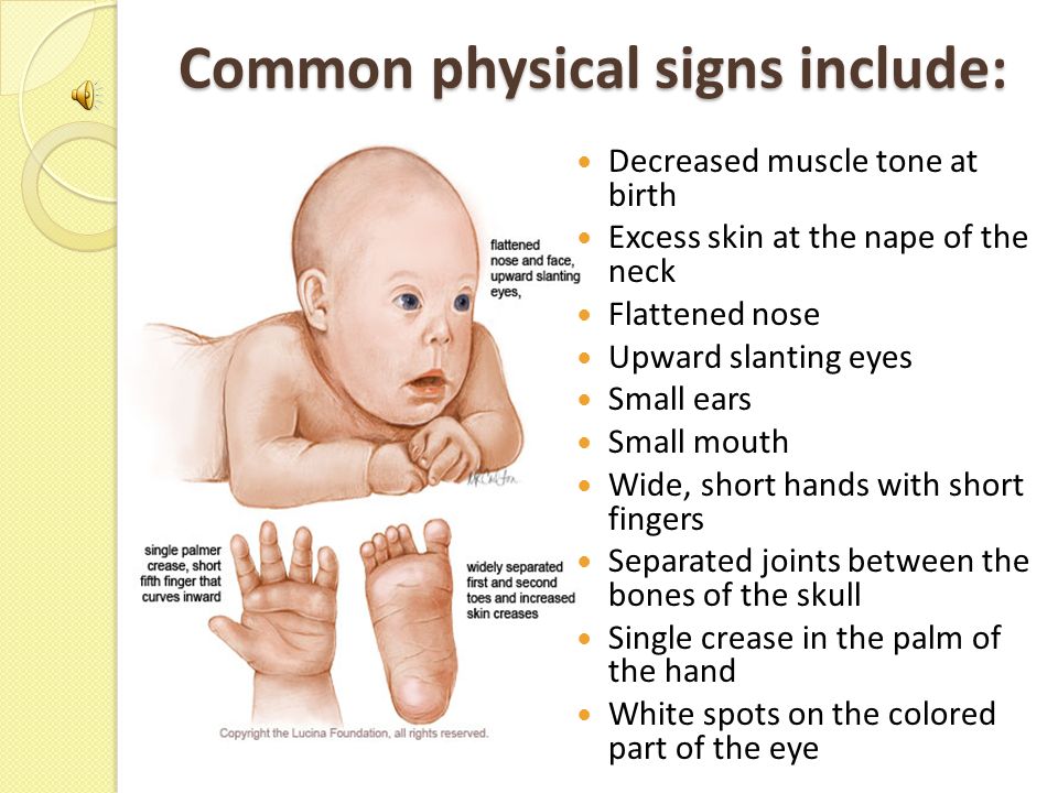 Of all the chromosomal abnormalities, this pathology is the most well-known for a wide range of people.
Down's syndrome is associated with many significant malformations. Such as, for example, congenital heart defects, duodenal atresia (a condition where part of the small intestine does not develop), an increased risk of developing acute leukemia (leukemia). About 50% of children with this condition are born with heart defects, the most common of which is a ventricular septal defect. Another disease that is often observed in such children is Hirschsprung's disease - an aganglionic area of the large intestine, leading to the development of intestinal obstruction. nine0003
Screening tests during pregnancy are used to assess the risk of having a baby with Down syndrome. These tests are painless and non-invasive (do not cause damage to the tissues of the pregnant woman and fetus), but do not accurately judge the presence or absence of congenital pathology. Nevertheless, their widespread use makes it possible to suspect risks and recommend invasive procedures.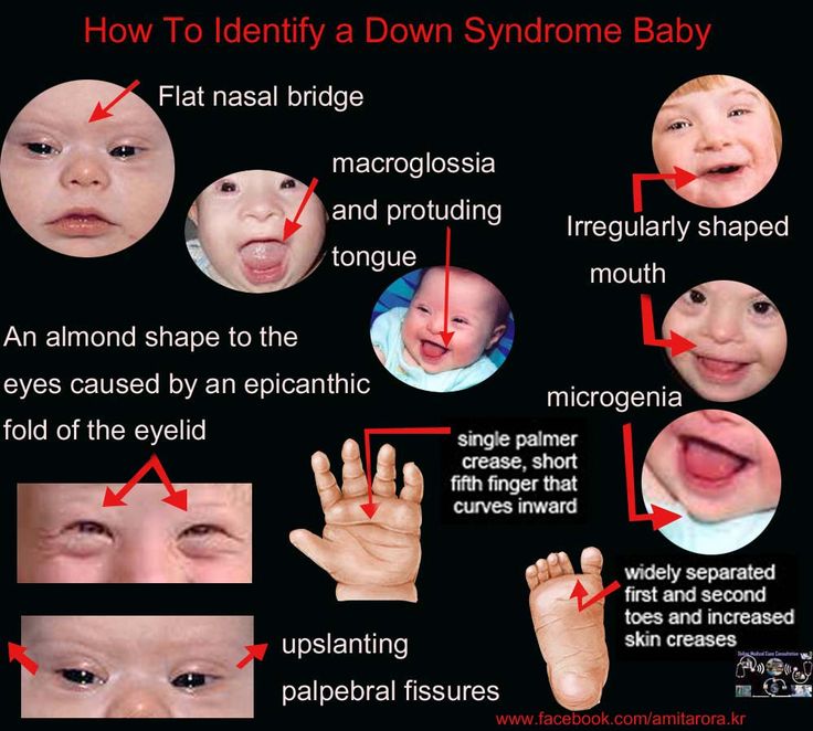 Invasive diagnostic methods include interventions performed under ultrasound guidance - amniocentesis, chordocentesis and chorionic villus sampling. nine0003
Fetal ultrasound is the main way to diagnose the likelihood of Down syndrome
Fetal ultrasound to detect developmental abnormalities is called ultrasound screening. Most often, ultrasound screening is combined with biochemical blood tests of a pregnant woman, and such a study is called combined screening. Depending on the choice of criteria, the probability of detecting Down syndrome in the second trimester is 60-91%. The use of color Doppler technology for ultrasound screening makes it possible to detect defects in the fetal heart and increases the accuracy of diagnosis. nine0003
Major ultrasound markers of Down syndrome
Ultrasound markers are the ultrasound measurements most likely to be associated with a high chance of having Down syndrome in the fetus. None of these markers is specific, that is, inherent only in this pathology.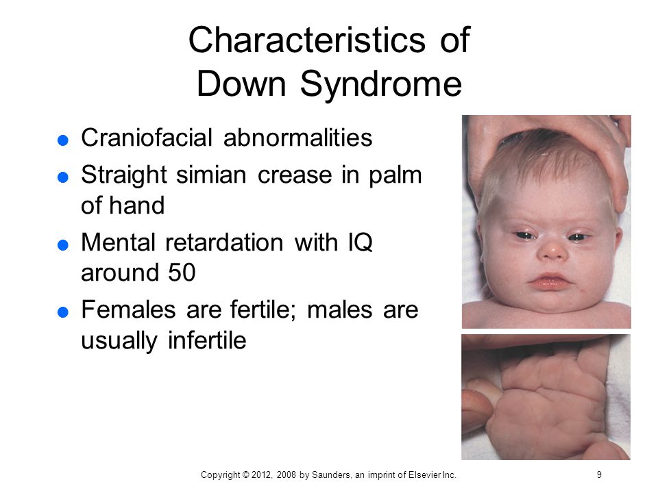 The following indicators are taken into account: the thickness of the collar space (75% of sensitivity), the absence of the nasal bone (58%), heart defects, short femurs and humeri, hyperechoic intestines, choroid plexus cysts, echogenic foci in the heart, signs of duodenal atresia . Of course, none of these markers is an absolute sign of a chromosomal abnormality. nine0003
Measuring the thickness of the nuchal space
The nuchal space (cervical fold) is the area between the folds of fetal tissues, which is transparent on ultrasound, located below the occiput in the area of neck formation. In children with chromosomal abnormalities, such as Down's syndrome or trisomy 18, fluid accumulates in this place. The measurement of this space is carried out from 11 to 14 weeks during the ultrasound of pregnancy in the first trimester. Measurements obtained at this time are the most significant for predicting the individual risk of developing fetal chromosomal abnormalities. The thickness of the collar space more than 3 mm is taken as an indicator of an increased likelihood of genetic defects in the fetus. The increase in the collar space does not mean that the fetus necessarily has Down syndrome, but if we take into account the gestational age of the fetus on which measurements were taken, the age of the mother and biochemical screening indicators, the probability of a correct diagnosis becomes higher than 80%. Although nuchal thickness screening is one of the most important tests for detecting chromosomal abnormalities, there are biases. The error arises due to violations of the measurement technique. This applies not only to measurements of the collar space, but also to the nasal bone. There are errors due to the human factor. The correct measurement technique and the experience of the ultrasound specialist are very important. nine0003
Measurements of the pelvis and structures of the fetal brain
In fetuses with trisomy 21, ultrasound may show cerebellar hypoplasia (reduction in size) and a decrease in the frontal lobe. These facts are used to assess the likelihood of Down syndrome. The combination of a shortened transverse size of the cerebellum and a decrease in the fronto-thalamic distance is of greater importance for the diagnosis of this chromosomal anomaly than measuring each distance of intracerebral structures separately. nine0003
In a fetus with Down's syndrome during ultrasound, the length of the iliac bones is clearly reduced and the angle between these bones is increased. This can be measured most clearly at the level of the middle of the sacrum in cross section.
Signs of Down's syndrome during ultrasound in the second trimester of pregnancy
When performing ultrasound of the fetus in the second trimester of pregnancy, such abnormalities can be detected as a violation of the formation of bones of the skeleton, expansion of the collar space by more than 5 mm, the presence of heart defects, expansion of the renal pelvis (pyeloectasia ), echogenicity of the intestine, cysts of the choroid plexus of the brain. Moreover, only heart defects, disorders of skeletal formation and expansion of the collar space are independent risk factors. nine0003
The use of only one of the measurements in assessing the probability of the presence of chromosomal abnormalities of the fetus, for example, determining the thickness of the collar space or measuring the iliac angle, is less accurate than taking into account the totality of measurements.
Another way to determine the risk of having a baby with Down syndrome
In addition to ultrasound signs, biochemical screening in the first and second trimester is used to determine the degree of risk. In this case, the blood of a pregnant woman is examined. The best results are obtained by combining ultrasonic and biochemical signs. If the combination of factors is threatening, a decision may be made to conduct invasive methods for diagnosing Down syndrome and other chromosomal abnormalities. Invasive techniques consist in obtaining particles of the skin or blood of the fetus, followed by genetic analysis. These methods are 100% reliable. At the same time, even complete well-being during screening in the first and second trimester using ultrasound and biochemical markers cannot guarantee the absence of chromosomal abnormalities in the fetus.