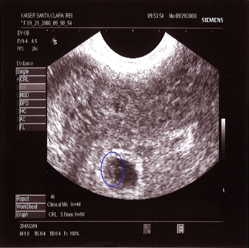
Transvag ultrasound at 4 weeks
Patience is key: Understanding the timing of early ultrasounds | Your Pregnancy Matters
×
What can we help you find?Refine your search: Find a Doctor Search Conditions & Treatments Find a Location
Appointment New Patient Appointment
or Call214-645-8300
MedBlog
Your Pregnancy Matters
November 20, 2018
Your Pregnancy Matters
Robyn Horsager-Boehrer, M. D. Obstetrics and Gynecology
The excitement newly pregnant women have to see how their baby is doing via an ultrasound can send them to an Ob/Gyn quickly. However, it’s important that they’re patient when their doctor recommends waiting until they are six weeks pregnant for their first ultrasound. Here’s why.
The first step women usually take after learning they’re pregnant is visiting with an Ob/Gyn. During this visit, an ultrasound is frequently done to confirm early pregnancy.
But an ultrasound doesn’t immediately show what women might expect. It’s typically not until a woman is six weeks pregnant that any part of the fetus is visible, which allows the doctor to determine whether a pregnancy will be viable.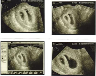
Because of this, it’s important that women understand what information their ultrasound can and cannot provide at certain times during their pregnancy. And they should be prepared to wait to learn more about their baby, if it’s too soon.
4 stages of early pregnancy and what we might see on ultrasound
An ultrasound is a routine part of prenatal care at six to nine weeks. The ultrasounds we might do prior to that, and the information those exams would reveal, generally occur in four stages:
- Stage One: If performed around the time a women’s menstrual period is expected, this ultrasound typically shows a fluffy, thick lining of the uterus that’s preparing for the fertilized egg to implant.
- Stage Two: This is usually at four to five weeks after a pregnant woman’s last period. The ultrasound commonly shows a small collection of fluid within the lining of the uterus that represents the early development of the gestational sac.

- Stage Three: This is usually about five and a half weeks after a pregnant woman’s last period. The ultrasound typically shows a gestational sac and within it we can see a 3-5 mm bubble-like structure, which is the yolk sac.
- Stage Four: Approximately six weeks after a pregnant woman’s last period, we can see a small fetal pole, one of the first stages of growth for an embryo, which develops alongside the yolk sac.
While these are the expected times to see the developing pregnancy with an ultrasound, not all pregnancies develop along the same timeline. Just like pregnancy tests, if there’s variability in the length of the menstrual cycle or when fertilization takes place, then what we see on ultrasound can change. It’s important that this ultrasound is performed vaginally for high-quality pictures.
If the first ultrasound doesn’t show a developing baby with a heartbeat, when should the next one be scheduled?
Newly pregnant women get anxious if we don’t see both a fetus and a heartbeat on the first ultrasound and frequently want to come back soon after for another look.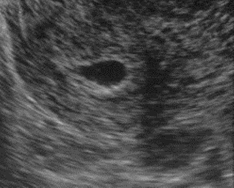 But it takes time to move through the early stages of pregnancy. And repeating an ultrasound still won’t be able to reassure the patient that the fetus is alive and growing, if we do it too soon.
The general recommendations are to wait two weeks if we only see a gestational sac and at least 11 days if a gestational and yolk sac are seen without a fetal pole. I prefer to wait two weeks for the next ultrasound in both of these scenarios. If we can see an early fetal pole but can’t see cardiac movement, then we repeat an ultrasound in one week. I know waiting is hard – but in my experience, it is much better to wait and get a definitive report on the status of your pregnancy than potentially have to come back multiple times.
Pregnancy loss can occur during any stage of pregnancy, though it’s most common in the first trimester. If we do an ultrasound and the length of the baby is more than 7mm, we should always see movement of the fetal heart. If we don’t, we know the pregnancy is not going to develop.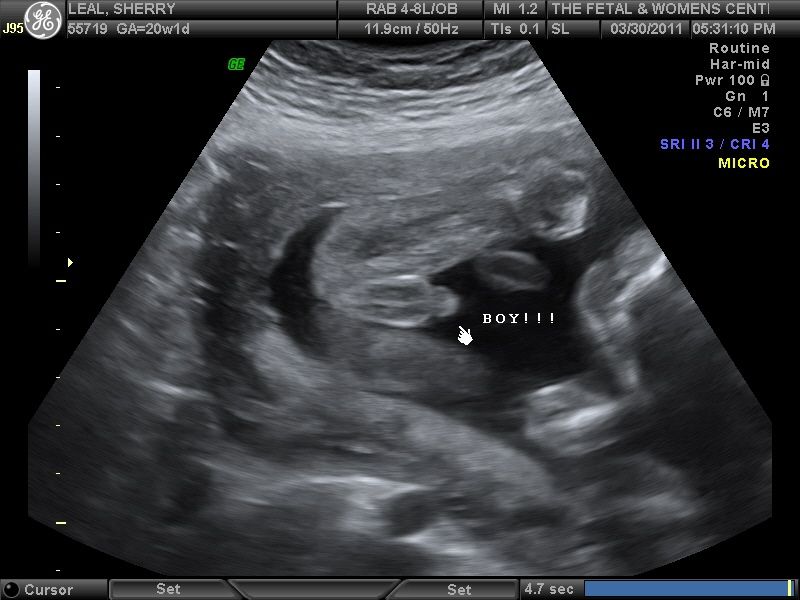
"It’s important that women understand the right time to visit their Ob/Gyn for an ultrasound, so they can know with more certainty what to expect during the exam."
– Robyn Horsager-Boehrer, M.D.
When might earlier ultrasounds be recommended?
If you have a history of tubal pregnancy, the most common being an ectopic pregnancy, or when the fertilized egg implants outside the uterus, or develop symptoms that are concerning for the possibility of a tubal pregnancy, your doctor might perform an ultrasound sooner. In this instance, the doctor is looking for any evidence of a pregnancy within the uterus, so a gestational sac with a yolk sac is reassuring and means that the chances of a tubal pregnancy are low. They also will likely want to take a close look at the fallopian tubes to see if there is any sign of a mass in the tube that might represent a growing tubal pregnancy.
Although it is hard to be patient, understanding the early timeline for a developing pregnancy will help you understand when first trimester ultrasounds should be performed.
Related reading: Helping you understand scary (but often harmless) pregnancy ultrasound findings
Stay on top of health care news. Subscribe to our blog today.
More in: Your Pregnancy Matters
Your Pregnancy Matters
- Robyn Horsager-Boehrer, M.D.
January 10, 2023
Your Pregnancy Matters
- Robyn Horsager-Boehrer, M.
 D.
D.
December 20, 2022
Your Pregnancy Matters
- Robyn Horsager-Boehrer, M.D.
December 13, 2022
Pediatrics; Your Pregnancy Matters
- Jessica Morse, M.
 D.
D.
December 6, 2022
Your Pregnancy Matters
- Shivani Patel, M.D.
November 22, 2022
Your Pregnancy Matters
- Robyn Horsager-Boehrer, M.D.
November 15, 2022
Your Pregnancy Matters
- Robyn Horsager-Boehrer, M.
 D.
D.
November 7, 2022
Mental Health; Your Pregnancy Matters
- Robyn Horsager-Boehrer, M.D.
October 11, 2022
Prevention; Your Pregnancy Matters
- Robyn Horsager-Boehrer, M.
 D.
D.
October 4, 2022
More Articles
© 2023 The University of Texas Southwestern Medical Center
Member of Southwestern Health Resources
Vaginal ultrasound | Pregnancy Birth and Baby
beginning of content3-minute read
Listen
A vaginal ultrasound is an ultrasound scan taken by a probe inserted into the vagina. It gives a clear picture of the fetus, cervix and placenta. It is also called an internal ultrasound or a transvaginal ultrasound.
What does a vaginal ultrasound test for?
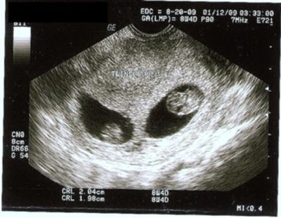
A vaginal ultrasound lets the doctor or ultrasound technologist (sonographer) see and measure the fetus. You might even discover if you are pregnant with twins or triplets.
It also allows the doctor or sonographer to look at your vagina, placenta, cervix, fallopian tubes, uterus and ovaries.
A vaginal ultrasound can be used as well as, or instead of, the standard abdominal ultrasound in which the probe is put on your belly.
Why is a vaginal ultrasound recommended?
A vaginal ultrasound will be recommended when your doctor or midwife wants a clearer diagnostic image than the one they can get through a standard abdominal ultrasound.
When is it used during pregnancy?
A vaginal ultrasound can confirm you are pregnant as it can detect the heartbeat very early in your pregnancy. It can also record the location and size of the fetus and determine if you are pregnant with 1 baby or more.
A vaginal ultrasound can also be used to diagnose problems or potential problems, including to:
- detect an ectopic pregnancy
- measure the cervix to determine the risk of a premature birth, which will allow any necessary interventions
- detect abnormalities in the placenta or cervix
- to determine the source of any bleeding
How is a vaginal ultrasound done?
A vaginal ultrasound is done by your doctor or sonographer in a hospital, clinic or consulting room. It uses a probe that is slightly wider than your finger.
You’ll probably be asked to empty your bladder (pee) before the ultrasound starts. If you’re using a tampon, you’ll need to take it out.
You’ll be asked to take off the lower half of your clothing and lie back on the examination table with your knees bent. There might be stirrups, or your hips might be slightly raised.
The probe is covered by a sheath, which is then covered in lubricating gel. It will be inserted slowly for about 5 to 8cm into your vagina. That usually doesn’t hurt, but you will feel pressure and it can be uncomfortable.
The probe will be moved around to get the best view of what is being examined. The examination usually takes 15 to 30 minutes.
If you are not comfortable having a male perform the examination, you can ask for a female sonographer. You can also ask for a female health worker to accompany you for support, or have a family member with you.
If you live in Western Australia, you must give written consent to a vaginal ultrasound.
When will I get the results?
Often you can see the ultrasound images on a monitor while you have your scan. If your specialist is there, they might discuss the results with you straight away.
If a specialist isn’t there, the sonographer is usually not allowed to discuss what they see with you. Your doctor or midwife will see the images after they have been processed. It usually takes a day or two to get the results.
Are there any risks involved?
Vaginal ultrasounds are safe for you and your baby, as long as your waters haven’t broken.
If you are allergic to latex, let the sonographer know so they can use a latex-free sheath on the probe.
There are no after-effects of the procedure, so you can get back to your normal activities, including driving yourself home if you wish.
Sources:
Australasian Society for Ultrasound in Medicine (Guidelines for the performance of first trimester ultrasound), Inside Radiology (Transvaginal ultrasound), Royal Australian and New Zealand College of Obstetricians and Gynaecologists (Screening in early pregnancy for adverse perinatal outcomes), The Royal Women's Hospital (Bleeding in early pregnancy)Learn more here about the development and quality assurance of healthdirect content.
Last reviewed: June 2020
Back To Top
Related pages
- Pregnancy checkups, screenings and scans
- Ultrasound scans during pregnancy
This information is for your general information and use only and is not intended to be used as medical advice and should not be used to diagnose, treat, cure or prevent any medical condition, nor should it be used for therapeutic purposes.
The information is not a substitute for independent professional advice and should not be used as an alternative to professional health care. If you have a particular medical problem, please consult a healthcare professional.
Except as permitted under the Copyright Act 1968, this publication or any part of it may not be reproduced, altered, adapted, stored and/or distributed in any form or by any means without the prior written permission of Healthdirect Australia.
Support this browser is being discontinued for Pregnancy, Birth and Baby
Support for this browser is being discontinued for this site
- Internet Explorer 11 and lower
We currently support Microsoft Edge, Chrome, Firefox and Safari. For more information, please visit the links below:
- Chrome by Google
- Firefox by Mozilla
- Microsoft Edge
- Safari by Apple
You are welcome to continue browsing this site with this browser. Some features, tools or interaction may not work correctly.
4th week fetal ultrasound
- Fetal development by week
- Our apparatus
- other types...
- Pregnancy management
- Fetometry data at various times
The cost of ultrasound in the first trimester up to 10 weeks is 450 UAH. The price includes biometrics according to protocols, 3D/4D visualization.
The cost of ultrasound in the first trimester for a period of 10 weeks 6 days is 550 hryvnia. The price includes prenatal screening, biometric protocols, 3D/4D visualization. nine0015
The cost of a comprehensive prenatal screening according to PRISCA (ultrasound + beta hCG + PAPP with the calculation of the individual risk of chromosomal pathologies (for example, Down syndrome or Edwards syndrome) - 1005 hryvnia.
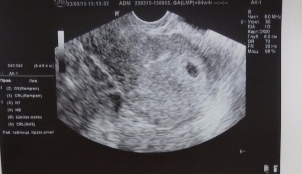
At ultrasound at 4 weeks of pregnancy you can see the presence of pregnancy in the uterine cavity. This is a small black circle a few millimeters in diameter called the fetal sac.In the uterus on ultrasound at 4 weeks of pregnancy, an expansion of the uterine vessels is noted.This is a normal vascular reaction associated with the need for more intensive nutrition of the growing blastocyst.The yellow body of pregnancy in the ovary continues to function actively, sending vital progesterone.Duplex scanning of the corpus luteum shows even more low-resistance blood flow. You may not yet suspect that you are pregnant, but your unborn child is going through big changes.The fertilized egg is now a rapidly growing ball of cells called the blastocyst.The blastocyst is getting deeper however, it grows into the wall of the uterus, the amniotic cavity begins to form, which will later become the environment for the intrauterine habitat of the fetus. The organization of the cells of the future placenta begins. The blastocyst will give rise to the tissues and organs of the baby and form the placenta. The embryo consists of three layers of cells - ectoderm (top layer), mesoderm (middle layer) and endoderm (inner layer). The ectoderm will form the baby's nervous system, skin, hair and nails, and eyes. The mesoderm will give rise to the development of the skeleton, connective tissue, hematopoietic system, heart, urogenital system, and most muscles. The endoderm will line the gastrointestinal tract and give rise to many internal organs (thyroid, liver, pancreas, etc.).
 From 4 weeks of pregnancy, it is especially important to avoid the use of drugs prohibited during pregnancy, if they are not prescribed for health reasons. With ultrasound at 4 weeks of gestation, it is impossible to reliably see the tissues of the embryo, but by the end of the fourth week of pregnancy at Ultrasound at 4 weeks of pregnancy a yolk sac is recorded - a ring of 2-3 mm. A week later, on this ring, you can see a tiny embryo that receives nutrients from the yolk sac.
The female body also undergoes changes. Some women practically do not feel them, some notice all possible manifestations at once.
Early symptoms of pregnancy: feeling of fluid retention, swelling (associated with an increased amount of the pregnancy hormone progesterone), mood swings, cramps, increased symptoms characteristic of premenstrual syndrome (PMS). Some women may note the presence of implantation bleeding. This is a scanty brown or pink discharge from the genital tract, which usually disappears on its own in 2-5 days.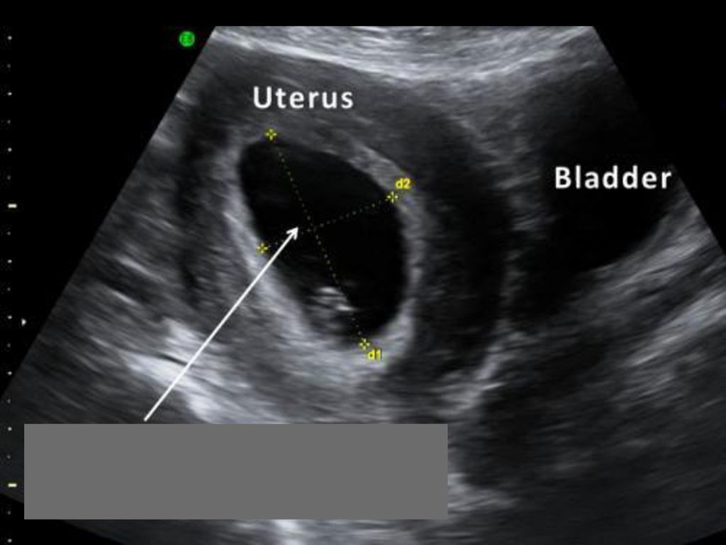 There is a heaviness in the lower abdomen, a feeling that menstruation is about to begin, but it does not begin. The breasts become more sensitive, the breasts may increase in size. nine0015
Ultrasound at 4 weeks of gestation can almost reliably determine the localization of the fetal sac and exclude ectopic pregnancy. Transvaginal ultrasonography at 4 weeks of gestation is preferred over transabdominal ultrasonography because transvaginal ultrasonography has a higher diagnostic accuracy in evaluating the uterine cavity. In the absence of a fetal egg in the uterine cavity with an ultrasound of the uterus and positive pregnancy tests, either a second ultrasound is performed after a week, and / or a blood test is done for beta-hCG (human chorionic gonadotropin) in dynamics to exclude an ectopic or non-developing pregnancy. nine0015
read more: 5th week of pregnancy
- Hydrotubation (echohydrotubation): study of the patency of the fallopian tubes (ultrasonic hysterosalpingoscopy)
- Transvaginal
- Ovaries
- Uterus
- Mammary glands
Female ultrasound
- Duplex Scan
- Cerebral vessels
- Neck vessels (duplex angioscanning of the main arteries of the head)
- Veins of the lower extremities
Vascular ultrasound
- Transrectal (trusion): prostate
- Scrotum (testicles) nine0003 Vessels of the penis
Male ultrasound
- Appendicitis
- Abdomen
- Gallbladder
- Stomach
- Intestines
- Bladder
- Soft tissue
- Pancreas
- Liver
- Kidney
- Joints
- Thyroid
- Echocardiography (ultrasound of the heart)
Organ ultrasound
- Varicose veins: ultrasound diagnosis of varicose veins
- Hypertension: Ultrasound diagnosis of hypertension
- Thrombosis: ultrasound diagnosis of vein thrombosis
- Ultrasound diagnosis of chronic pancreatitis
- for kidney stones
- for cholecystitis
Ultrasound diagnostics of diseases
- Hip joints in newborns (if hip dysplasia is suspected)
Pediatric ultrasound
Early pregnancy ultrasound
Two stripes on the test is always a complete surprise for a woman, even if she already knows about her position in her soul.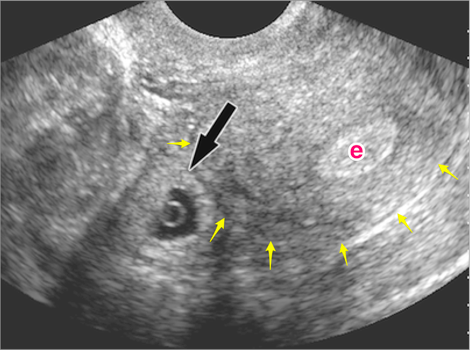 After the first confirmation, the girl most often wants to see the baby with her own eyes and make sure that he is developing normally. This can be done using an instrumental method such as ultrasound. Usually, expectant mothers, especially expecting their first child, have a lot of questions.
How to watch out for ectopic and missed pregnancies
Often, pregnant women at the very beginning are afraid of two conditions - an ectopic or missed pregnancy. Especially experienced by those who have already experienced it once.
In an ectopic pregnancy, the fertilized egg does not reach the uterus and is implanted in other places - most often these are the fallopian tubes. If nothing is done, the embryo will grow and burst the tube. In this case, the woman can not be saved. Therefore, an ectopic pregnancy should be terminated without hesitation. Since the embryo is not removed separately, the entire tube is cut out. This can be done twice in a lifetime, after which pregnancy occurs only after artificial replanting using IVF. An ectopic pregnancy can be suspected indirectly by the level of β-hCG, but only the presence of a fetal egg in the uterus on ultrasound clearly refutes it. nine0015
But the experiences of the expectant mother do not end there. Frozen pregnancy is impossible to predict. In some cases, doctors shrug their shoulders, and why this happened remains unknown. It is dangerous to wait for a non-developing egg to come out by itself for a long time, since it can begin to decompose, and the uterus can become inflamed. Up to 6 weeks, the gynecologist can offer a less traumatic medical interruption, and after - classic abortive methods. It is possible to unambiguously identify a frozen one only by ultrasound - it shows that the fetal egg is smaller than it should be for its term or a heartbeat is not heard. At random, such a diagnosis is never made, because the life of the unborn child depends on it, and most often an ultrasound is prescribed again after a few days. If it turns out that the dimensions have not changed, and there is still no heartbeat, then - alas! nine0015
Why you need early ultrasound
Ultrasound diagnostics is a safe method that has no contraindications. But it is pointless to do it before 4-5 weeks, the fetal egg cannot be seen so early. In this case, a transvaginal probe is used for ultrasound. In public clinics, the first ultrasound diagnosis is carried out as part of the first screening at 12 weeks, and earlier it is prescribed only if the woman herself or her doctor is worried about something:
- a woman complains of pain in the lower part of the abdominal region and bloody “daub”;
- expectant mother has serious chronic diseases;
- pregnancy occurred, although the couple was protected by a spiral; 90,003 patients took dangerous and heavy drugs, had an infection, and were irradiated. Although the principle of “all or nothing” usually works up to 4 weeks, doctors recommend being examined;
- in the anamnesis, the future mother herself and her relatives had multiple pregnancies; nine0004
- the embryo was transplanted using IVF;
- physician suspects hydatidiform mole, ectopic or miscarriage;
- the patient was diagnosed with various formations in the uterus and ovaries;
- if there is a risk of developing genetic abnormalities (for example, there have already been interrupted pregnancies due to malformations, chromosomal disorders).
