Prenatal exposure to radiation is most dangerous during
Radiation and Pregnancy: Information for Clinicians
This overview provides physicians with information about prenatal radiation exposure as an aid in counseling pregnant women.
How to use this document
This information is for clinicians. If you are a patient, we strongly advise that you consult with your physician to interpret the information provided, as it may not apply to you. Information on radiation exposure during pregnancy for members of the public can be found on the Health Information for Specific Groups webpage.
CDC recognizes that providing information and advice about radiation to expectant mothers falls into the broader context of preventive healthcare counseling during prenatal care. In this setting, the purpose of the communication is always to promote health and long-term quality of life for the mother and child.
This page is also available as a PDF pdf icon[365 KB]
Radiation exposure to a fetus
Most of the ways a pregnant woman may be exposed to radiation, such as from a diagnostic medical exam or an occupational exposure within regulatory limits, are not likely to cause health effects for a fetus. However, accidental or intentional exposure above regulatory limits may be cause for concern.
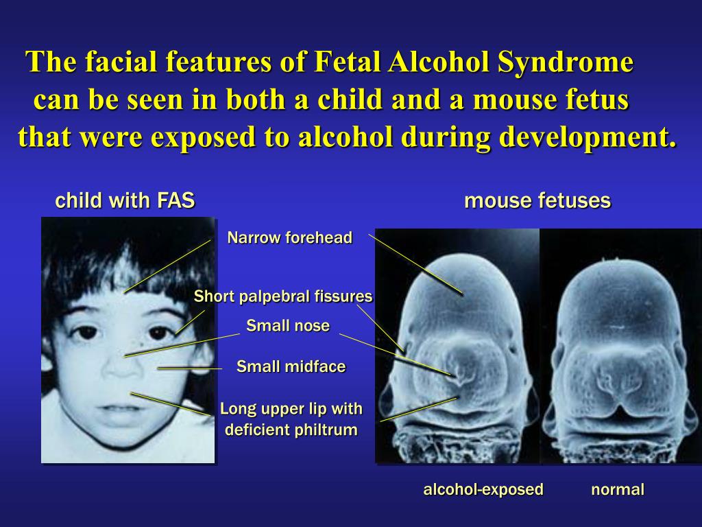
Although radiation doses to a fetus tend to be lower than the dose to the mother, due to protection from the uterus and surrounding tissues, the human embryo and fetus are sensitive to ionizing radiation at doses greater than 0.1 gray (Gy). Depending on the stage of fetal development, the health consequences of exposure at doses greater than 0.5 Gy can be severe, even if such a dose is too low to cause an immediate effect for the mother. The health consequences can include growth restriction, malformations, impaired brain function, and cancer.
Estimating the Radiation Dose to the Embryo or Fetus
Health effects to a fetus from radiation exposure depend largely on the radiation dose. Estimating the radiation dose to the fetus requires consideration of all sources external and internal to the mother’s body, including the following:
- Dose from an external source of radiation to the mother’s abdomen.
- Dose from inhaling or ingesting a radioactive substance that enters the bloodstream and that may through the placenta.
- Dose from radioactive substances that may concentrate in maternal tissues surrounding the uterus, such as the bladder, and that could irradiate the fetus.
Most radioactive substances that reach the mother’s blood can be detected in the fetus’ blood. The concentration of the substance depends on its specific properties and the stage of fetal development. A few substances needed for fetal growth and development (such as iodine) can concentrate more in the fetus than in corresponding maternal tissue.
Consideration of the dose to specific fetal organs is important for substances that can localize in specific organs and tissues in the fetus, such as iodine-131 or iodine-123 in the thyroid, iron-59 in the liver, gallium-67 in the spleen, and strontium-90 and yttrium-90 in the skeleton.
Radiation experts can assist in estimating the radiation dose to the embryo or fetus
Hospital medical physicists and health physicists are good resources for expertise in estimating the radiation dose to the fetus.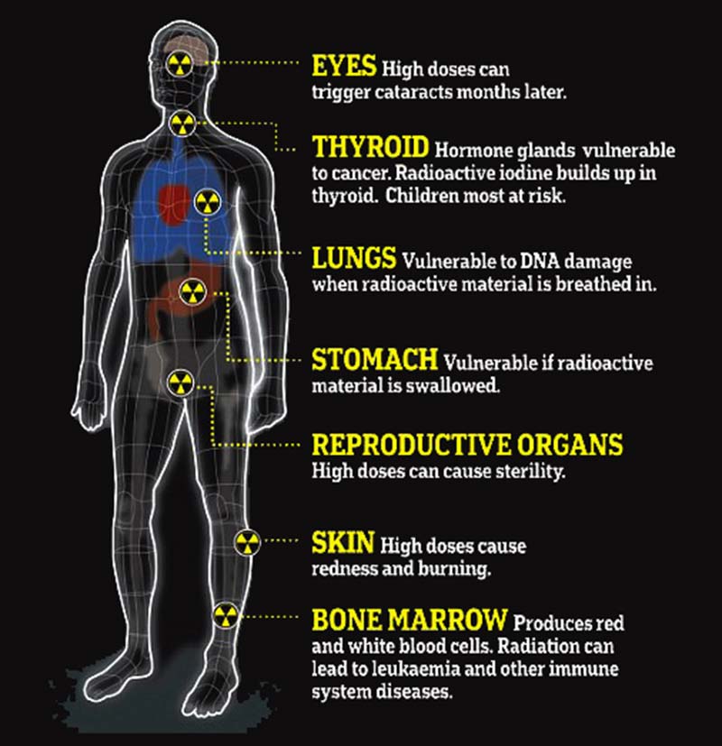 In addition to the hospital or clinic’s specialized staff, physicians may access resources from or contact the following organizations for assistance in estimating fetal radiation dose.
- The National Council on Radiation Protection and Measurementsexternal icon’ Report No. 174, “Preconception and Prenatal Radiation Exposure: Health Effects and Protective Guidance” [NCRP2013] provides detailed information for assessing fetal doses from internal uptakes.
- The International Commission on Radiological Protection’s “Publication 84: Pregnancy and Medical Radiation”external icon [ICRP2000] provides fetal dose estimations from medical exposures to pregnant women.
- The Conference of Radiation Control Program Directorsexternal icon maintains a list of state Radiation Control/Radiation Protection program contact information.
- The Health Physics Societyexternal icon maintains a list of active certified Health Physicists.
- The American Association of Physicists in Medicineexternal icon provides information resources.
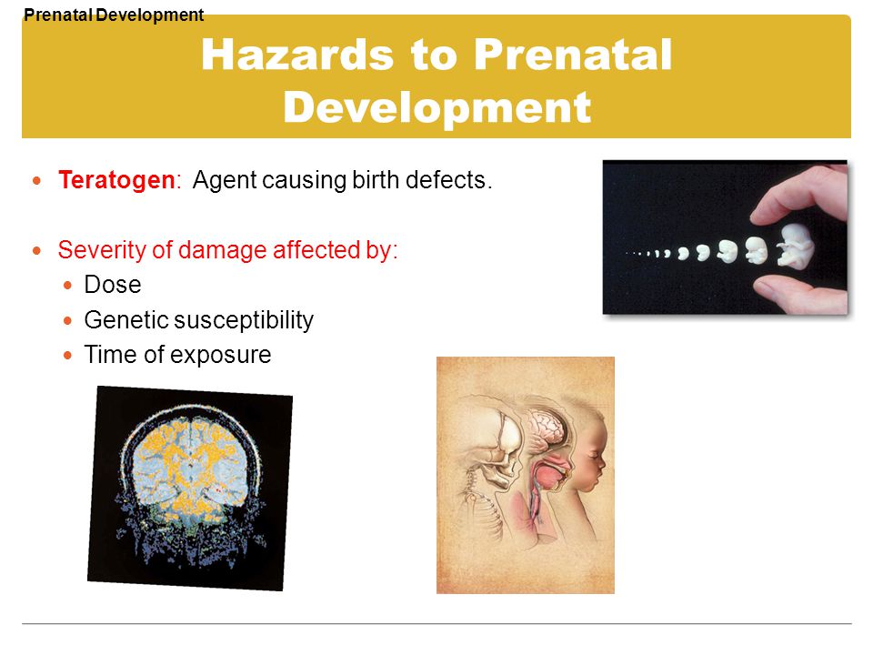
Once the fetal radiation dose is estimated, potential health effects can be assessed.
Potential Health Effects of Prenatal Radiation Exposure (Other Than Cancer)
Table 1 summarizes the potential non-cancer health risks of concern. This table is intended to help physicians advise pregnant women who may have been exposed to radiation, not as a definitive recommendation. The indicated doses and times post-conception are approximations.
| Acute Radiation Dose* to the Embryo/Fetus | Time Post Conception (up to 2 weeks) | Time Post Conception (3rd to 5th weeks) | Time Post Conception (6th to 13th weeks) | Time Post Conception (14th to 23rd weeks) | Time Post Conception (24th week to term) |
|---|---|---|---|---|---|
<0.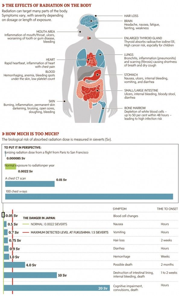 10 Gy 10 Gy (10 rads) | Noncancer health effects NOT detectable | ||||
| 0.10–0.50 Gy (10–50 rads) | Failure to implant may increase slightly, but surviving embryos will probably have no significant (non-cancer) health effects. | Growth restriction possible | Growth restriction possible | Noncancer health effects unlikely | |
| > 0.50 Gy (50 rads) The expectant mother may be experiencing acute radiation syndrome in this range, depending on her whole-body dose. | Failure to implant will likely be high, depending on dose, but surviving embryos will probably have no significant (non- cancer) health effects. | Probability of miscarriage may increase, depending on dose. Probability of major malformations, such as neurological and motor deficiencies, increases. Growth restriction is likely | Probability of miscarriage may increase, depending on dose. Growth restriction is likely. | Probability of miscarriage may increase, depending on dose. Growth restriction is possible, depending on dose. (Less likely than during the 6th to 13th weeks post conception) Probability of major malformations may increase | Miscarriage and neonatal death may occur, depending on dose. § |
| 8th to 25th Weeks Post Conception: The most vulnerable period for intellectual disability is 8th to 15th weeks post conception Severe intellectual disability is possible during this period at doses > 0.5 Gy Prevalence of intellectual disability (IQ<70) is 40% after an exposure of 1 Gy from 8th to 15th week Prevalence of intellectual disability (IQ<70) is 15% after an exposure of 1 Gy from 16th to 25th week Table adapted from Table 1.1. of the National Council on Radiation Protection and Measurements’ Report No. | |||||
Gestational age and radiation dose are important determinants of potential non-cancer health effects. The following points are of particular note.
- During the first 2 weeks post-conception, the health effect of concern from an exposure of ≥ 0.1 Gy is the possibility of death of the embryo. Because the embryo is made up of only a few cells, damage to one cell, the progenitor of many other cells, may cause the death of the embryo, and the blastocyst may fail to implant in the uterus. Embryos that survive, however, are unlikely to exhibit congenital abnormalities or other non-cancer health effects, no matter what dose of radiation they received.
- In all stages post-conception, radiation-induced non-cancer health effects are not detectable for fetal doses below about 0.10 Gy.
Carcinogenic Effects of Prenatal Radiation Exposure
Radiation exposure to an embryo/fetus may increase the risk of cancer in the offspring, especially at radiation doses > 0.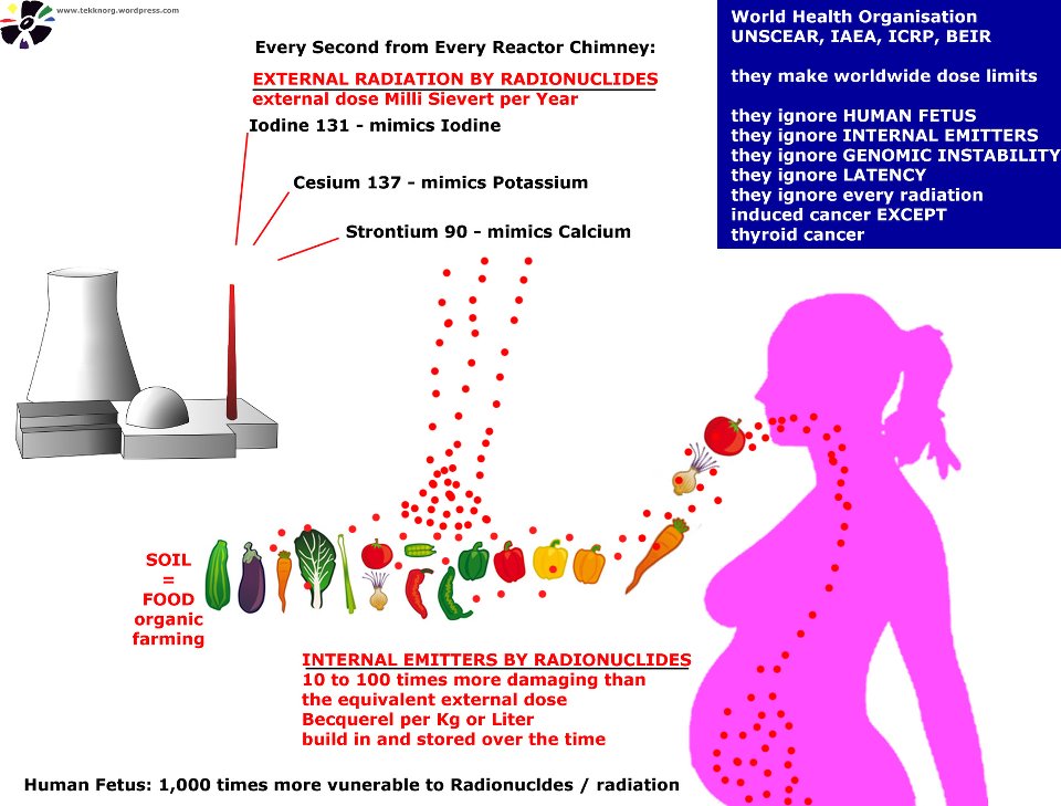 1 Gy, which are well above typical doses received in diagnostic radiology. However, attempting to quantify cancer risks from prenatal radiation exposure presents many challenges. These challenges include the following:
- The primary data for the risk of developing cancer from prenatal exposure to radiation come from the lifespan study of the Japanese atomic bomb (A-bomb) survivors. [Preston et al. 2008]. The analysis of that cohort includes cancer incidence data only up to the age of 50 years. This precludes making lifespan risk estimates as a result of prenatal radiation exposure.
- From the Japanese lifespan study [Preston et al. 2008], it can be concluded that for those exposed
in early childhood (birth to age 5 years), the theoretical risk of an adult-onset cancer by age 50 is approximately ten-fold greater than the risk for those who received prenatal exposure. Therefore, the risk following prenatal exposure may be considerably lower than for radiation exposure in early childhood
[NCRP2013].
- No reliable epidemiological data are available from studies to determine which stage of pregnancy is the most sensitive for radiation-induced cancer in the offspring [NCRP2013].
The lifespan study of the Japanese A-bomb survivors is continuing as the cohort ages. Future analyses of the accumulating data should provide a better understanding of the lifetime risk of cancer from prenatal and early childhood radiation exposure.
References
ICRP2000] International Commission on Radiological Protection. 2000. Valentin, J. (2000). Pregnancy and medical radiation. Oxford: Published for the International Commission on Radiological Protection.
[NCRP2013] National Council on Radiation Protection and Measurements. 2013. Preconception and Prenatal Radiation Exposure Health Effects and Protective Guidance. (2013). Bethesda: National Council on Radiation Protection & Measurements.
Preston DL, Cullings H, Suyama A, Funamoto S, Nishi N, Soda M, Mabuchi K, Kodama K, Kasagi F, Shore RE.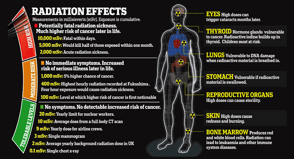 2008. Solid cancer incidence in atomic bomb survivors exposed in utero or as young children. J Natl Cancer Inst 100(6):428-436.
[UNSCEAR2013] United Nations Scientific Committee on the Effects of Atomic Radiation. 2013. Sources, effects and risks of ionizing radiation. Vol. II, Scientific Annex B: Effects of radiation exposure of children.
For more information on medical management and other topics on radiation emergencies:
Radiation Emergency Assistance Center/Training Siteexternal icon (REAC/TS) is a program uniquely qualified to teach medical personnel, health physicists, first responders and occupational health professionals about radiation emergency medical response.
Radiation Emergency Medical Management (REMM)external icon provides guidance to health care providers (primarily physicians) about clinical diagnosis and treatment of radiation injury during radiological and nuclear emergencies.
Conference of Radiation Control Program Directorsexternal icon
Health Physics Societyexternal icon
International Commission on Radiological Protectionexternal icon
National Council on Radiation Protection and Measurementsexternal icon
American Association of Physicists in Medicineexternal icon
Radiation Exposure In Pregnancy - StatPearls
Ilsup Yoon; Todd L. Slesinger.
Author Information
Last Update: May 8, 2022.
Introduction
Choosing the most appropriate imaging modality for pregnancy patients is a common clinical question encountered daily. The general principle for imaging during pregnancy is similar to imaging for the general population, with the goal of radiation exposure being as low as reasonably achievable (ALARA). What is unique during pregnancy is that fetus radiation exposure is an essential factor in deciding optimal imaging studies. Understanding of consequences of radiation exposure on a fetus, degrees of fetal radiation exposure by each imaging modality, and techniques on reducing fetal radiation exposure is vital in choosing the best diagnostic imaging modality. While it is crucial to minimize fetal radiation exposure as much as possible, it is essential to remember that diagnostic studies should not be avoided for fear of radiation exposure, especially when these studies can dramatically change patient management. This activity will discuss the consequences of radiation exposure on a fetus, degrees of radiation exposure by each modality, and techniques of reducing fetal radiation exposure. Solid understanding of how each imaging modality contributes to fetal radiation dose will significantly help in choosing the most appropriate imaging study that provides the best diagnostic information at the lowest level of radiation exposure.
The consequence of radiation exposure in fetuses is mostly based on observations rather than based on scientific research. Ethical issues prohibit researching on the fetus. Therefore, most of the data on the impact of radiation on the fetus derives from observations of patients who suffered Japan’s Hiroshima bombing and the Chernobyl nuclear power plant disaster.[1][2] Based on the observations made from the victims of the high level of radiation exposure, the consequences of radiation exposure can categorize into four broad groups, including pregnancy loss, malformation, developmental delay or retardation, and carcinogenesis.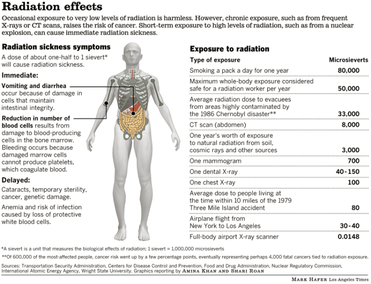 [1] Pregnancy loss most often happens when radiation exposure happens during early gestation (less than two weeks).[2] Malformations of body parts and developmental delays occur during the organogenesis period (2 weeks to 8 weeks) and are dependent on the radiation dose.[2] Below the threshold level of radiation exposure, there is minimal disruption of organogenesis. Above the threshold, the degree of malformation is related to the dose of the radiation. Lastly, carcinogenesis is considered a stochastic effect. In other words, cancer can develop at any level of radiation exposure. However, the probability of developing cancer increases with the increase in the dose of radiation.
In the United States, the background radiation exposure for the whole body per year is estimated to be 3.1 mSv (310 mrem). United States Nuclear Regulation Commission (USNRC) also recommends total fetus exposure during pregnancy to be less than 5.0 mSv (500 mrem). The fetus radiation dose below 50 mGy is considered safe and not cause any harm.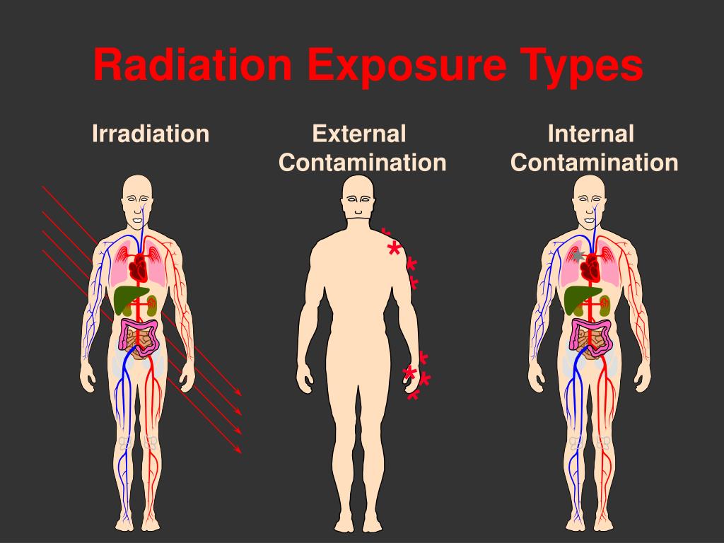 According to the Center for Disease Control (CDC), radiation dose between 50 mGy to 100 mGy is regarded inconclusive in terms of impact on the fetus. Doses above 100 mGy, especially doses above 150 mGy, are viewed as the minimum amount of dosage at which negative fetal consequences will occur, based on observation. The majority of the diagnostic studies performed during the pregnancy are below the threshold level.
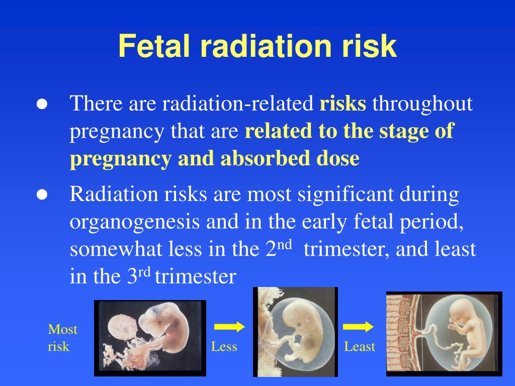
The effect of radiation exposure during pregnancy also depends on the gestational age of the fetus. The embryo/fetus is most susceptible to radiation during organogenesis (2 to 7 weeks gestational age) and in the first trimester. The fetus is more resistant to the radiation during the second and third trimester. Dose between 0.05 to 0.5 Gy is generally considered safe for the fetus during the second and third trimester while it is considered potentially harmful during the 1st-trimester fetus. Even though the fetus is more resistant to the radiation during the second and third trimester, a high dose of radiation (greater than 0. 5 Gy or 50 rad) may result in adverse effects including miscarriage, growth reduction, IQ reduction, and severe mental retardation. Therefore, clinicians and radiologists should counsel the pregnant patient regardless of the gestational age.[3]
Occupational radiation exposure for a pregnant employee should be monitored to make sure the total amount of radiation exposure is under the regulatory limit. According to the National Council of Radiation Protection and Measurement (NCRP), the total dose equivalent to the embryo/fetus should not exceed 500 mRem during the length of the pregnancy. It should not exceed 50mRem in any month during pregnancy.[4]
The practice decision on selecting the most appropriate imaging modality for pregnancy should have their basis in the expert opinions of the treating clinician. Nonetheless, the American College of Radiology does provide recommendations on the appropriateness of imaging modalities in accessing common clinical conditions. Each imaging modality is categories into usually appropriate, may be appropriate, and usually not appropriate. For instance, for a pregnant patient presenting with right lower abdominal pain concerning for appendicitis, ultrasound and MRI are usually appropriate imaging modality. CT abdomen and pelvis with or without contrast is categorized as may be appropriate. Abdominal radiograph, Tc-99m WBC scan, and fluoroscopy contrast enema are considered usually not appropriate. The American College of Radiology appropriateness criteria provides the current practice policies and guidelines in the US regarding imaging in pregnant patients.
Anatomy
Understanding the anatomic location of the uterus is essential in understanding why a particular type of study contributes to a higher radiation dose. The uterus is located within the female pelvis. Therefore, studies that are further away from the pelvis contribute to less radiation dose than the studies that aim at the pelvis. Additionally, the uterus is located superior and anterior aspect of the pelvis during the pregnancy. X-ray beams that project in a posterior to anterior (PA) direction contribute to less radiation than the beam projected in anterior to posterior (AP) direction because, in PA projection, the X-ray gets attenuated before reaching anteriorly located uterus.
Plain Films
A single plain radiograph does not contribute to a significant radiation dose to the fetus. The estimated radiation dose to the fetus varies and ranges from 0.001 mGy to 10 mGy depending on the type of the study.[5] The highest radiation dose is the lumbar spine radiograph, which has a maximal fetal radiation dose of 10 mGy.[5] Nonetheless, even fetus radiation exposure for the lumbar spine radiograph is significantly lower than the threshold limit of the safe radiation exposure dose of 50 mGy, radiation level that is considered safe and without significant harm.
Regardless of the radiation dose, it is vital to reduce radiation exposure to the fetus as much as possible. Plain films are obtained more frequently than computed tomography (CT), and radiation doses from multiple plains films can easily accumulate. Therefore, it is essential to employee dose reducing techniques as often as possible. When performing plain film examination of non-pelvis structure, pelvic lead apron should always be utilized to reduced unnecessary fetus radiation.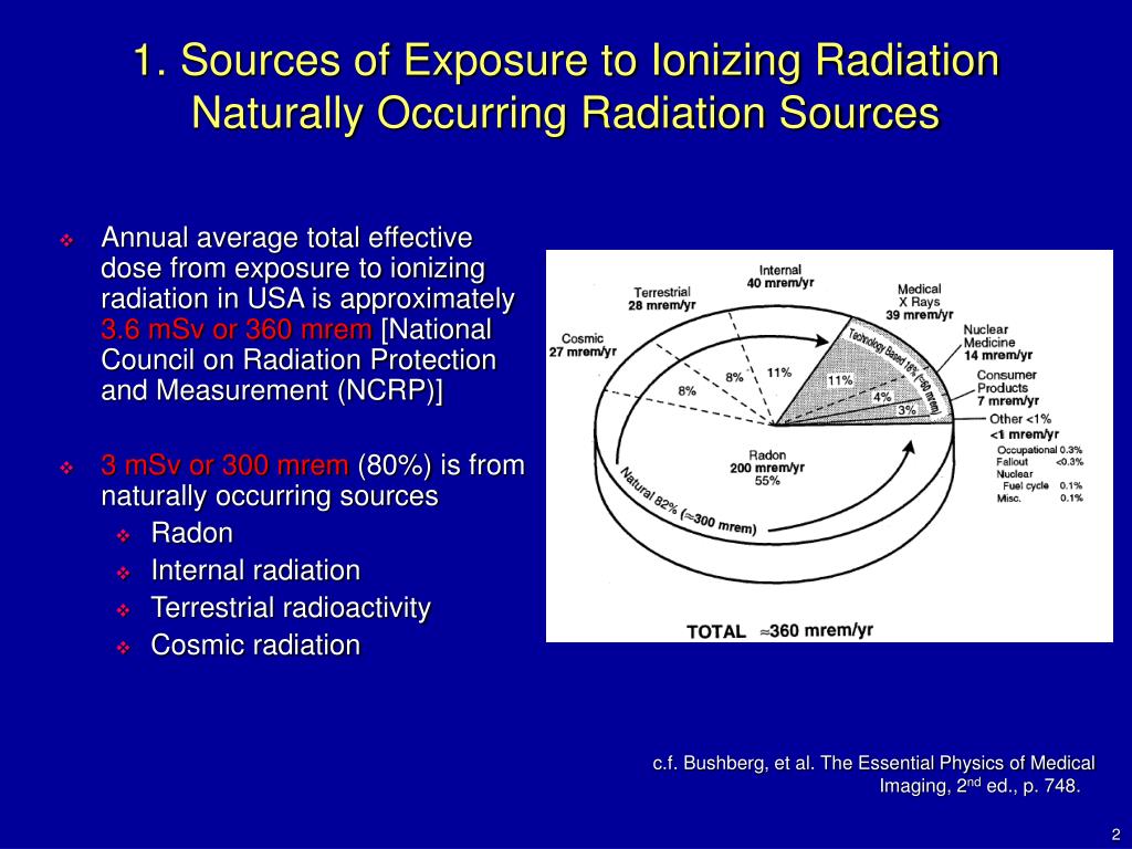 PA projection of the pelvis also contributes to the lower dose than the AP projection. Technologists should optimally position patients before taking a radiograph to reduce the number of repeat examinations from unsatisfactory views. Lastly, clinicians should obtain radiographs only when it is helping clinical management.
Computed Tomography
On the other hand, CT contributes to a significant amount of fetal radiation. The amount of fetus radiation exposure again varies by the type of study with CT pelvis contributing the highest amount of fetal radiation of 50 mGy.[5] This dose is right at the limit above which there is a documented negative impact to the fetus.
Because CT contributes to much higher fetal radiation, it is essential always to be considerate of other options when contemplating utilizing CT on pregnancy patients. Other imaging modalities, including MRI, plain radiograph, ultrasound, and nuclear medicine studies, should be considered first before performing CT.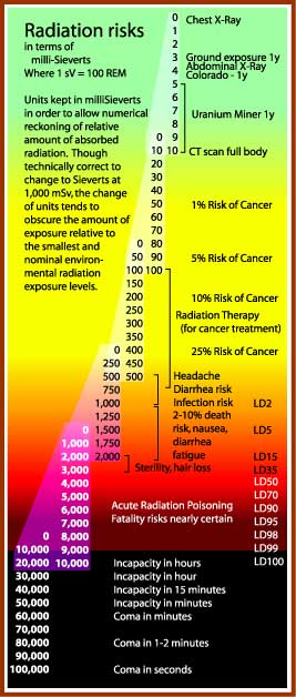 Common clinical presentations like appendicitis first require evaluation with MRI instead of CT.[6] Right upper quadrant ultrasound should be utilized if there is a clinical concern for cholecystitis.[7] Renal ultrasound should merit consideration before CT for nephrolithiasis and collecting system obstruction. If CT is the first indicate study of choice as in traumatic pregnancy patients to evaluate for intra-abdominal trauma, it is crucial to optimize CT setting to minimize the dose. Using wide pitch and narrow collimation can reduce the radiation dose. Besides, CT protocols should be optimized to minimize unnecessary radiation exposure. Clinicians should only perform additional delayed imaging when there is a clinical indication. Unnecessary multiphasic protocols should get simplified into a single-phase protocol.
When obtaining CT images of body parts outside of the abdomen and pelvis, the scattered radiation exposure to the fetus is minimal. Therefore, a shield does not significantly reduce the radiation exposure on the fetus during the CT scan.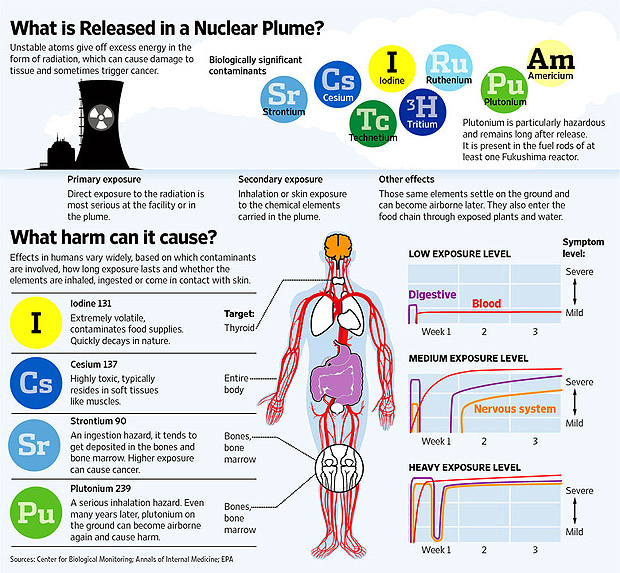 A lead shield may be an unnecessary precaution. However, it does minimally reduce the dose from scatter radiation and may provide patients a sense of reassurance and protection. The use of a lead shield is up to the discretion of the institution and provider.[8]
Magnetic Resonance
MRI uses a magnetic field to generate diagnostic images and does not contribute to ionizing radiation.
Ultrasonography
Ultrasound uses sound waves to generate diagnostic images and does not contribute to ionizing radiation.
Nuclear Medicine
In nuclear medicine, radiopharmaceuticals are injected into patients. These radiopharmaceuticals are distributed throughout the body and emit radiation at the target location. Radiation energies from these radiotracers then convert into diagnostic images. The overall radiation exposure to the fetus depends on how much radiotracer gets delivered to or near the fetus. Fetal radiation exposure in nuclear medicine depends on multiple variables, including maternal excretion and uptake of the radiopharmaceutical, fetal distribution of the radiopharmaceutical, placental permeability of the radiopharmaceutical, tissue affinity of the radiopharmaceutical, the half-life of the radiotracer, the dose of the radiotracer, and type of radiation emitted from the radiotracer.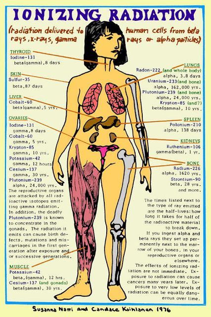 Generally, for nuclear medicine studies utilizing a radiopharmaceutical that gets excreted through the kidneys, patients are encouraged to hydrate and urinate to maximize the urinary excretion of radiopharmaceuticals.
Specific clinical scenarios worth mentioning for nuclear medicine study is pregnancy patient with concern for pulmonary embolism. The first imaging modality should be an ultrasound of lower extremity to look for deep venous thrombosis. If there is still clinical suspicion for pulmonary embolism, CT pulmonary angiogram (CTPA) is preferable to the ventilation-perfusion (VQ) scan. The fetal dose of VQ is much higher than CTPA, even though the maternal dose is much lower. Due to the lower fetal dose, CTPA is the preferred test of choice.[9]
The thyroid iodine scan is not an option during pregnancy. Iodine 121 and 131 are taken up by fetus thyroid and therefore contraindicated.[10]
Angiography
Fetal radiation exposure from angiography and fluoroscopy should only be for emergent clinical settings.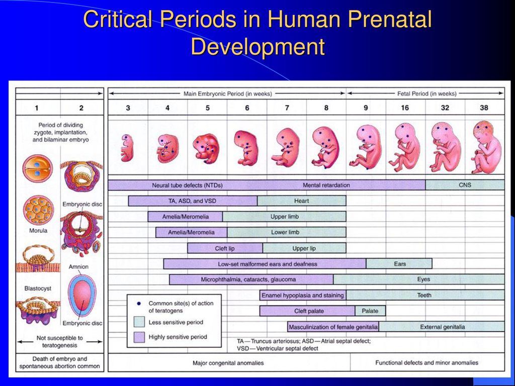 A physician performing fluoroscopy should utilize essential dose reducing techniques including pulse fluoroscopy instead of continuous fluoroscopy, last image hold rather than full exposure, and colimitation to appropriate field of view. Magnification increases the radiation dose and should be used only if necessary.
Patient Positioning
Appropriate patient positioning is vital in producing a diagnostic quality of images and prevent repeat examination that increases fetal radiation. Technologists should optimally position patients before imaging to obtain appropriate views for the exam. Obtaining diagnostic imaging at the first attempt eliminates the need for a repeat examination and significantly reduces unnecessary radiation exposure to the fetus.
Clinical Significance
The plain film, CT, nuclear medicine studies, and fluoroscopy uses ionizing radiation to obtain diagnostic images. A high level of radiation has adverse effects on the fetus; therefore, referring clinicians should consider alternative imaging modalities for pregnancy patients.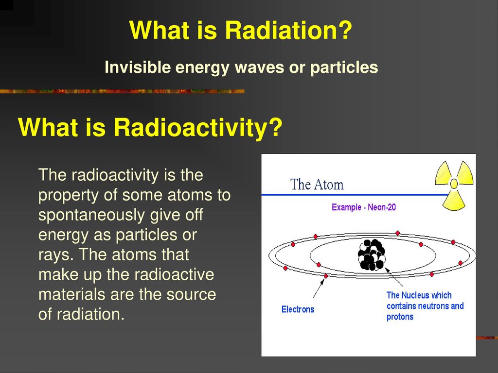 If diagnostic studies that expose radiation to the fetus are clinically required, it should be performed without delay but must take place in a way that minimizes radiation exposure to the fetus. Solid understanding of how each imaging modality contributes to fetal radiation exposure, techniques to reducing radiation exposure in the fetus, and adverse consequences of high radiation exposure is critical in providing the most appropriate imaging studies for pregnant patients.
Review Questions
Access free multiple choice questions on this topic.
Comment on this article.
References
- 1.
Brent RL. Saving lives and changing family histories: appropriate counseling of pregnant women and men and women of reproductive age, concerning the risk of diagnostic radiation exposures during and before pregnancy. Am J Obstet Gynecol. 2009 Jan;200(1):4-24. [PubMed: 19121655]
- 2.
De Santis M, Cesari E, Nobili E, Straface G, Cavaliere AF, Caruso A.
 Radiation effects on development. Birth Defects Res C Embryo Today. 2007 Sep;81(3):177-82. [PubMed: 17963274]
- 3.
Williams PM, Fletcher S. Health effects of prenatal radiation exposure. Am Fam Physician. 2010 Sep 01;82(5):488-93. [PubMed: 20822083]
- 4.
McCollough CH, Schueler BA, Atwell TD, Braun NN, Regner DM, Brown DL, LeRoy AJ. Radiation exposure and pregnancy: when should we be concerned? Radiographics. 2007 Jul-Aug;27(4):909-17; discussion 917-8. [PubMed: 17620458]
- 5.
Tremblay E, Thérasse E, Thomassin-Naggara I, Trop I. Quality initiatives: guidelines for use of medical imaging during pregnancy and lactation. Radiographics. 2012 May-Jun;32(3):897-911. [PubMed: 22403117]
- 6.
Vu L, Ambrose D, Vos P, Tiwari P, Rosengarten M, Wiseman S. Evaluation of MRI for the diagnosis of appendicitis during pregnancy when ultrasound is inconclusive. J Surg Res. 2009 Sep;156(1):145-9. [PubMed: 19560166]
- 7.

Wallace GW, Davis MA, Semelka RC, Fielding JR. Imaging the pregnant patient with abdominal pain. Abdom Imaging. 2012 Oct;37(5):849-60. [PubMed: 22160283]
- 8.
Uzoigwe CE, Middleton RG. Occupational radiation exposure and pregnancy in orthopaedics. J Bone Joint Surg Br. 2012 Jan;94(1):23-7. [PubMed: 22219242]
- 9.
Pahade JK, Litmanovich D, Pedrosa I, Romero J, Bankier AA, Boiselle PM. Quality initiatives: imaging pregnant patients with suspected pulmonary embolism: what the radiologist needs to know. Radiographics. 2009 May-Jun;29(3):639-54. [PubMed: 19270072]
- 10.
Jain C. ACOG Committee Opinion No. 723: Guidelines for Diagnostic Imaging During Pregnancy and Lactation. Obstet Gynecol. 2019 Jan;133(1):186. [PubMed: 30575654]
Ionizing radiation, health effects and protective measures
Ionizing radiation, health effects and protective measures- Healthcare issues »
- A
- B
- B
- G
- D
- E
- and
- 9000 O
- P
- R
- C
- T
- in
- Ф
- x
- C hours
- Sh
- K
- U
- I
- Popular Topics
- Air pollution
- Coronavirus disease (COVID-19)
- Hepatitis
- Data and statistics »
- Newsletter
- The facts are clear
- Publications nine0005
- Find the country »
- A
- B
- B
- G
- D
- E
- and
- L
- N
- O
- P
- With
- T
- U
- F
- x
- Sh
- e
- i
- i
004 b
- WHO in countries »
- Reporting
- Regions »
- Africa
- America
- Southeast Asia
- Europe
- Eastern Mediterranean
- Western Pacific
- Media Center
- Press releases
- Statements
- Media messages nine0005
- Comments
- Reporting
- Online Q&A
- Developments
- Photo reports
- Questions and answers
- Update
- Emergencies "
- News "
- Disease Outbreak News
- WHO Data »
- Dashboards »
- COVID-19 Monitoring Dashboard
- Highlights "
- About WHO »
- General director
- About WHO
- WHO activities
- Where does WHO work?
- Governing Bodies »
- World Health Assembly
- Executive committee
- Main page/
- Media Center / nine0004 Newsletters/
- Read more/
- Ionizing radiation, health effects and protective measures
\n
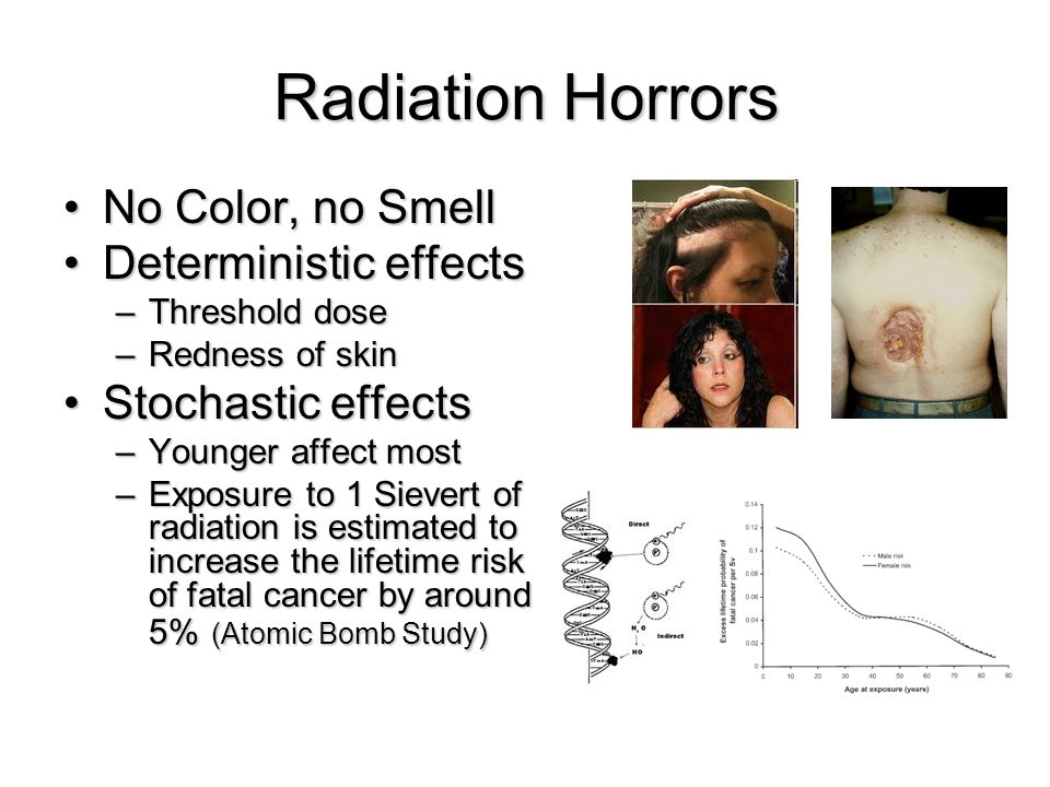
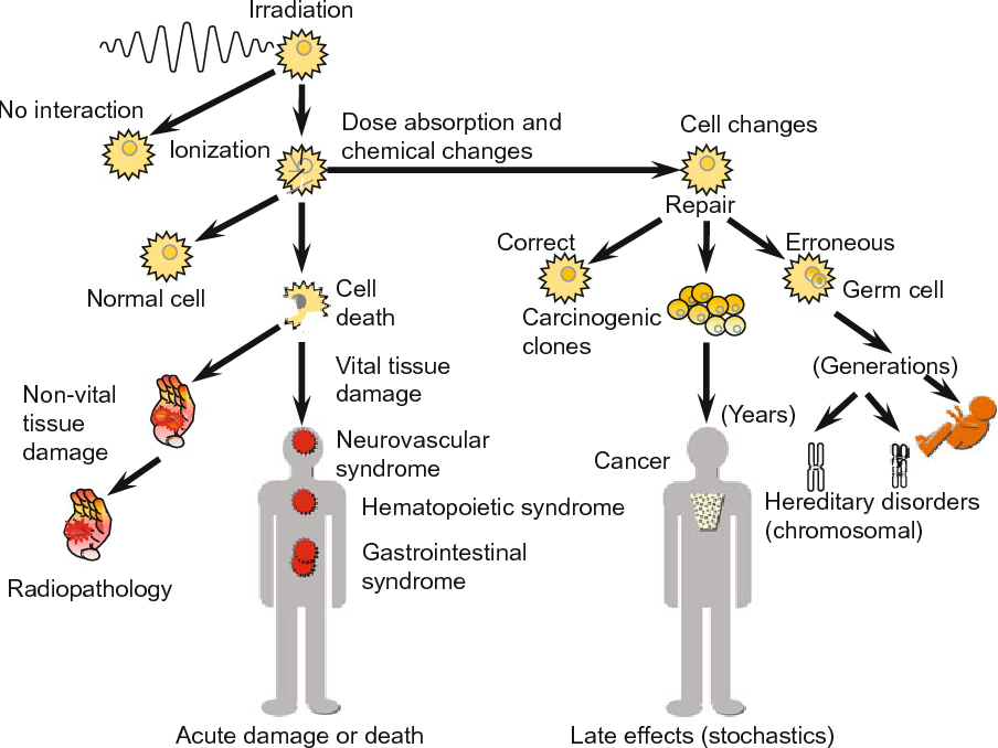
\nAbove certain thresholds, exposure to radiation may impair tissue and/or organ function and may cause acute reactions such as reddening of the skin, hair loss, radiation burns, or acute radiation syndrome. These reactions are stronger at higher doses and higher dose rates. For example, the threshold dose for acute radiation syndrome is approximately 1 Sv (1000 mSv). nine0285
\n
\nIf the dose is low and/or for a long period of time (low dose rate), the risk associated with this is significantly reduced, since in this case the probability of repair of damaged tissues increases. However, there is a risk of long-term consequences, such as cancer that may take years or even decades to appear. Effects of this type do not always appear, but their probability is proportional to the radiation dose. This risk is higher in the case of children and adolescents, as they are much more sensitive to the effects of radiation than adults. nine0285
\n
\nEpidemiological studies in exposed populations, such as atomic bomb survivors or radiotherapy patients, have shown a significant increase in the likelihood of cancer at doses above 100 mSv.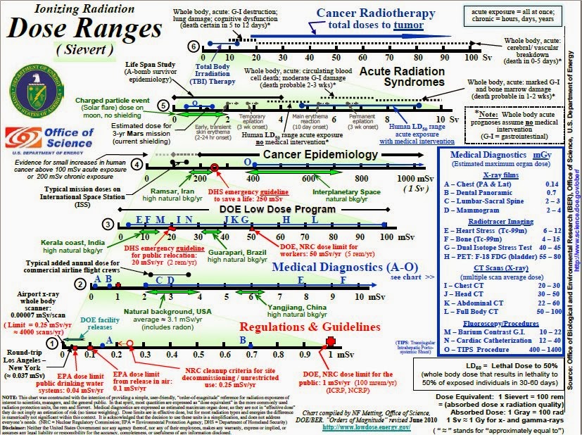 In some cases, more recent epidemiological studies in humans exposed as children for medical purposes (Childhood CT) suggest that the likelihood of cancer may be increased even at lower doses (in the range of 50-100 mSv) . nine0285
\n
\nPrenatal exposure to ionizing radiation can cause fetal brain damage at high doses in excess of 100 mSv between 8 and 15 weeks of gestation and 200 mSv between 16 and 25 weeks of gestation. Human studies have shown that there is no radiation-related risk to fetal brain development before 8 weeks or after 25 weeks of gestation. Epidemiological studies suggest that the risk of developing fetal cancer after exposure to radiation is similar to the risk after exposure to radiation in early childhood. nine0285
\n
WHO activities
\n
\nWHO has developed a radiation program to protect patients, workers and the public from the health risks of radiation exposure in planned, existing and emergency exposures. This program, which focuses on public health aspects, covers activities related to exposure risk assessment, management and communication.
\n
\nIn line with its core function of \"setting, promoting and monitoring norms and standards\", WHO collaborates with 7 other international organizations to revise and update international standards for basic safety related to radiation (SBB). WHO adopted new international PRSs in 2012 and is currently working to support the implementation of PRSs in its Member States. nine0285
\n
","datePublished":"2016-04-29T09:30:00.0000000+00:00","image":"https://cdn.who.int/media/images/default -source/imported/radiation/radiation-africa630x420-jpg.jpg?sfvrsn=e8581c1b_10","publisher":{"@type":"Organization","name":"World Health Organization: WHO","logo": {"@type":"ImageObject","url":"https://www.who.int/Images/SchemaOrg/schemaOrgLogo. jpg","width":250,"height":60}},"dateModified ":"2016-04-29T09:30:00.0000000+00:00","mainEntityOfPage":"https://www.who.int/ru/news-room/fact-sheets/detail/ionizing-radiation-health -effects-and-protective-measures","@context":"http://schema.org","@type":"Article"}; nine0285
Key Facts
- Ionizing radiation is a form of energy released by atoms in the form of electromagnetic waves or particles.
- People are exposed to natural sources of ionizing radiation such as soil, water, plants, and man-made sources such as X-rays and medical devices.
- Ionizing radiation has numerous useful applications, including medicine, industry, agriculture, and scientific research. nine0005
- As the use of ionizing radiation increases, so does the potential for health hazards if it is used or restricted inappropriately.
- Acute health effects such as skin burn or acute radiation syndrome may occur when the radiation dose exceeds certain levels.
- Low doses of ionizing radiation may increase the risk of longer term effects such as cancer.
What is ionizing radiation? nine0303
Ionizing radiation is a form of energy released by atoms in the form of electromagnetic waves (gamma or x-rays) or particles (neutrons, beta or alpha). The spontaneous decay of atoms is called radioactivity, and the excess energy that results from this is a form of ionizing radiation. Unstable elements formed during decay and emitting ionizing radiation are called radionuclides.
All radionuclides are uniquely identified by the type of radiation they emit, the energy of the radiation, and their half-life. nine0285
Activity, used as a measure of the amount of radionuclide present, is expressed in units called becquerels (Bq): one becquerel is one decay per second. The half-life is the time required for the activity of a radionuclide to decay to half its original value. The half-life of a radioactive element is the time it takes for half of its atoms to decay. It can range from fractions of a second to millions of years (for example, the half-life of iodine-131 is 8 days, and the half-life of carbon-14 is 5730 years).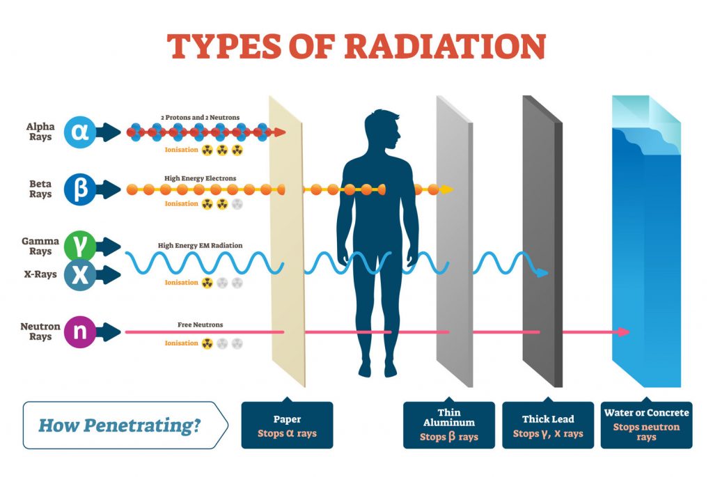 nine0285
Radiation sources
People are exposed to natural and artificial radiation every day. Natural radiation comes from numerous sources, including over 60 naturally occurring radioactive substances in soil, water and air. Radon, a naturally occurring gas, is formed from rocks and soil and is the main source of natural radiation. Every day people inhale and absorb radionuclides from air, food and water.
Humans are also exposed to natural radiation from cosmic rays, especially at high altitudes. On average, 80% of the annual dose that a person receives from background radiation is from naturally occurring terrestrial and space sources of radiation. The levels of such radiation vary in different rheographic zones, and in some areas the level can be 200 times higher than the global average.
Humans are also exposed to radiation from man-made sources, from nuclear power generation to the medical use of radiation diagnosis or treatment. Today, the most common artificial sources of ionizing radiation are medical devices, such as x-ray machines, and other medical devices. nine0285
Exposure to ionizing radiation
Exposure to radiation can be internal or external and can occur in a variety of ways.
Internal exposure to ionizing radiation occurs when radionuclides are inhaled, ingested, or otherwise enter the circulation (eg, injection, injury). Internal exposure stops when the radionuclide is excreted from the body, either spontaneously (with feces) or as a result of treatment. nine0285
External contamination can occur when radioactive material in the air (dust, liquid, aerosols) is deposited on skin or clothing. Such radioactive material can often be removed from the body by simple washing.
Exposure to ionizing radiation may also occur as a result of external radiation from a suitable external source (eg, such as exposure to radiation emitted by medical x-ray equipment). External exposure stops when the radiation source is closed, or when a person goes outside the radiation field.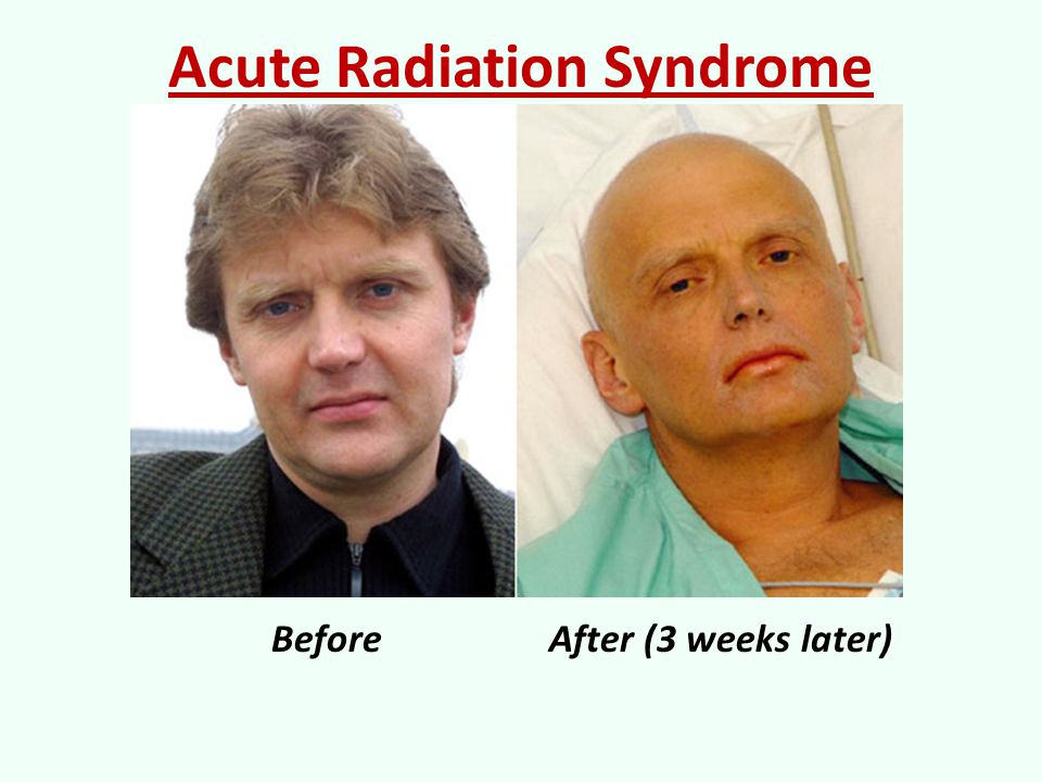 nine0285
People can be exposed to ionizing radiation in a variety of settings: at home or in public places (public exposure), in their workplace (occupational exposure), or in health care settings (patients, caregivers and volunteers).
Exposure to ionizing radiation can be classified into three types of exposure.
The first case is planned exposure, which is due to the intentional use and operation of radiation sources for specific purposes, for example, in the case of the medical use of radiation for the diagnosis or treatment of patients, or the use of radiation in industry or for scientific research purposes. nine0285
The second case is existing sources of exposure where radiation exposure already exists and for which appropriate control measures need to be taken, such as exposure to radon in homes or workplaces, or exposure to natural background radiation in environmental conditions.
The last case is exposure to emergencies caused by unexpected events requiring prompt action, such as nuclear incidents or malicious acts. nine0285
Medical uses of radiation account for 98% of total radiation dose from all artificial sources; it accounts for 20% of the total impact on the population. There are 3,600 million diagnostic radiological examinations, 37 million nuclear procedures and 7.5 million therapeutic radiotherapy procedures worldwide each year.
Health effects of ionizing radiation
Radiation damage to tissues and/or organs depends on the received radiation dose or absorbed dose, which is expressed in grays (Gy). nine0285
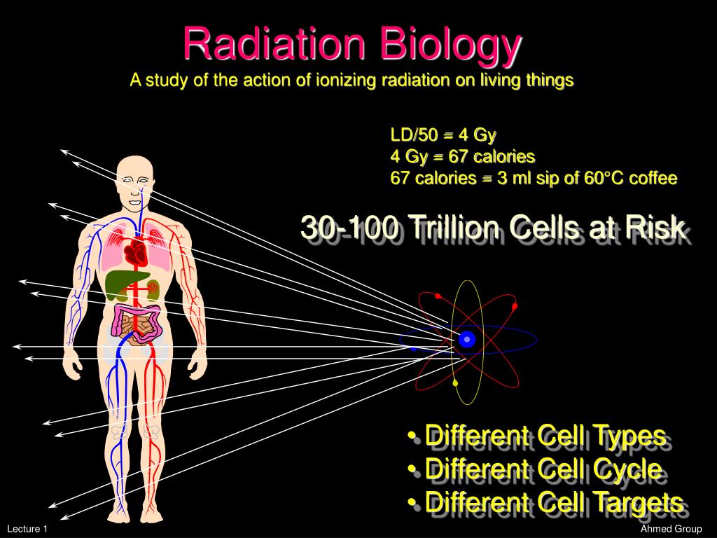
Effective dose is used to measure ionizing radiation in terms of its potential to cause harm. Sievert (Sv) is a unit of effective dose, which takes into account the type of radiation and the sensitivity of tissues and organs. It makes it possible to measure ionizing radiation in terms of the potential for harm. Sv takes into account the type of radiation and the sensitivity of organs and tissues.
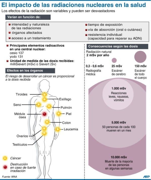
Sv is a very large unit, so it is more practical to use smaller units such as millisievert (mSv) or microsievert (µSv).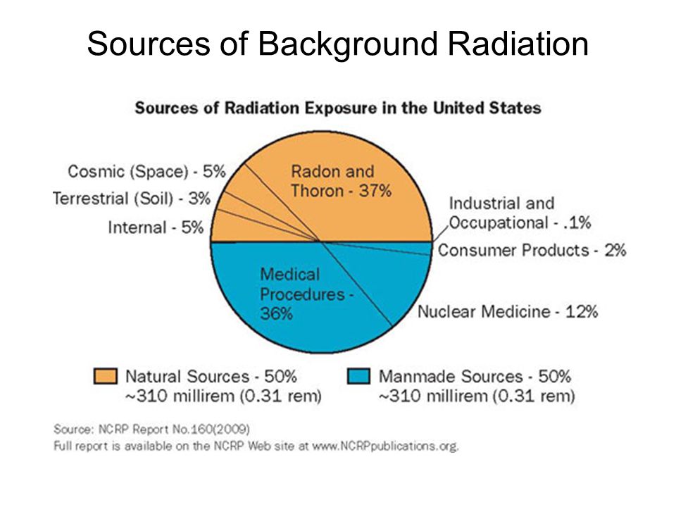 One mSv contains 1000 µSv, and 1000 mSv equals 1 Sv. In addition to the amount of radiation (dose), it is often useful to show the release rate of that dose, such as µSv/hour or mSv/year. nine0285
Above certain thresholds, exposure may impair tissue and/or organ function and may cause acute reactions such as reddening of the skin, hair loss, radiation burns, or acute radiation syndrome. These reactions are stronger at higher doses and higher dose rates. For example, the threshold dose for acute radiation syndrome is approximately 1 Sv (1000 mSv).
If the dose is low and/or a long period of time is applied (low dose rate), the resulting risk is significantly reduced, since in this case the likelihood of repair of damaged tissues increases. However, there is a risk of long-term consequences, such as cancer that may take years or even decades to appear. Effects of this type do not always appear, but their probability is proportional to the radiation dose. This risk is higher in the case of children and adolescents, as they are much more sensitive to the effects of radiation than adults.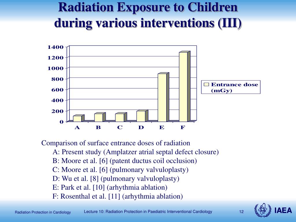 nine0285
Epidemiological studies in exposed populations, such as atomic bomb survivors or radiotherapy patients, have shown a significant increase in the likelihood of cancer at doses above 100 mSv. In some cases, more recent epidemiological studies in humans exposed as children for medical purposes (Childhood CT) suggest that the likelihood of cancer may be increased even at lower doses (in the range of 50-100 mSv) . nine0285
Prenatal exposure to ionizing radiation can cause fetal brain damage at high doses in excess of 100 mSv between 8 and 15 weeks of gestation and 200 mSv between 16 and 25 weeks of gestation. Human studies have shown that there is no radiation-related risk to fetal brain development before 8 weeks or after 25 weeks of gestation. Epidemiological studies suggest that the risk of developing fetal cancer after exposure to radiation is similar to the risk after exposure to radiation in early childhood. nine0285
WHO activities
WHO has developed a radiation program to protect patients, workers, and the public from the health hazards of radiation in planned, existing, and emergency exposures. This program, which focuses on public health aspects, covers activities related to exposure risk assessment, management and communication.
Under its core function of "setting, promoting and monitoring norms and standards", WHO is collaborating with 7 other international organizations to revise and update international standards for basic radiation safety (BRS). WHO adopted new international PRSs in 2012 and is currently working to support the implementation of PRSs in its Member States. nine0285
- Consequences of the Chernobyl accident for health
Radiation pathology of the thyroid gland lecture 2. Iodine blockade in case of accidents at nuclear production | Kasatkina
Introduction
- Nuclear production as a source of increased danger
Currently, there are more than 400 nuclear power plants (NPPs) operating on the planet, and more than 100 are under construction. In addition, there are a large number of individual nuclear reactors. K 19In the 1990s, 46 power units operated at 15 nuclear power plants on the territory of the former USSR, 111 reactors in the USA, and 12 more are under construction.
Hundreds of tons of uranium oxide are loaded into nuclear reactors. Therefore, during the generation of atomic energy, they accumulate a huge amount of radioactive substances (RS), which are formed during the physical decay of the nuclei of fuel atoms. Reactors are, first of all, a potential source of radiation hazard and the ingress of radioactive substances contained in them into the environment and the human body. nine0285
By 1987, 284 serious nuclear accidents at nuclear power plants were registered in the world, which were accompanied by the release of radioactive substances into the environment. The largest among them are in Northern England (Windscale, 1957), in the USA (Three Mile Island, 1979) and in the USSR (Chernobyl, 1986).
Along with this, incidents also periodically occur in radiochemical production, more than 250 of them occurred at Soviet enterprises alone, and the most severe of them are those associated with the occurrence of a self-sustaining chain reaction.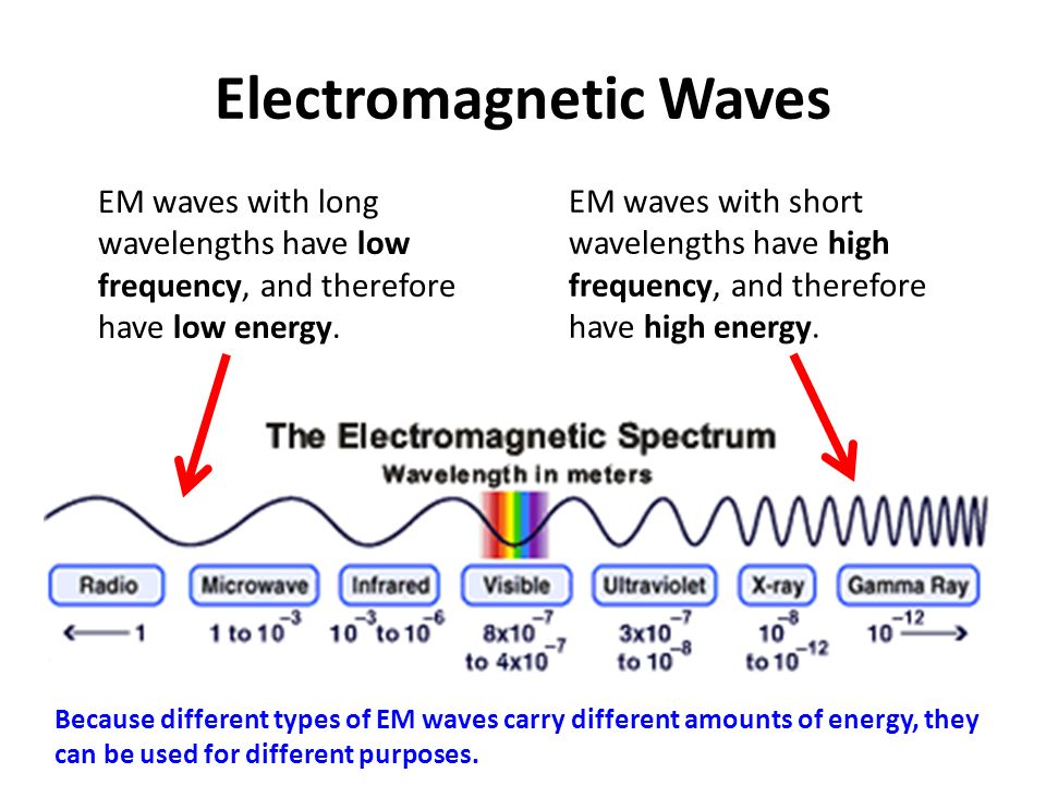 K 19In 94, there were 9 such incidents in the USA, 7 in Russia (the most significant at the Mayak production association in Chelyabinsk-65, the Siberian Chemical Plant in Tomsk-7 and the mining and chemical plant in Krasnoyarsk-26).
The catastrophes themselves, as well as the number of victims from them, are unpredictable in advance, neither in place nor in time. This excludes the possibility for healthcare organizations, especially in cases of mass catastrophe, to provide planned and complete preparation for a specific type and size of disaster. Life shows that a certain medical preparedness for radiation damage in the event of a possible nuclear accident must be maintained not only at the state level, but also in the regions, at national economy facilities. There is no doubt about the relevance of individual training of doctors on the issues of radiation safety of the population. nine0285
The purpose of this lecture is to familiarize a wide range of doctors with the relevant recommendations on radiation protection of the human thyroid gland, which are adapted to the conditions of domestic healthcare practice and use the modern progressive experience of a number of international national organizations.
- The concept of a radiation accident
According to the classification of the World Health Organization, nuclear accidents are classified as catastrophes (along with meteorological, topological, tellurgical and tectonic). From a biomedical standpoint radiation accident is the release of radioactive substances outside the nuclear power reactor in excess of the established norms, as a result of which an increased radiation hazard can be created, which poses a threat to human life and health. Such an unexpected situation will entail an external exposure to radiation on personnel or part of the population, as well as exposure to the ingestion of radioactive substances in doses that exceed the maximum allowable and determine the radiation risk of early and long-term consequences that are unfavorable to human health. nine0285
In recent decades, the contingent of professionals who have contact with diverse sources of radiation has continued to expand.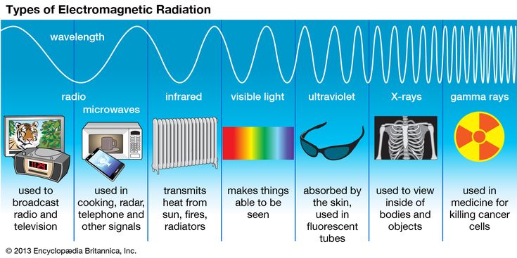 In developed countries, their share among the population increased 5 times over 20 years (from 0.1% in 1972 to 0.5% in 1992). And by the beginning of the 3rd millennium, their numbers are predicted to double.
At the same time, the experience accumulated as a result of the accident at the Chernobyl nuclear power plant demonstrates the potential danger of radiation catastrophes not only for a narrow circle of professional contingent, but also for the many millions of people living surrounded by sources of "peaceful atom" (even at a distance of hundreds of kilometers from them). Therefore, in case of an emergency at a nuclear facility, the readiness of medical personnel to provide large-scale qualified medical care for radiation protection of people is of particular importance. nine0285
- Characteristics of radioactive fallout during accidents at nuclear power plants and zones of radioactive contamination
The inherent characteristics of radiation accidents are the suddenness of the phenomenon itself, loss of control over the radiation source (uncontrolled chain reaction), possible formation of radioactive contamination centers or additional exposure of various categories of people in excess of the established standards.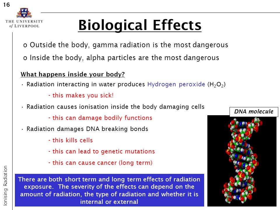
Nuclear accidents are divided into 3 types according to the distribution boundary of released radioactive substances and radiation consequences. nine0285
- Local accident is an accident, the radiation consequences of which are limited to one building or structure, and in which the exposure of personnel and contamination of the building or structure above the levels envisaged during normal operation are possible.
- Local accident - radiation consequences are limited to the buildings and territory of the NPP, exposure of personnel and contamination of buildings and structures located on the territory of the plant is possible, above the levels established for normal operation. nine0005
- General accident — radiation consequences extend beyond the territory of the NPP and lead to public exposure and environmental pollution above the established levels.
The degree of contamination of the area during an accident at a nuclear power plant depends on the amount of radioactive material exiting the reactor, its type (RBMK or VVER), the height of the radioactive material release into the atmosphere, the distance from the accident site (the length and width of the radioactive trace), the category of stability and wind speed, etc.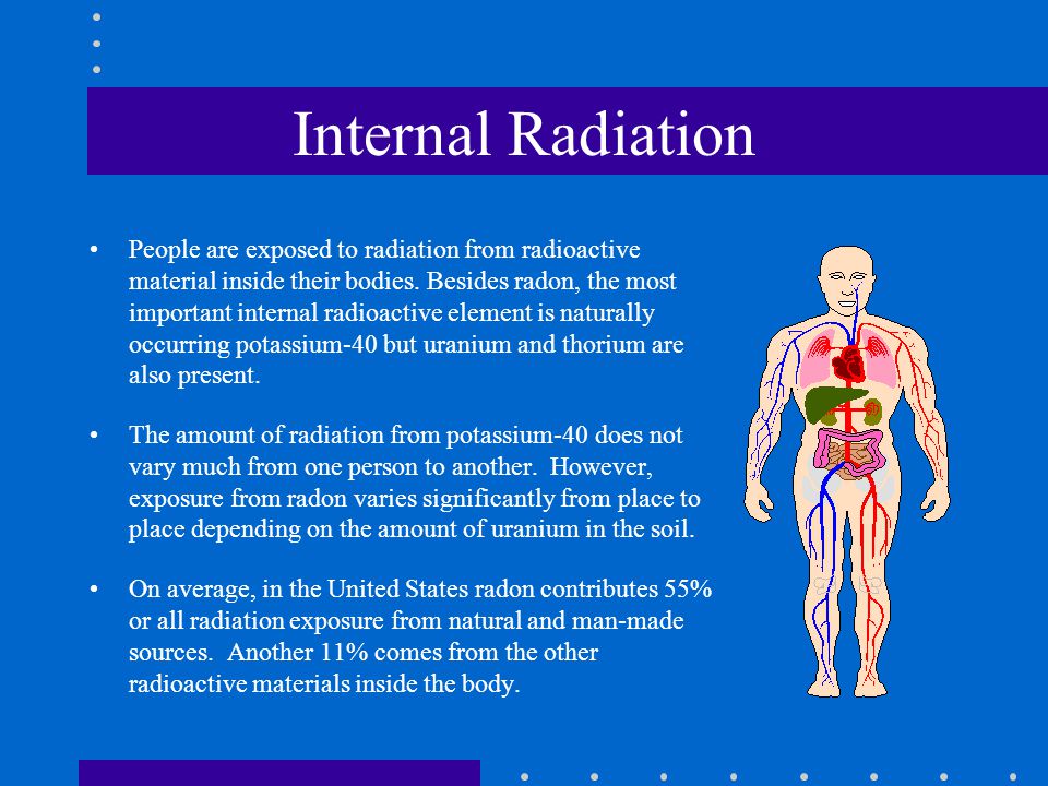 meteorological factors ("dry/wet" fallout). The radiation characteristics of the areas of contamination of the area are given in Table. nineteen0285
The thyroid gland is a critical organ for radiation exposure
The short-lived isotopes of iodine ( 13, 1— 135 1) are the most dangerous among radioactive emissions. Entering the body, they are quickly included in the same metabolic chains as stable J 27 I. During the decay of radioactive iodine, p-particles directly affect molecules and cells, causing a damaging effect on them and causing a pathological process. nine0433 The critical organ for radiation exposure to radioisotopes of iodine is the thyroid gland. In this organ, small in volume and weight (from 1 g in newborns to 25 g in adult men), which is also richly vascularized and intensively supplied with blood, they accumulate much faster and in the largest amount compared to all other tissues of the human body.
30% of the iodine entering the systemic circulation is absorbed precisely by the thyroid tissue, which it leaves for a long time - with a biological half-life of 120 days. Due to the short half-life
Table 1
Radiation characteristics of zones of radioactive contamination of the area during accidents at nuclear power plants
| Zone name | Zone index | Radiation dose for the 1st year after the accident, rad | Radiation dose rate via 1 h after accident mrad/h | |||
| outer border | inner border | middle zone | outer border | inner border | ||
| Radiation hazard zone | M | 5 | 50 | 16 | 14 | 140 |
| Moderate pollution zone | nine0286 A | 50 | 500 | 160 | 140 | 1400 |
| Heavy pollution area | B | 500 | 1500 | 866 | 1400 | 4200 |
| Dangerous contamination zone | B | 1500 | 5000 | 2740 | 4200 | 14 000 |
| Area of extremely dangerous pollution | G | 5000 | - | 9000 | 14 000 | - nine0492 |
yes radioactive iodine ( | 3, 1 - 135 1) - from 2 hours to 8 days, it creates the possibility of its intense effect on thyrocytes in a short period of time. The remaining 70% of iodine is evenly distributed in other organs, mainly in an organically bound form, from where it disappears 10 times faster - with a half-retention period of 12 days.
Of great importance are the data on the direct dependence of the intensity of incorporation of iodine radioisotopes on the iodine supply of man, ie, on the initial—before the radiation accident—intrathyroid content of stable iodine. In countries with sufficient iodine intake (more than 150 mcg/day), uptake of 131 1 after 24 hours is 19-22%. And in the states belonging to the zones of mild and moderate iodine deficiency (iodine supply is less than 100 and 50 mcg/day, respectively), the iodine storage function of the thyroid gland, which has "depleted" trace element reserves, is increased by 2-3 times; is 40-60%.It should be noted that most of the territories of the Russian Federation are natural foci of iodine deficiency, where a reliable supply of iodized products and antistrumine prophylaxis have not been carried out over the past decades. 0285
0285
Thyroid doses are expressed in rads (or centigrays), they largely depend on the age of the victim, the physiological state of the body and the routes of intake of radioactive iodine isotopes.
In an adult (over 18 years of age), at the time of oral intake of 1 mCi 131 1, an irradiation dose of 1.8 rad is formed, with inhalation - 1.1 rad. The total dose for the absorption of such activity by the thyroid gland (formed from the moment of admission to complete excretion) will be approximately 6 rad. In pregnant women, due to increased absorption of iodine, the dose is approximately 1.5 times higher. In children and adolescents, the dose upon receipt of the same number of isotopes will be greater due to the lower mass of the organ (in a child under the age of 1 year - 7.5 times, 1-3 years - 5.2 times, 3-7 years - 3.2 times, 7-9years - 2.2 times, 9-13 years old - 1.6 times, in adolescents 13-15 years old - 1.3 times, 15-18 years old - 1.2 times). At the prenatal stage of development (in fetuses at the 14-35th week of gestation), the dose is 1.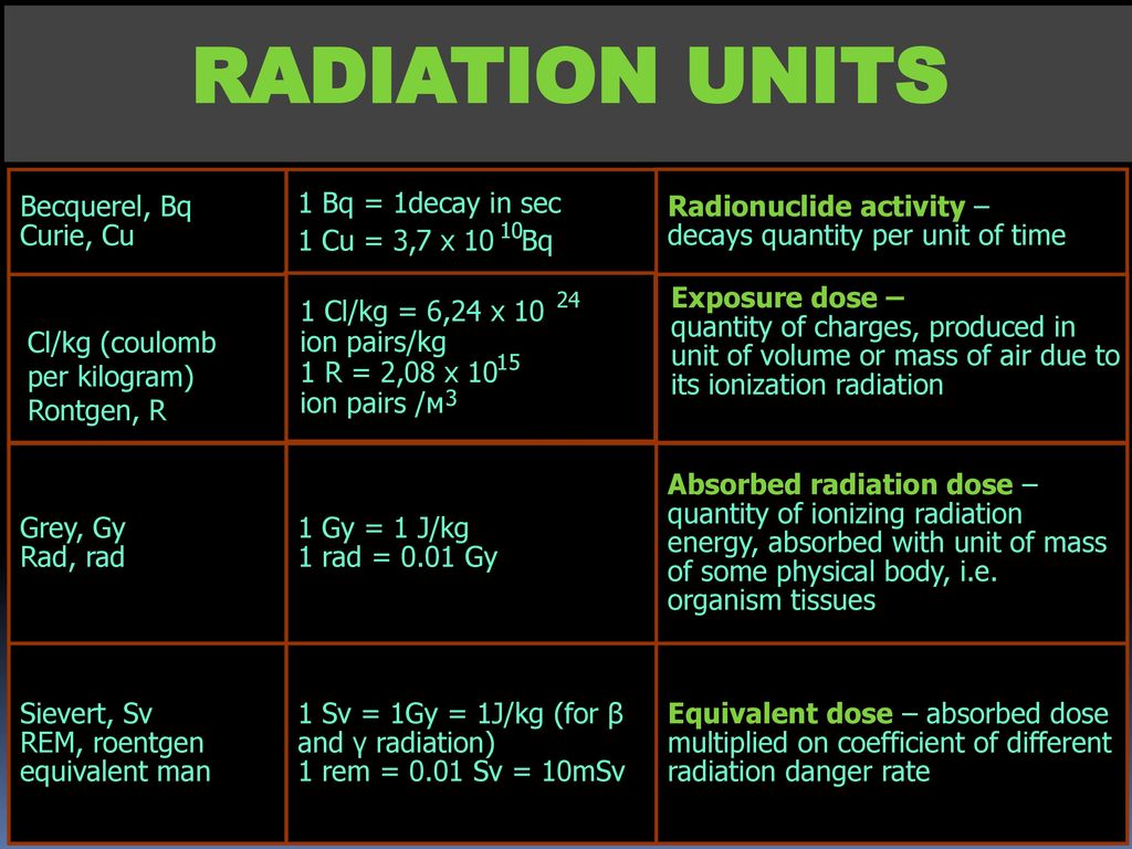 5-3 times higher than the maternal dose.
Thus, regardless of the density of radioactive fallout in a particular area, among the population exposed to radiation, there are critical groups of people, who are able to form the highest possible doses in the thyroid gland and therefore have the greatest risk of developing a variety of thyroid pathologies. These are prenatally irradiated, children and adolescents under 18 years of age, pregnant women and nursing mothers. Such a radiobiological approach substantiates the need for differentiated measures for the radiation protection of the thyroid gland in the population from different critical (age) groups. For these persons, special maximum permissible levels of exposure are provided, more stringent criteria are presented for rationing the content of radionuclides in food, and special recommendations have been developed regarding the procedure for evacuation from the source of contamination and drug blockade of the thyroid gland, which prevents its saturation with radioactive iodine. nine0285
Classification of radiation pathology of the thyroid gland
The specific effect of radioactive isotopes of iodine on the thyroid gland can be realized in the early stages. i.e., in the first weeks or months after the onset of exposure (acute injury), or later, throughout life (long-term effects).
Acute damage to in the form of manifest primary hypothyroidism leads to radiation death of thyroid cells, which is possible with the absorption of a very large dose of radioactive iodine, called thyroidectomy. It amounts to several thousand rads (25-30 Gy) and is formed when the population lives in zones C and D of radioactive contamination of the area (zones of dangerous and extremely dangerous contamination; see Table 1). nine0285
Long-term effects of (in the long-term after exposure) are manifested in two ways, which depends on the dose of ionizing radiation.
- th variant is represented by stochastic (probabilistic, non-somatic, mutagenic) effects in the form of radiation-induced thyroid tumors (including malignant ones).
 The possibility of their development is probable under irradiation (conditionally) in any dose above zero.
 - th option is deterministic (non-stochastic) diseases in the form of autoimmune thyroiditis and primary hypothyroidism; they manifest, as previously thought, only when the absorbed dose exceeds a threshold level corresponding to approximately 200 rad.
Living in zones M, A and B (zones of radiation hazard, moderate and severe pollution; see Table 1) presents a radiation hazard of formation of long-term consequences for the thyroid gland.
According to the latest information, the timing of the clinical debut of long-term effects of radiation exposure on the thyroid gland is approximately estimated at 5-50 years after the incorporation of radioactive iodine. nine0285
Thus, exposure of thyroid cells to radioactive iodine can lead even very late after the accident (up to 50 years or more) to the development of neoplasms of the thyroid gland (including cancer), autoimmune thyroiditis, and hypothyroidism.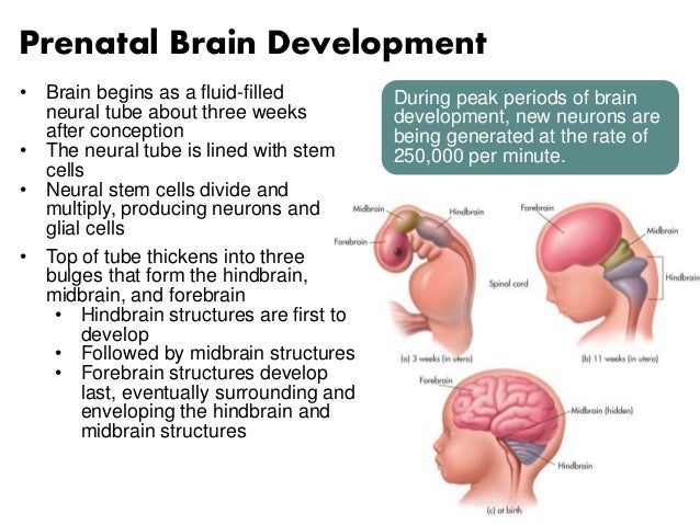
Radiation protection of the thyroid gland
By radioprotective drugs, administered to the human body, means chemical or biochemical preparations designed to reduce or block the intake or subsequent deposition of PB in the body; acceleration of removal from the body of radionuclides that have entered it; weakening the pathological consequences of radiation effects on the human body. nine0285
The most acceptable from a practical point of view, pathogenetically justified in the light of the predicted risk and subject to planning as a preventive, emergency or extremely urgent protective measure is the use of stable iodine preparations. The radioprotective effect of stable iodine is associated with its competitive interaction with radioactive iodine. The incorporation of the latter is blocked under conditions of maximum oversaturation of the thyroid gland with pharmacological doses of iodine preparations. In addition, the prophylactic use of these drugs reduces the vascularization of the thyroid tissue, reduces the intensity of blood flow through its vessels, and ultimately further limits the flow of radioactive iodine into it. Iodine blockade is carried out with a potential or actual release of radioactive iodine into the atmosphere from nuclear power reactors. nine0285
With a single (single) inhalation intake of radioactive iodine (due to inhalation), the thyroid dose reaches a maximum within 1-2 days, and half of this dose is formed in the first 6 hours. Therefore,
iodine (130 mg KI or 170 mg KIO 3 equivalent to 100 mg iodine) in the blockade of the flow of radioactive iodine into the thyroid gland of adults (with a single inhalation intake of 131 1)
Conditions for the use of stable iodine preparations
Reduced uptake 131 1.%
Early before admission
| in 48 hours | 46 |
| in 36 hours | 45 |
| in 24 hours | 89 |
| nine0286 for 8 hours | 94 |
| in 6 hours | 99 |
| Simultaneously with intake of 131 1 After intake of 131 1 in the body: | 89-97 |
| after 2 hours | 68-90 |
| after 6 hours | 50 |
| after 8 hours | 0 |
it is important that stable iodine prophylaxis be administered as soon as possible after a radioactive iodine release or (ideally) before a release.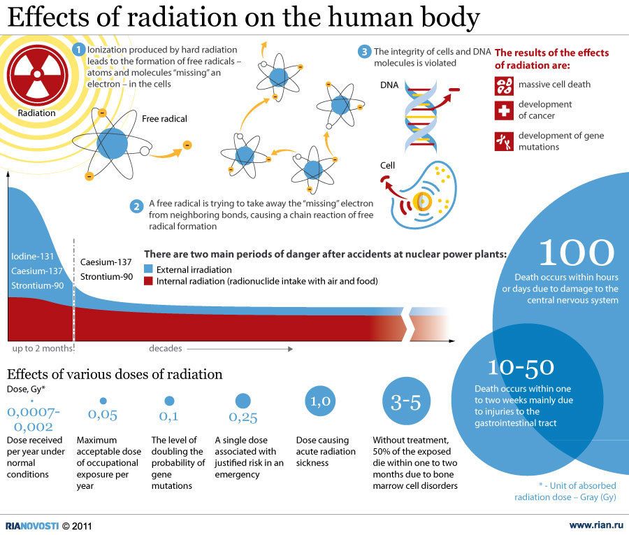 In this case, the age and physiological characteristics of a particular person are necessarily taken into account.
The most effective is the advance (with a potential threat of release) and early (immediately after the start of the accident) administration of iodine preparations (Table 2). Early (during the day before the start of inhalation of radioactive iodine) iodine blockade of the thyroid gland reduces the incorporation of radioactive iodine by 95%, reception of stable iodine simultaneously with the intake of radioisotopes - by 97%, within 6 hours after the start of exposure - by 50%. It is generally accepted that taking stable iodine later than 12 hours gives a slight effect, and after 1 day it becomes practically ineffective.
With chronic inhalation intake of isotopes (Table 3), daily intake of 100 mg of iodine for 7-10 days reduces the absorption of radioactive iodine by 96%. The use of more than 100 mg daily doses of iodine is impractical because of the risk of side effects with a similar protective effect.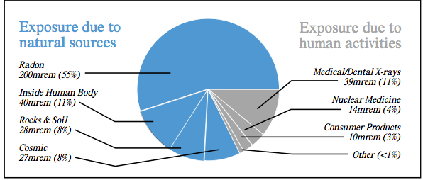 nine0285
The schemes of blockade of the thyroid gland with stable iodine preparations recommended by international experts of the IAEA in case of release of radioactive iodine into the atmosphere are given in Table. 4 (Series of publications on security. - 1981. - No. 55; 1988 - No. 88). When planning this protection measure, the duration of taking iodine preparations is limited, based on the conditions so that the maximum total (course) dose does not exceed 1 g.
inhalation of radioactive iodine, but the effectiveness of these measures is calculated only for the inhalation intake of isotopes into the body. nine
To prevent the intake of radioactive iodine through another equally dangerous - oral route (through the digestive organs) a whole range of non-drug restrictive measures is taken, aimed at
Table 3
from its daily dose in the blockade of the entry of radioactive iodine into the thyroid gland of adults (with chronic inhalation intake of 13 *1 within 14 days)
| Daily dose of KI for 7 days, mg | Capture reduction 131 1% |
| 10 | 87 |
| 100 | 96 |
| 200 | 94 |
| 200 (every other day) nine0492 | 95 |
Recommended doses of iodine preparations (130 mg KI or 170 mg KIO 3 equivalent to 100 mg iodine) for the release of radioactive iodine into the atmosphere 1
| Contingent | 1st day after the start of the accident | 2-10 days after the start of the accident |
| Adults and children over 1 year old | nine0509 50 mg | |
| Children under 1 year old | 50 mg | 50 mg |
1 Planning for off-site protective actions in cases of radiation accidents at nuclear installations.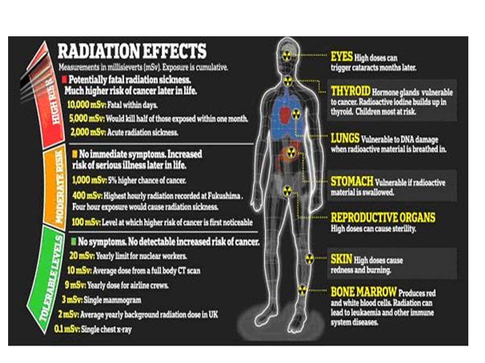 A series of publications on security. Vienna: IAEA. - 1981. No. 55.
temporary ban on the consumption of certain categories of contaminated food (mainly dairy, vegetable, etc.) or water from certain sources of water supply along with the reorganization of the system for providing the population with food and drinking water. The decision to introduce such prohibitive measures falls within the competence of the relevant non-medical commissions and is made in accordance with the regulations in force in an emergency situation according to the criteria for temporarily permissible levels of contamination of food products. nine0285
Therefore, the medical services are responsible for organizing timely rational radioprotective prophylaxis among various population groups by blocking the thyroid gland with stable iodine preparations. In this case, it should be borne in mind that in the case of rapid evacuation (within 6 hours after the start of the accident), the use of iodine blockade is not necessary, since evacuation itself is the best way to protect a person from radiation.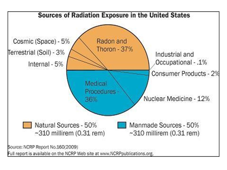 However, evacuation is realistic for a rather limited contingent of victims and will cover only people living in zone G (a zone of extremely dangerous pollution). In other areas of radioactive fallout, thyroid exposure can be avoided only with the help of iodine blockade of the organ. nine0285
Special attention should be paid to pregnant women in this program because such high doses of iodine, although safe for the rest of the population, during the gestational period can harm the development of the fetus in general and its thyroid gland in particular. Freely penetrating the placenta, iodine is absorbed in large quantities by the fetal thyroid gland, as a result of which it is able to significantly suppress its function and further development (the Wolff-Chaikoff effect). Pharmacological doses of inorganic iodine significantly inhibit the oxidation of iodine in fetal thyrocytes, inhibit its organization and block the release of thyroid hormones from the gland. In this regard, the radiation protection of pregnant women includes, along with the introduction of a blocking dose of iodine, an additional intake of potassium perchlorate.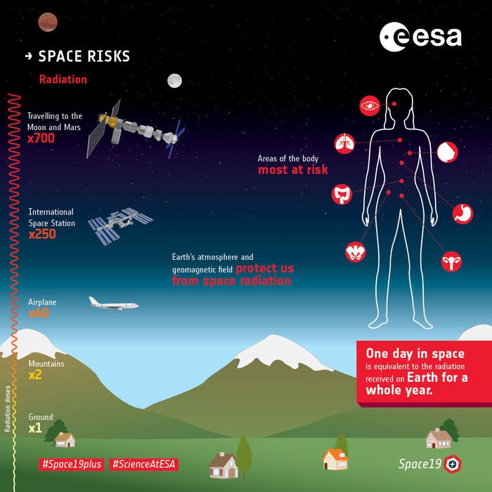 This compound interferes with the process of iodine accumulation, competitively inhibits the "iodine pump", reduces the capture of any iodine isotopes (including stable iodine), which subsequently ensures its accelerated excretion. nine0285
Specific actions of doctors and the public to carry out radioprotective measures should begin after the decision of the relevant emergency and civil defense services on the threat in connection with an emergency in nuclear production.
The main way to alert the public about actions in the event of dangerous situations is the transmission of an alarm message over wired broadcasting networks (through apartment and outdoor loudspeakers), as well as through local broadcasting stations and television. To attract the attention of the population in emergency cases, sound sirens and other signaling means are turned on before the transmission of information. nine0285
After such a special notification, health authorities and institutions should start iodine prophylaxis as a matter of urgency.
Emergency Iodine Prophylaxis Algorithm
Potassium iodide (KI) tablets are used. For an emergency situation, a standard packing from the reserves of civil defense AI-2 is provided (first-aid kit indi-
Fig. 1. Individual first-aid kit AI-2. nine0433 "Radioprotective agent No. 2" - KI tablets at a dose of 125 mg (Fig. 1). In the absence of a first-aid kit, it is permissible to replace 1 tablet of this remedy with 125 tablets of antistrumine (1 mg each) or with an equivalent amount of iodine in the form of aqueous solutions of K.I, and in extreme cases - alcohol tinctures of iodine.
For children under 2 years of age, 1/3 tablet (40 mg) of radioprotective agent No. 2, dissolved in 100 ml of milk (known to be pure) or nutritional mixture, 1 time per day, or 1-2 drops of 5% iodine tincture 3 times a day. For children under 1 year of age, it is desirable to refuse breastfeeding at this time, since radioactive iodine is easily concentrated in mother's milk.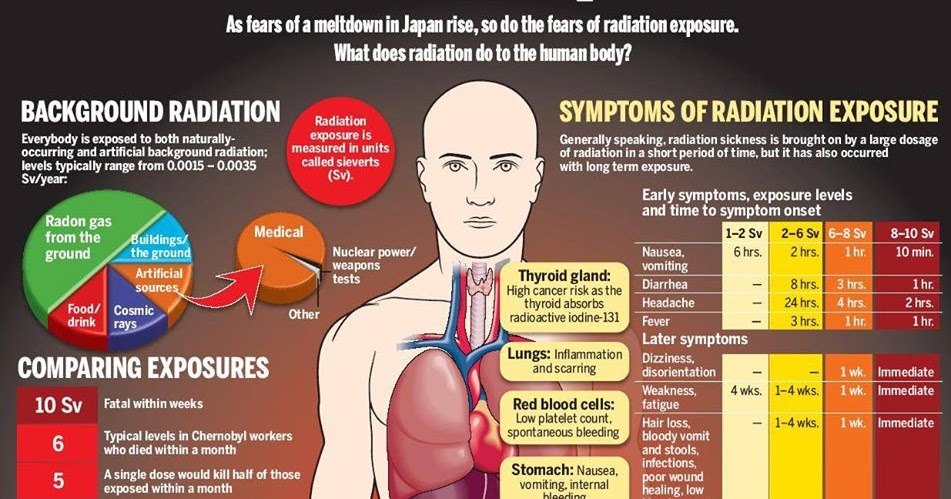 nine0434
Children over 2 years of age and adults are shown to take a whole tablet of a radioprotective drug once a day or replace it with a three-time intake of 3-5 drops of 5% iodine tincture after meals; iodine tincture is washed down with sweet tea, jelly, milk.
For pregnant women, the "adult" regimen is recommended in combination with the additional intake of potassium perchlorate at a dose of 750 mg once a day.
Iodine prophylaxis is carried out until the direct threat of the intake of radioactive isotopes of iodine into the body is eliminated (but not more than 7 days). nine0285
Side effects of stable iodine preparations
The risk to public health associated with the use of stable iodine preparations is generally low. However, it is not possible to exclude individual adverse reactions in a small subset of the population with iodine hypersensitivity or diseases in which stable iodine can exacerbate them.
There are 2 types of adverse reactions to iodine preparations: 1) intrathyroid - effects that appear in the thyroid gland itself. Potentially possible formation of autoimmune thyroiditis (or manifestation of its previously hidden, subclinical forms), toxic adenoma of the thyroid gland (the "Iodine-Basedow" phenomenon in nodular goiter or thyroid autonomy, especially in older people with a long stay before the accident in conditions of iodine deficiency). endemia) or recurrence of diffuse toxic goiter; 2) extrathyroid - effects developing from other organs. As shown by the unique experience in the history of modern radiobiology of iodine blockade of the thyroid gland among the population of Poland, their frequency does not exceed 5% (Table 5).
In a special study aimed at identifying side effects after taking a blocking dose of KI, during which 17,100 respondents of different age and gender were interviewed, the same frequency of adverse reactions was noted in children and adults. The nature of the symptoms was also similar. But in children, more often than other complications and more often than in adults, side effects from the gastrointestinal tract (vomiting, abdominalgia, diarrhea) were noted. Adults often noted a rash on the skin (like "iodism"), cephalalgia and difficulty breathing. Side effects of iodine preparations were found mainly at doses that significantly exceeded those recommended. nine0285
Table 5
Extrathyroid side effects of prophylactic iodine blockade performed in Poland April 29-May 2, 1986
| Symptom | Number of examined, % | |
| children | adults | |
| No complications | 95.4 | 95.5 |
| Side effects | 4.6 | 4.5 |
| from the gastrointestinal tract | 2.93 | 1.60 |
| including: | ||
| vomiting | 2.38 | 0. |
| stomach pain | 0.36 | 0.63 |
| diarrhea | 0.19 | 0.12 |
| skin rash | 1.07 | 1.24 |
| headache | 0.18 | 0.69 |
| shortness of breath nine0492 | 0.11 | 0.63 |
| other | 0.35 | 0.20 |
| Mumps | 0 | 0 |
| Conjunctivitis | 0 | 0 |
As a rule, all adverse reactions were mild and did not require medical attention. Only 1/5 of the people who had complications turned to doctors for advice. Due to exacerbation of bronchopulmonary obstruction, only 2 out of 5,000 adults surveyed were hospitalized: they voluntarily underwent prophylaxis, despite a aggravated allergic history (hypersensitivity to iodine). Thus, radioprotective blockade of the thyroid gland with stable iodine preparations should generally be considered safe. nine0285
Information about the effectiveness and long-term effects of iodine blockade of the thyroid gland in connection with the accident at the Chernobyl nuclear power plant.
The above recommendations have a serious radiobiological substantiation, they are based on the materials of in-depth scientific study. But until recently, we did not know to what extent these experimental and theoretical ideas are realized in their practical application. Only the events associated with the Chernobyl accident gave rise to large-scale radiation protection measures in certain regions, including the iodine blockade. An analysis of its results (early and remote), information about the effectiveness and errors of this experiment, undoubtedly, should become a lesson for all mankind in the event of a radiation accident in the future. nine0285
Despite the fact that in the spring of 1986 the territories of many European countries were exposed to radiation pollution, preventive measures to protect the thyroid gland were carried out only in 2 countries. According to official statements, in certain regions of 3 republics of the former USSR (Belarus, Ukraine, Russia), which surround the nuclear power plant, 5.4 million people (including 1.7 million children) were covered with iodine prophylaxis. On a larger scale, this program was carried out in Poland, where 17.5 million inhabitants received stable iodine (95% of the child population and 23% of adults). Currently, information on the radioprotective effect of these measures on the formation of the total dose in the thyroid gland has been accumulated only by Polish scientists. Their experience has shown that even a milder regimen of iodine prophylaxis (compared to that recommended by the IAEA) is quite effective in protecting against exposure to radioactive iodine. Thus, a single dose of KI at a fairly late date (4-7 days) after the onset of the accident, even at a half dose of the above formula, made it possible to reduce the incorporation of radioactive iodine 29.04, 30.04 and 01.05.86 by 40, 25 and 12% compared with persons who did not receive a radioprotective agent.
 As a result, in 90% of children, the dose load on the thyroid gland did not exceed 5 rad.
Four years after the events described, 12,000 Polish children were assessed in a pilot study for thyroid status based on reliable hormonal markers. It turned out that the functional parameters of the thyroid gland in children who received the blockade, and those who remained without it, practically did not differ. At the same time, it has not been established from
Fig. 2. Thyroid status in children of the city of Volkhov after iodine blockade in 1986 (n = 60) and in control (n - 110).
1 - iodine blockade; 2 - control.
a - frequency of goiter; b - ultrasonic volume; c - level of thyroid-stimulating hormone; g - thyroglobulin level; e - antibodies to thyroglobulin.
changes in the structure of thyroid pathology in adult residents of Poland, regardless of the radioprotective tactics during the accident. In other words, the Polish experience made it possible to establish at least 2 very important facts on a huge population of people: I) stable iodine really reduces the thyroid absorption of radioactive iodine, due to which the dose of radiation to the gland is significantly reduced; the effectiveness of the blockade is the higher, the earlier after the start of the accident it is carried out; 2) a single intake of a large amount of iodine very rarely causes minor side reactions outside the thyroid gland, and on the part of the thyroid system itself, in the years following the blockade, adverse reactions that could be associated with exposure to excessive intake of a stable element have not been established. nine0285
Domestic experience in studying the consequences of iodine blockade in the population of Russia, unfortunately, is much more modest. The updated information on the real volume of medical measures taken is very remarkable: the results of special interviews with the population of the most contaminated areas suggest that only a small number of their inhabitants (5-20%) actually took iodine.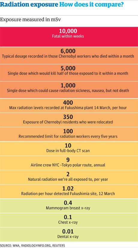 On the other hand, we have our own observations on the thyroid status of children who have completed a course of iodine prophylaxis, compared with peers who did not receive iodine. These materials were presented by us at the III All-Russian Congress of Endocrinologists. Complementing the data of Polish scientists, our information in connection with a longer catamnesis - 9years after the accident at the Chernobyl nuclear power plant are of particular interest.
It turned out that although the terms of pharmacological protection of the population of Russia were even more inadequate (1 month after the start of the accident), and it is almost impossible to evaluate the dependence of the dose on the blockade after 10 years, nevertheless, some results attract special attention (Fig. 2) .
- Morphological and functional parameters of the thyroid status of children who received radioprotective iodine blockade after 9years, they differ by half the frequency and size of goiter, lower levels of thyroid stimulating hormone and thyroglobulin in the blood serum (closer to the physiological adaptive norm than the corresponding indicators in children from the comparison group).

 174, “Preconception and Prenatal Radiation Exposure: Health Effects and Protective Guidance” [NCRP2013].
 85
85 