Of a pelvic bone
Anatomy, Bony Pelvis and Lower Limb, Pelvic Bones - StatPearls
Matthew D. Burgess; Forshing Lui.
Author Information
Last Update: July 25, 2022.
Introduction
The bones of the pelvis are a critical part of the central portion of the skeleton. They serve as a transition from the axial skeleton and the appendicular skeleton of the lower body, serving as an attachment point for some of the strongest muscles in the human body while withstanding the forces generated by them. The curved nature of the pelvic bone creates a closed structure, itself lined with various muscles and housing various blood supplies, lymphatic structures, nerves, and organs, including the intestines, urinary bladder, and internal sex organs.
Structure and Function
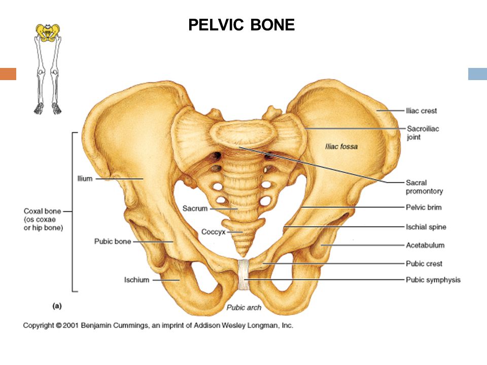
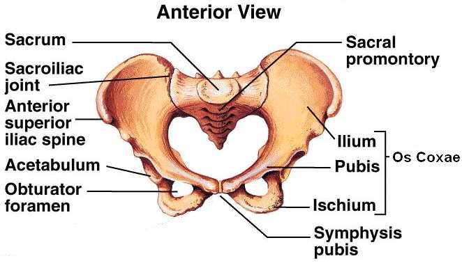
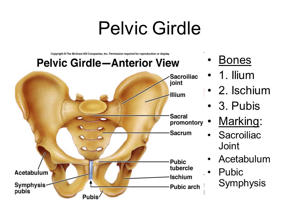
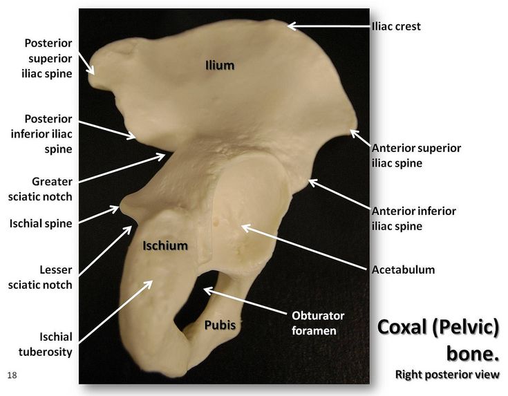
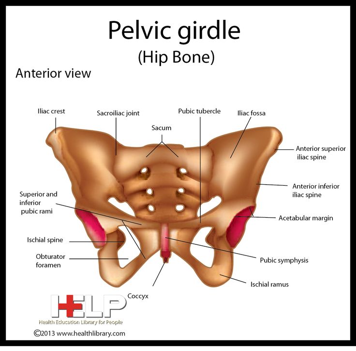
The pelvis is comprised of the large hip bone, the os coxae, on each side. The os coxae itself is composed of the ilium (the flat superior protuberance), ischium (the curved anterior protuberance), and pubis bones (the curved inferior protuberance). The os coxae attach anteriorly to each other at the pubic symphysis and posteriorly to each side of the sacrum, creating the sacroiliac joint. These three os coxae bones meet each other at the acetabulum, a medial structure that serves as an attachment point for the head of the femur. The pubis and ischium also articulate inferiorly at the ramal epiphyses, with their curving nature leaving an opening between them known as the obturator foramen. This foramen allows the obturator nerve to leave the pelvic cavity. Each of the pelvic bones also has many landmarks (i.e., tuberosities, notches) unique to the specific bone. The superior edge of the ilium is named the crest of the ilium, or, more commonly, the iliac crest, with a tuberosity below it on the ilium’s anterior edge known as the anterior inferior iliac spine. The ilium also has a posterior-inferior landmark known as the greater sciatic notch on it, with a subsequent lesser sciatic not being on the posterior-inferior side of the ischium.
 On the anterior side of the same edge of the ischium is the tuberosity of the ischium.[1][2]
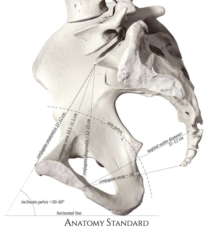
The primary function of the pelvic bones is to transfer the load of the upper body onto the lower limbs while standing or walking. This function takes place through the connection of the axial and appendicular skeletons at the sacroiliac joint. When a man stands upright, the center of gravity lies in the center of the body. The pelvis transmits the weight to the femur and both lower extremities. The rigid structure of these bones also protects the organs that lie within their confinements. Such organs include the bladder, rectum, urethra, and uterus in females.[3]
Embryology
Pelvic bones begin their development as mesenchymal tissue of the embryonic lower limb buds. This mesenchyme begins extending out in three directions, corresponding to each of the three bones of the os coxae. The ischial and pubic masses migrate around the obturator nerve, fusing inferiorly to form the obturator foramen.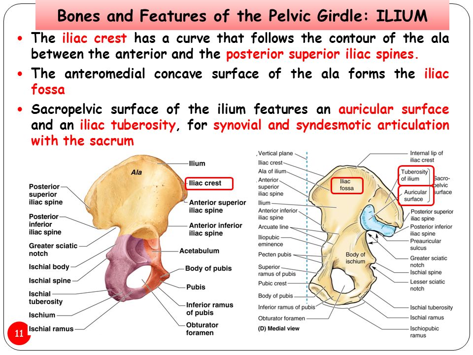 Each pubic mass migrates medially to meet at the future pubic symphysis, while interactions with the primitive spinal column draw the iliac mass toward the sacrum derived from notochord mesenchyme. These mesenchymal masses then undergo endochondral ossification, with ilium, ischium, and pubis beginning primary ossification at third, fourth-to-fifth, and fifth-to-sixth months of gestation, respectively. Each bone’s primary center of ossification is near the future acetabulum, with the ossification process fanning out superiorly, inferiorly, and anteriorly in the direction of each bone. Sacral ossification begins in the third month, with each of the vertebrae having three centers of ossification. Secondary ossification occurs during the first year of life. Secondary ossification centers are at the articulation of the three pelvic bones in the acetabulum, the crest of the ilium, anterior inferior iliac spine, tuberosity of the ischium, symphysis pubis, and ramal epiphyses. These growth plates close during mid-puberty.
 [1]
[1]
Blood Supply and Lymphatics
Many blood vessels are contained within the cavity created by the pelvic bones. These vessels include the common iliac artery, external and internal iliac artery, superior and inferior gluteal artery, obturator artery, superior vesical artery, and internal pudendal artery, along with their accompanying veins. Some of these arteries and their branches, such as the internal iliac, remain within the pelvic cavity while others travel out, such as the gluteals and external iliac, to supply regions such as the buttocks and lower limbs, respectively. The gonadal and uterine arteries will also run through and into the pelvic cavity, respectively, depending upon sex. Major lymph nodes in this region include the obturator, common iliac, external and internal iliac, hypogastric, superior rectal, presacral, presacral, promontory, and perirectal nodes. Nodes with the corresponding vasculature appear in pairs, one being on the anatomical left and the other on the anatomical right.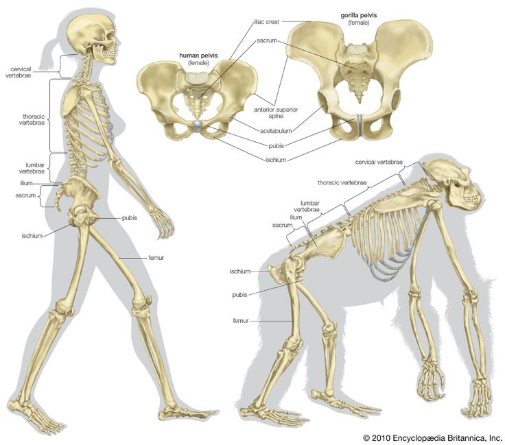 Pelvic lymph nodes are a common site of prostatic and gynecologic cancer metastasis.[4][5][6][7]
Nerves
The pelvic bones, and the girdle made by them, house nerves from the lumbar and sacral plexuses. Nerves of the lumbar plexus contained in the pelvis include the genitofemoral, lateral femoral cutaneous, obturator, and femoral nerves. The genitofemoral pierces through the psoas muscle posterior-to-anterior to reach the genital region on the anterior face of the pelvis. The lateral femoral cutaneous nerve leaves the spine and transverses the pelvis exiting under the anterior brim of the crest of the ilium to reach the thigh. The obturator nerve descends medially to the psoas to leave the pelvis through the obturator foramen. The femoral nerve runs between the psoas and the iliacus, emerging from the pelvis above the pubis to continue into the anterior thigh.[2][8]
Sacral plexus nerves of the pelvis include the sciatic, superior, and inferior gluteal, posterior femoral, and pudendal nerves.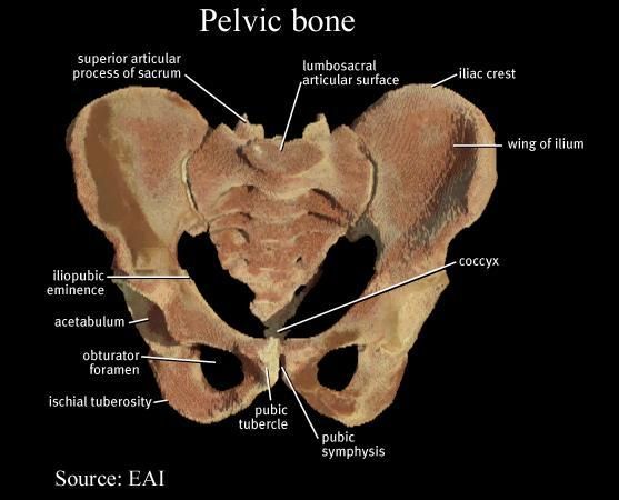 The first four of these nerves all leave the pelvis traveling under the greater sciatic notch, serving innervations on the posterior gluteal and thigh regions. The superior gluteal nerve gives innervations to the gluteus minimus and medius, while the inferior gluteal innervated the gluteus maximus, which extends the hip. The pudendal nerve travels within the pelvic girdle, sending interventions to various sphincters and genitalia structures. This nerve commonly gets injured during the vaginal delivery of children.[9]
Muscles
The muscles around the pelvis are essential in maintaining the stability of the pelvic girdle, maintaining an erect posture, and assisting the movement of the trunk and lower extremities.
The internal iliac crest serves as the origin of the Iliacus muscle, which travels inferiorly and meets with the psoas, which originates above the pelvis but travels down through it, to make the iliopsoas. This muscle eventually exits the pelvis and inserts itself upon the femur.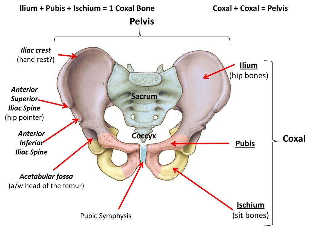 The piriformis is another muscle that originates inside of the pelvis and inserts externally on the femur.[10]
The pubic symphysis serves as an attachment point for both abdominal muscles, including the rectus abdominis, internal and external obliques, and transversus abdominis, as well as muscles of the adductor compartment of the leg, including the adductor brevis/longus/magnus, gracilis, and pectineus. Attached along the inferior edges of the pubis running posteriorly to the sacrum are the pelvic floor muscles, which include the puborectalis, iliococcygeal, ischiococcygeus, pubococcygeus, external anal sphincter muscles, and levator ani. The obturator internus muscles also have insertions on the internal side of the pubis and travel on the pelvic floor muscles, eventually leaving the greater sciatic notch and inserting on the posterior surface of the femur. The obturator externus travels from the anterior face of the pubis posteriorly to insert on the backside of the femur as well.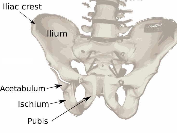 The gluteal muscles, quadratus femoris, and semitendinosus muscles all also have attachments on the pelvic bones. The inguinal ligament, a common anatomical landmark involved with a hernia, runs from the anterior portion of the iliac spine to the front of the pubic symphysis.[11][12][13][14]
Physiologic Variants
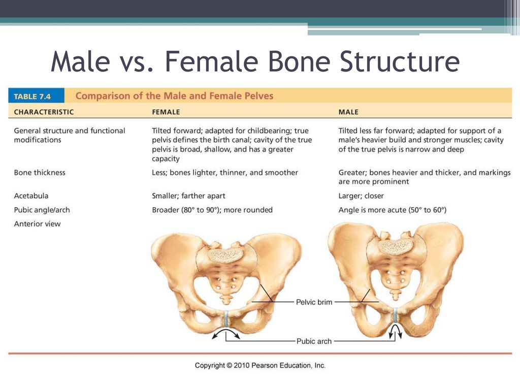
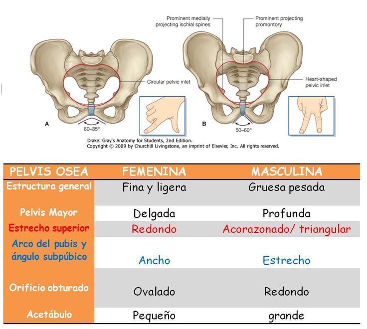
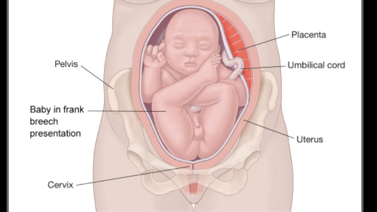
The pelvis in females is very different from that of males; this is very important for females to provide an adequate birth canal for the large head of the fetus during birth. To achieve these means, the female pelvis is generally broader and shallower (gynecoid shape). The birth canal has to be rounded and more spacious. The sciatic notch is much wider with shorter symphysis pubis, and the pubic bones have a much broader angle with each other. The female coccyx is more mobile, and the sacrum is shorter and less curved.
Clinical Significance
A weakness of muscles of the pelvis, such as in limb-girdle muscular dystrophy, will result in an abnormal waddling type of gait.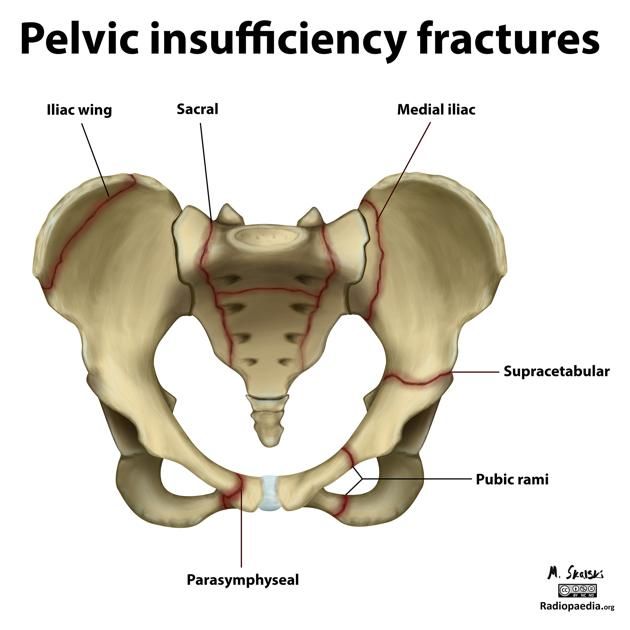 A unilateral weakness of the hip abductor muscles or subluxation of a hip joint will cause a Trendelenburg gait. An analysis of how a patient walks can give clues to the underlying problems related to the structure and function of the pelvis.
Surgeries performed on pelvic structures may be complicated by excessive bleeding or hematoma formation. Surgery may also be complicated by infection and abscess formation. In the pelvis, hematoma or abscess usually remains localized in the retroperitoneal space just in front of the iliopsoas muscles. The presentation is often related to compression of the femoral plexus or the femoral nerve in the retroperitoneal space. The patient will present with postoperative weakness of the hip flexors and knee extensors with or without the involvement of the obturator muscles, depending on whether it is compression of the femoral plexus or the femoral nerve.
A female with a male type (android) of the pelvis will encounter problems during childbirth, which is one of the most prevalent indications for a caesarian section. [15]
[15]
Review Questions
Access free multiple choice questions on this topic.
Comment on this article.
Figure
Pelvis, Male, Ilium, Arcuate Line, Pubis, Ischium, Pubis Arch, Sacrum,. Contributed by Gray's Anatomy Plates
Figure
Pelvis, Female, Pubic Arch, Brim of lesser Pelvis,. Contributed by Gray's Anatomy
Figure
Compilation of 6 images detailing anatomy of female pelvic cavity. Contributed by Gray's Anatomy Plates (Public Domain)
Figure
Pelvic nerves. Image courtesy S Bhimji MD
Figure
Pelvic bones. Image courtesy O.Chaigasame
References
- 1.
Verbruggen SW, Nowlan NC. Ontogeny of the Human Pelvis. Anat Rec (Hoboken). 2017 Apr;300(4):643-652. [PubMed: 28297183]
- 2.
Navarro-Zarza JE, Villaseñor-Ovies P, Vargas A, Canoso JJ, Chiapas-Gasca K, Hernández-Díaz C, Saavedra MÁ, Kalish RA. Clinical anatomy of the pelvis and hip. Reumatol Clin.
 2012 Dec-2013 Jan;8 Suppl 2:33-8. [PubMed: 23228531]
- 3.
DeLancey JO. What's new in the functional anatomy of pelvic organ prolapse? Curr Opin Obstet Gynecol. 2016 Oct;28(5):420-9. [PMC free article: PMC5347042] [PubMed: 27517338]
- 4.
Spratt DE, Vargas HA, Zumsteg ZS, Golia Pernicka JS, Osborne JR, Pei X, Zelefsky MJ. Patterns of Lymph Node Failure after Dose-escalated Radiotherapy: Implications for Extended Pelvic Lymph Node Coverage. Eur Urol. 2017 Jan;71(1):37-43. [PMC free article: PMC5571898] [PubMed: 27523595]
- 5.
Ohashi H, Kikuchi S, Aota S, Hakozaki M, Konno S. Surgical anatomy of the pelvic vasculature, with particular reference to acetabular screw fixation in cementless total hip arthroplasty in Asian population. J Orthop Surg (Hong Kong). 2017 Jan 01;25(1):2309499016685520. [PubMed: 28498719]
- 6.
Lawton CA, Michalski J, El-Naqa I, Buyyounouski MK, Lee WR, Menard C, O'Meara E, Rosenthal SA, Ritter M, Seider M.
 RTOG GU Radiation oncology specialists reach consensus on pelvic lymph node volumes for high-risk prostate cancer. Int J Radiat Oncol Biol Phys. 2009 Jun 01;74(2):383-7. [PMC free article: PMC2905150] [PubMed: 18947938]
- 7.
Charoenkwan K, Kietpeerakool C. Retroperitoneal drainage versus no drainage after pelvic lymphadenectomy for the prevention of lymphocyst formation in women with gynaecological malignancies. Cochrane Database Syst Rev. 2017 Jun 29;6(6):CD007387. [PMC free article: PMC6353272] [PubMed: 28660687]
- 8.
O'Rahilly R, Müller F, Meyer DB. The human vertebral column at the end of the embryonic period proper. 4. The sacrococcygeal region. J Anat. 1990 Feb;168:95-111. [PMC free article: PMC1256893] [PubMed: 2182589]
- 9.
Xiaoqiang L, Xuerong Z, Juan L, Mathew BS, Xiaorong Y, Qin W, Lili L, Yingying Z, Jun L. Efficacy of pudendal nerve block for alleviation of catheter-related bladder discomfort in male patients undergoing lower urinary tract surgeries: A randomized, controlled, double-blind trial.
 Medicine (Baltimore). 2017 Dec;96(49):e8932. [PMC free article: PMC5728874] [PubMed: 29245259]
- 10.
Cho JY, Moon H, Park S, Lee BJ, Park D. Isolated injury to the tibial division of sciatic nerve after self-massage of the gluteal muscle with massage ball: A case report. Medicine (Baltimore). 2019 May;98(19):e15488. [PMC free article: PMC6531083] [PubMed: 31083184]
- 11.
Lee SC, Endo Y, Potter HG. Imaging of Groin Pain: Magnetic Resonance and Ultrasound Imaging Features. Sports Health. 2017 Sep/Oct;9(5):428-435. [PMC free article: PMC5582693] [PubMed: 28850315]
- 12.
Otcenasek M, Krofta L, Baca V, Grill R, Kucera E, Herman H, Vasicka I, Drahonovsky J, Feyereisl J. Bilateral avulsion of the puborectal muscle: magnetic resonance imaging-based three-dimensional reconstruction and comparison with a model of a healthy nulliparous woman. Ultrasound Obstet Gynecol. 2007 Jun;29(6):692-6. [PubMed: 17523155]
- 13.
Nacion AJD, Park YY, Yang SY, Kim NK.
 Critical and Challenging Issues in the Surgical Management of Low-Lying Rectal Cancer. Yonsei Med J. 2018 Aug;59(6):703-716. [PMC free article: PMC6037599] [PubMed: 29978607]
- 14.
Reiner CS, Weishaupt D. Dynamic pelvic floor imaging: MRI techniques and imaging parameters. Abdom Imaging. 2013 Oct;38(5):903-11. [PubMed: 22349892]
- 15.
Abitbol MM. The shapes of the female pelvis. Contributing factors. J Reprod Med. 1996 Apr;41(4):242-50. [PubMed: 8728076]
Anatomy, Function of Bones, Muscles, Ligaments
What is the female pelvis?
The pelvis is the lower part of the torso. It’s located between the abdomen and the legs. This area provides support for the intestines and also contains the bladder and reproductive organs.
There are some structural differences between the female and the male pelvis. Most of these differences involve providing enough space for a baby to develop and pass through the birth canal of the female pelvis.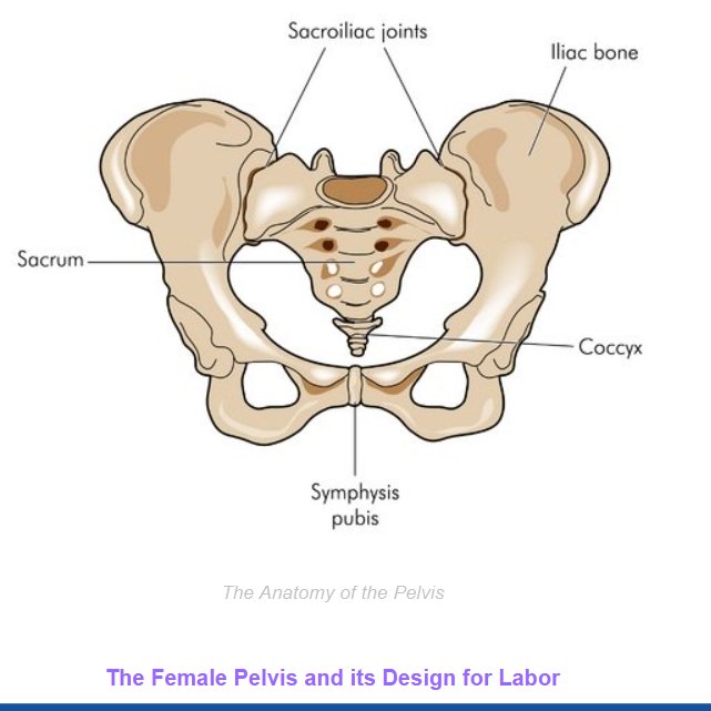 As a result, the female pelvis is generally broader and wider than the male pelvis.
Below, learn more about the bones, muscles, and organs of the female pelvis.
Female pelvis anatomy and function
Female pelvis bones
Hip bones
There are two hip bones, one on the left side of the body and the other on the right. Together, they form the part of the pelvis called the pelvic girdle.
The hip bones join to the upper part of the skeleton through attachment at the sacrum. Each hip bone is made of three smaller bones that fuse together during adolescence:
- Ilium. The largest part of the hip bone, the ilium, is broad and fan-shaped. You can feel the arches of these bones when you put your hands on your hips.
- Pubis. The pubis bone of each hip bone connects to the other at a joint called the pubis symphysis.
- Ischium. When you sit down, most of your body weight falls on these bones.
 This is why they’re sometimes called sit bones.
The ilium, pubis, and ischium of each hip bone come together to form the acetabulum, where the head of the thigh bone (femur) attaches.
Sacrum
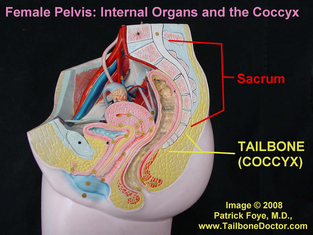
The sacrum is connected to the lower part of the vertebrae. It’s actually made up of five vertebrae that have fused together. The sacrum is quite thick and helps to support body weight.
Coccyx
The coccyx is sometimes called the tailbone. It’s connected to the bottom of the sacrum supported by several ligaments.
The coccyx is made up of four vertebrae that have fused into a triangle-like shape.
Female pelvis muscles
Levator ani muscles
The levator ani muscles are the largest group of muscles in the pelvis. They have several functions, including helping to support the pelvic organs.
The levator ani muscles consist of three separate muscles:
- Puborectalis. This muscle is responsible for holding in urine and feces.
 It relaxes when you urinate or have a bowel movement.
 - Pubococcygeus. This muscle makes up most of the levator ani muscles. It originates at the pubis bone and connects to the coccyx.
- Iliococcygeus. The iliococcygeus has thinner fibers and serves to lift the pelvic floor as well as the anal canal.
Coccygeus
This small pelvic floor muscle originates at the ischium and connects to the sacrum and coccyx.
Female pelvis organs
Uterus
The uterus is a thick-walled, hollow organ where a baby develops during pregnancy.
During the reproductive years, the lining of the uterus sheds every month during menstruation if you don’t become pregnant.
Ovaries
There are two ovaries located on either side of the uterus. The ovaries produce eggs and also release hormones, such estrogen and progesterone.
Fallopian tubes
The fallopian tubes connect each ovary to the uterus.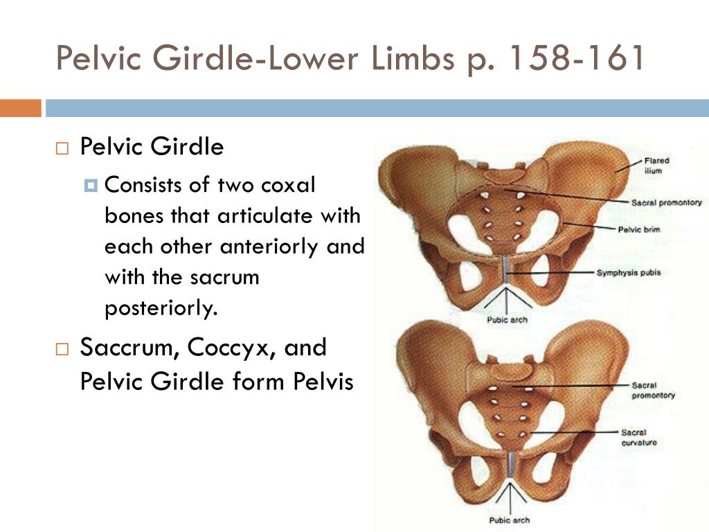 Specialized cells in the fallopian tubes use hair-like structures called cilia to help direct eggs from the ovaries toward the uterus.
Cervix
The cervix connects the uterus to the vagina. It’s able to widen, allowing sperm to pass into the uterus.
In addition, thick mucus produced in the cervix can help to prevent bacteria from reaching the uterus.
Vagina
The vagina connects the cervix to the exterior female genitalia. It’s also called the birth canal, as the baby passes through the vagina during delivery.
Rectum
The rectum is the lowest part of the large intestine. Feces collects here until exiting through the anus.
Bladder
The bladder is the organ that collects and stores urine until it’s released. Urine reaches the bladder through tubes called ureters that connect to the kidneys.
Urethra
The urethra is the tube that urine travels through to exit the body from the bladder.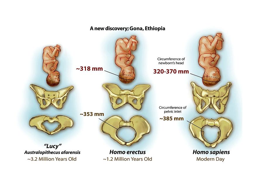 The female urethra is much shorter than the male urethra.
Female pelvis ligaments
Broad ligament
The broad ligament supports the uterus, fallopian tubes, and ovaries. It extends to both sides of the pelvic wall.
The broad ligament can be further divided into three components that are linked to different parts of the female reproductive organs:
- mesometrium, which supports the uterus
- mesovarium, which supports the ovaries
- mesosalpinx, which supports the fallopian tubes
Uterine ligaments
Uterine ligaments provide additional support for the uterus. Some of the main uterine ligaments include:
- the round ligament
- cardinal ligaments
- pubocervical ligaments
- uterosacral ligaments
Ovarian ligaments
The ovarian ligaments support the ovaries. There are two main ovarian ligaments:
- the ovarian ligament
- the suspensory ligament of the ovary
Female pelvis diagram
Explore this interactive 3-D diagram to learn more about the female pelvis:
Female pelvis conditions
The pelvis contains a large number of organs, bones, muscles, and ligaments, so many conditions can affect the entire pelvis or parts within it.
Some conditions that can affect the female pelvis as a whole include:
- Pelvic inflammatory disease (PID). PID is an infection that occurs in the female reproductive system. While it’s often caused by a sexually transmitted infection, other infections can also cause PID. Untreated PID can lead to complications, such as infertility or ectopic pregnancy.
- Pelvic organ prolapse. Pelvic organ prolapse occurs when the muscles in the pelvis can no longer support its organs, such as the bladder, uterus, or rectum. This can cause one or more of these organs to press down on the vagina. In some cases, this can cause a bulge to form outside of the vagina.
- Endometriosis. Endometriosis occurs when the tissue that lines the inside walls of the uterus (endometrium) begins to grow outside of the uterus. The ovaries, fallopian tubes, and other tissues in the pelvis are typically affected by the condition. Endometriosis can lead to complications, including infertility or ovarian cancer.
Symptoms of a pelvic condition
Some common symptoms of a pelvic condition can include:
- pain in the lower abdomen or pelvis
- a feeling of pressure or fullness in the pelvis
- unusual or foul-smelling vaginal discharge
- pain during sex
- bleeding in between periods
- painful cramping during or before periods
- pain during bowel movements or when urinating
- a burning feeling when urinating
Tips for a healthy pelvis
The female pelvis is a complex, important part of the body. Follow these tips to keep it in good health:
Stay on top of your reproductive health
See a gynecologist for a yearly health screen. Things like pelvic exams and Pap smears can aid in identifying pelvic conditions or infections early.
You can get a free or low-cost pelvic exam at your local Planned Parenthood clinic.
Practice safe sex
Use barriers — such as condoms or dental dams — during sexual activity, especially with a new partner, to avoid infections that could lead to PID.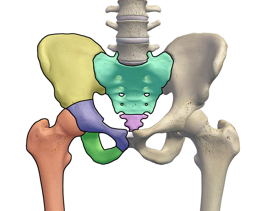
Try pelvic floor exercises
These types of exercises can help to strengthen the muscles in the pelvis, including those around the bladder and vagina.
Stronger pelvic floor muscles can aid in preventing things like incontinence or organ prolapse. Here’s how to get started.
Never ignore unusual symptoms
If you’re experiencing anything unusual in your pelvic area, such as bleeding between periods or unexplained pelvic pain, make an appointment with your doctor. Left untreated, some pelvic conditions can have lasting impacts on your health and fertility.
Pelvic dysfunction and posture disorder
Pelvic dysfunction and posture disorder
By: Admin | Tags: | Comments: 0 | April 3, 2018
All three pelvic bones converge in the so-called acetabulum, which is why intraosseous dysfunctions of any pelvic bone will inevitably affect the ligaments of the hip joint, the biomechanics of the entire pelvis, spine, and also the lower extremities. The pelvic bones provide support for the spine. Only their correct location can provide a person with an even posture.
In addition, the pelvis provides coordinated movement to the lower limbs and torso (they move in tandem). In the case when the pelvis is located normally, a person can perform various movements, twisting, tilting without pain and discomfort. Displacement (skew) of the pelvis from normal positions provokes dysfunctional disorders of the spine, due to the fact that there is a change in the axis of distribution of loads during movement.
For example, we all know that axle misalignment in a car causes rapid wheel wear. A similar situation occurs in the spine: there is an excessive load on certain areas, which leads to rapid wear of the structures of the spine.
Pelvic tilt also causes pain and dysfunction in the neck, pain in the neck radiating to the shoulders and arms. Pelvic dysfunction can contribute to carpal tunnel syndrome and other limb problems. That is why this pathology requires detailed consideration and treatment by specialists.
Types of pelvic dysfunctions
The most common problem that specialists have to deal with is recurrent cases of blockage of the sacroiliac joint.
Types of pelvic dysfunction also include:
- anterior and posterior rotation of the pelvic bone,
- superior pelvic displacement,
- opening/closing of the pelvic bone,
- lower/superior displacement of the pubic bone;
- dysfunctions of anterior/posterior, unilateral/multilateral sacral torsion, etc.
Causes of pelvic dysfunction
Pelvic dysfunction and postural disorders can develop for various reasons
These can be:
- Birth injuries;
- Complications after rickets, bone tuberculosis, poliomyelitis;
- Sequelae of past spinal fractures;
- Systemic connective tissue diseases;
- Presence of flat feet;
- Prolonged stay in the wrong position.
Incorrect posture can be caused by malformations of intrauterine development, which cause vertebral dystrophy and various deformities of the spine and pelvic bones in adulthood. But pathologies caused by weakness of the muscular corset or individual muscle groups have become much more widespread. They, in turn, can occur with prolonged abnormal position of the pelvis. Many of us sit incorrectly during study or work, which contributes to the increased development of some muscles and the weakening of others. There is an uneven effect on the pelvic bones, they are bent. Bad habits can exacerbate this situation, such as slouching your back or crossing one leg over the other while sitting. All this leads to such unpleasant phenomena as the curvature of the pelvic bones, scoliosis, kyphosis, lordosis and other diseases.
An improperly fitted bed also increases the risk of pelvic dysfunction and postural distortion. A soft mattress, in which a person literally drowns during sleep, can become a Procrustean bed, after which the limbs will change their length and position.
In some cases, the cause of the development of pathologies are spinal injuries, improperly selected shoes, too high heels and many other factors.
Consequences of pelvic dysfunction and poor posture
Violation of the physiological position of the spine can lead to:
- osteochondrosis, pain in the back and lower back;
- the development of diseases of the digestive system;
- visual impairment;
- diseases of the cardiovascular system;
- decrease in general immunity;
- frequent fatigue;
- appearance of degenerative changes in the spine,
- herniated disc,
- scoliosis,
- osteoarthritis,
- spinal stenosis,
- sciatica, etc.
How to treat pelvic dysfunction?
The most important thing here is early diagnosis and early treatment. In advanced cases, one has to resort to serious operations that cannot always completely eliminate the defect, and the person remains sick or disabled for life.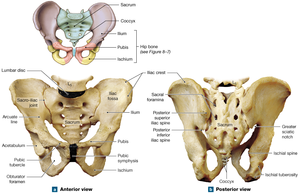
Only qualified medical assistance can break the vicious circle and restore the correct position of the pelvis and spine.
In the early stages of the development of the disease, the following methods are used in combination:
- Special gymnastics;
- Therapeutic swimming in the pool;
- Observance of the correct position of the body and back during study and work;
- Use of orthopedic pillows and mattresses during sleep;
- Therapeutic massage.
But, unfortunately, classical methods of treatment are not always effective. That is why, more and more often, official medicine is turning to osteopathy - a direction that successfully copes with the treatment of diseases of the musculoskeletal system.
Correction and strengthening of the spine with the help of osteopathic techniques gives a tangible positive result. Many experts agree that today it is these methods that allow you to get the most tangible results.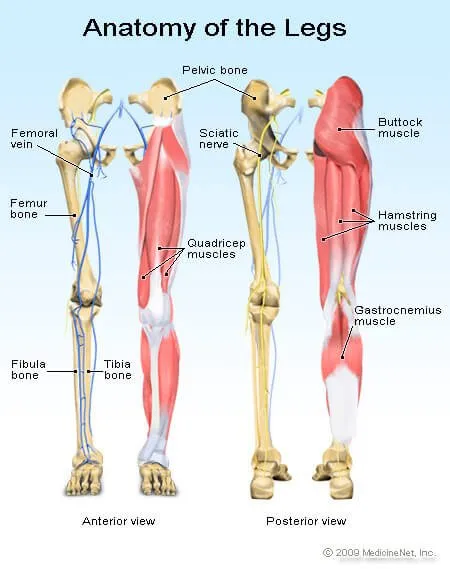
Having studied in detail the work of all systems and organs, the osteopath receives an objective picture of his condition and only after that determines the tactics of subsequent treatment.
Qualified specialists perfectly master and apply in practice various therapeutic techniques, with the help of which the normal axis of the spine is restored, muscles are strengthened, clamps, blocks of pathological changes are removed.
Osteopathy ensures the activation of self-healing processes in the body. With this method, you can eliminate the "root of the problem", and not stop the consequences. Sessions are especially useful for babies to eliminate the consequences of birth injuries, to strengthen the correct position of the spine. During the sessions, the specialist aims to eliminate the symptoms of the disease and find its causes, as well as to heal the patient's body as a whole.
In the complex, special physical exercises, acupuncture and many other techniques and methods can be used to restore the correct position of the pelvis and the natural mobility of the spine without surgical interventions.
A timely visit to an osteopath will help you avoid pain, correct your posture, and once and for all forget about the untidy sensations that accompany spinal curvature.
The advantage of osteopathy is its accessibility. Today, everyone who needs posture correction and the treatment of hip dysfunction can easily receive the necessary assistance by undergoing treatment sessions with specialists who practice the application of this effective method.
Biologists have found that whales use the pelvis for sex - Gazeta.Ru
Biologists have found that whales use the pelvis for sex - Gazeta.Ru | NewsDoctors found that one workout increases the level of anti-cancer proteins 13:54
The Kremlin announced the initial task of the SVO in Ukraine 13:54
19FortyFive: Russia defeated the West in an economic war and ruined its plans 13:53
Developer explains why push notifications are disabled on iPhone 13:52
Producer Rudchenko predicted Oksimiron's victory in a rap battle against Morgenstern 13:52
'Wonder Woman' Director Commented on the Freeze of the New Film in the Franchise 13:50
Shot: Sellers on Wildberries pay models for reviews with intimate photos 13:49
Gynecologists explained why drinking coffee after pregnancy 13:49
Investors withdrew almost $1.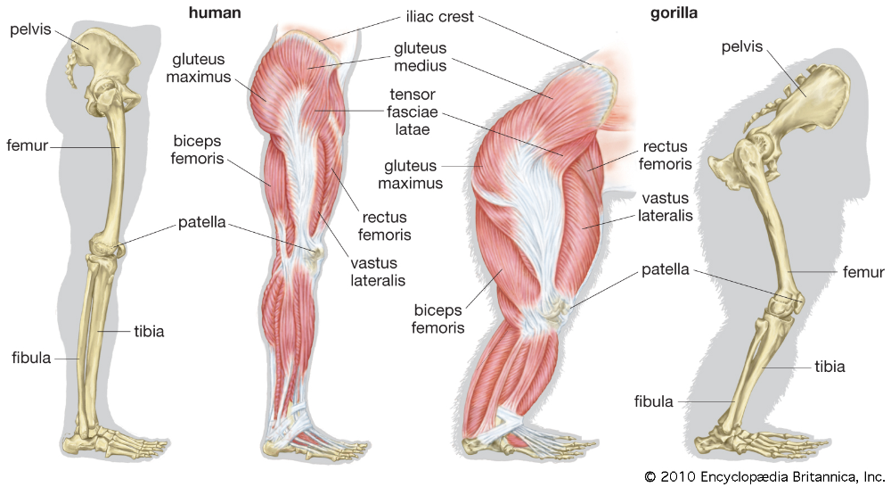 5 billion from Binance in one day as... 13:48
Lera Kudryavtseva told how her husband reacted to the 15-year difference... 13:47
Science
All cetaceans have a vestigial pelvic bone, a reminder that their ancestors once lived on land and were able to walk. Scientists have wondered why this bone has not disappeared over 40 million years of whale evolution, and found that it plays an important role in their sexual intercourse. Biologists wrote about their discovery in the journal Evolution .
Once considered useless, the whale's pelvic bone is attached to the muscles that control the movement of the penis. So with the loss of limbs in the ancestors of the whales, she received a new role, no less important.
Biologists have studied hundreds of pelvic bones from museum collections of marine mammal skeletons. By comparing the size of these bones with the size of the testicles, they concluded that the pelvic bones of cetaceans were the object of sexual selection.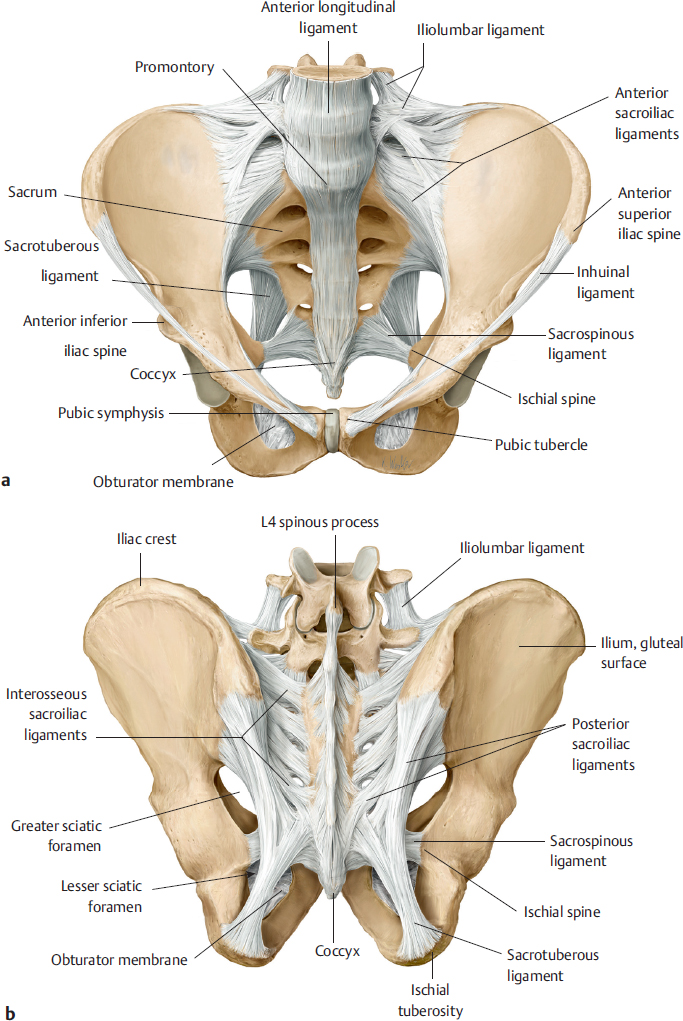
Animals that practice promiscuity, when males compete for more females, develop larger testes and a larger pelvic bone.
Scientists see this as an example of vestigial organs taking on a new role in the body. For example, the human appendix has been shown to be important for the immune system.
Subscribe to Gazeta.Ru in News, Zen and Telegram.
To report a bug, select the text and press Ctrl+Enter
News
Zen
Telegram
Picture of the day
Russian military operation in Ukraine. Day 294
Online broadcast of the special military operation in Ukraine — Day 294
Belarus remains the leader among all debtors to Russia. And took a new loan
RIA Novosti: Belarus' debt to Russia increased to $8.5 billion in 2021
The suspects are a welder, a foreman and a supervisor.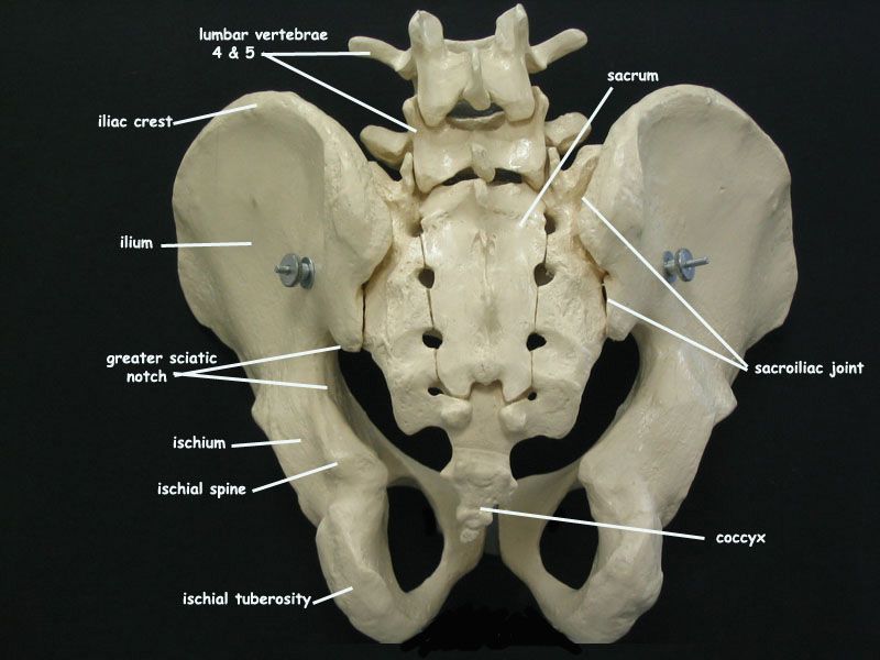 How the fire in the Mega-Khimki shopping center is being investigated
Investigative Committee announced the arrest of a third suspect in the case of a fire at the OBI warehouse in Khimki
Peskov: there were no proposals for a Christmas or New Year truce with Ukraine
The President of Latvia proposed the creation of a "Riga Tribunal" for Ukraine
Deputy Leonov criticized the idea of the Ministry of Finance to combine CHI and VHI
The Pope urged to celebrate Christmas modestly and give the money saved to Ukrainians
News and materials
Doctors have found that one workout increases the level of anti-cancer proteins
The Kremlin announced the initial task of the SVO in Ukraine
19FortyFive: Russia defeated the West in an economic war and ruined its plans
Developer explains why push notifications are disabled on iPhone
Producer Rudchenko predicted Oksimiron's victory in a rap battle against Morgenstern
'Wonder Woman' director comments on freeze of new
filmShot: Sellers on Wildberries pay models for reviews with intimate photos
Gynecologists explained why drinking coffee after pregnancy
Investors withdrew almost $1.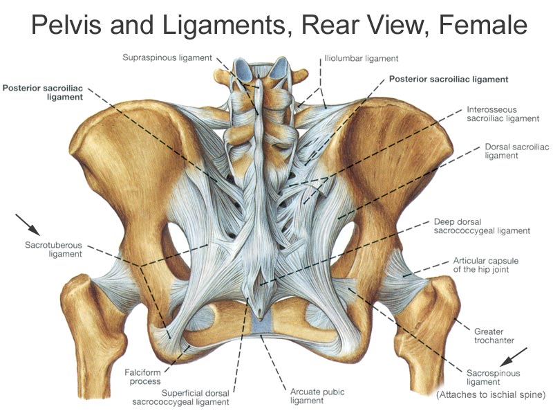 5 billion from Binance in one day amid growing anxiety in the crypto market
Lera Kudryavtseva told how her husband reacted to the 15-year age difference with TV presenter
43-year-old Hi-Fi star Tatyana Tereshina posed topless
"Wednesday" star Christina Ricci finalizes her divorce from her first husband, whom she accused of abuse
Saudi Defense Minister visits UK M777 howitzer manufacturer
Portugal player involved in sex scandal
In Kuzbass, a "black sky" regime was announced in Kemerovo, Novokuznetsk and Prokopyevsk
Two concerts of Valery Meladze canceled in Novosibirsk
The Kremlin assessed the damage from Lithuania's steps towards the Kaliningrad region
In Tomsk, an 8-year-old boy got tangled in a blanket and ended up in intensive care
All news
United States charged five Russians with secretly purchasing electronics for the Russian defense sector
Five Russians face up to 30 years in prison in the United States on charges of purchasing military electronics 10:51
Ovechkin scored 800 NHL goals and set a unique achievement
Ovechkin scored a hat-trick against Chicago and became the first star of the day in the NHL
“You forbade us to leave with threats”: star couples who stayed together after breaking up
Kristina Asmus and Garik Kharlamov and four more couples who did not leave after the breakup
“After another vaccination, Lenya stopped responding to the name”: stories of mothers of special children
Russian women told how their children communicate with speech problems
"Most Effective Defensive Weapon System".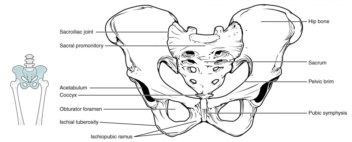 US Ready to Deliver Patriot to Ukraine
CNN: United States may announce delivery of Patriot air defense systems to Ukraine this week
"Operations in an extremely sensitive environment." What British Marines are doing in Ukraine
The general revealed the role of British Marines in "high-risk operations" in Ukraine
Kurt Cobain, Freddie Mercury and Princess Diana. What dead celebrities would look like now
The deputies undertook to protect the Russian language from foreign borrowings
The draft law on the protection of the Russian language from foreign words was adopted in the first reading
"We are for nuclear deterrence." How a mock-up rocket reached the US Embassy in Moscow
Traffic police stopped a car in Moscow with a mock-up of the Sarmat rocket and the inscription "To Washington"
"Her Truth": what a drama about the journalists who exposed Harvey Weinstein turned out to be
Review of the drama "Her Truth" with Carey Mulligan and Zoe Kazan about the exposure of Harvey Weinstein
"Very tired." Arestovich spoke about the state of Zelensky
Adviser to the head of the Office of the President of Ukraine Arestovich spoke about Zelensky's severe fatigue
"Transition to the Air Force of Tomorrow". How American hypersonic is catching up with Russia 13.12.2022, 17:24
"Out of the question." The Kremlin responded to Zelensky's proposal to withdraw troops
Peskov said that the withdrawal of Russian troops from the special operation zone in Ukraine is impossible

