Down syndrome fetuses
Facts about Down Syndrome | CDC
What is Down Syndrome?
Down syndrome is a condition in which a person has an extra chromosome. Chromosomes are small “packages” of genes in the body. They determine how a baby’s body forms and functions as it grows during pregnancy and after birth. Typically, a baby is born with 46 chromosomes. Babies with Down syndrome have an extra copy of one of these chromosomes, chromosome 21. A medical term for having an extra copy of a chromosome is ‘trisomy.’ Down syndrome is also referred to as Trisomy 21. This extra copy changes how the baby’s body and brain develop, which can cause both mental and physical challenges for the baby.
Even though people with Down syndrome might act and look similar, each person has different abilities. People with Down syndrome usually have an IQ (a measure of intelligence) in the mildly-to-moderately low range and are slower to speak than other children.
Some common physical features of Down syndrome include:
- A flattened face, especially the bridge of the nose
- Almond-shaped eyes that slant up
- A short neck
- Small ears
- A tongue that tends to stick out of the mouth
- Tiny white spots on the iris (colored part) of the eye
- Small hands and feet
- A single line across the palm of the hand (palmar crease)
- Small pinky fingers that sometimes curve toward the thumb
- Poor muscle tone or loose joints
- Shorter in height as children and adults
How Many Babies are Born with Down Syndrome?
Down syndrome remains the most common chromosomal condition diagnosed in the United States. Each year, about 6,000 babies born in the United States have Down syndrome. This means that Down syndrome occurs in about 1 in every 700 babies.1
Types of Down Syndrome
There are three types of Down syndrome. People often can’t tell the difference between each type without looking at the chromosomes because the physical features and behaviors are similar.
- Trisomy 21: About 95% of people with Down syndrome have Trisomy 21.2 With this type of Down syndrome, each cell in the body has 3 separate copies of chromosome 21 instead of the usual 2 copies.
- Translocation Down syndrome: This type accounts for a small percentage of people with Down syndrome (about 3%).2 This occurs when an extra part or a whole extra chromosome 21 is present, but it is attached or “trans-located” to a different chromosome rather than being a separate chromosome 21.
- Mosaic Down syndrome: This type affects about 2% of the people with Down syndrome.
 2 Mosaic means mixture or combination. For children with mosaic Down syndrome, some of their cells have 3 copies of chromosome 21, but other cells have the typical two copies of chromosome 21. Children with mosaic Down syndrome may have the same features as other children with Down syndrome. However, they may have fewer features of the condition due to the presence of some (or many) cells with a typical number of chromosomes.
Causes and Risk Factors
- The extra chromosome 21 leads to the physical features and developmental challenges that can occur among people with Down syndrome. Researchers know that Down syndrome is caused by an extra chromosome, but no one knows for sure why Down syndrome occurs or how many different factors play a role.
- One factor that increases the risk for having a baby with Down syndrome is the mother’s age. Women who are 35 years or older when they become pregnant are more likely to have a pregnancy affected by Down syndrome than women who become pregnant at a younger age.
 3-5However, the majority of babies with Down syndrome are born to mothers less than 35 years old, because there are many more births among younger women.6,7
Diagnosis
There are two basic types of tests available to detect Down syndrome during pregnancy: screening tests and diagnostic tests. A screening test can tell a woman and her healthcare provider whether her pregnancy has a lower or higher chance of having Down syndrome. Screening tests do not provide an absolute diagnosis, but they are safer for the mother and the developing baby. Diagnostic tests can typically detect whether or not a baby will have Down syndrome, but they can be more risky for the mother and developing baby. Neither screening nor diagnostic tests can predict the full impact of Down syndrome on a baby; no one can predict this.
Screening Tests
Screening tests often include a combination of a blood test, which measures the amount of various substances in the mother’s blood (e. g., MS-AFP, Triple Screen, Quad-screen), and an ultrasound, which creates a picture of the baby. During an ultrasound, one of the things the technician looks at is the fluid behind the baby’s neck. Extra fluid in this region could indicate a genetic problem. These screening tests can help determine the baby’s risk of Down syndrome. Rarely, screening tests can give an abnormal result even when there is nothing wrong with the baby. Sometimes, the test results are normal and yet they miss a problem that does exist.
Diagnostic Tests
Diagnostic tests are usually performed after a positive screening test in order to confirm a Down syndrome diagnosis. Types of diagnostic tests include:
- Chorionic villus sampling (CVS)—examines material from the placenta
- Amniocentesis—examines the amniotic fluid (the fluid from the sac surrounding the baby)
- Percutaneous umbilical blood sampling (PUBS)—examines blood from the umbilical cord
These tests look for changes in the chromosomes that would indicate a Down syndrome diagnosis.
Other Health Problems
Many people with Down syndrome have the common facial features and no other major birth defects. However, some people with Down syndrome might have one or more major birth defects or other medical problems. Some of the more common health problems among children with Down syndrome are listed below.8
- Hearing loss
- Obstructive sleep apnea, which is a condition where the person’s breathing temporarily stops while asleep
- Ear infections
- Eye diseases
- Heart defects present at birth
Health care providers routinely monitor children with Down syndrome for these conditions.
Treatments
Down syndrome is a lifelong condition. Services early in life will often help babies and children with Down syndrome to improve their physical and intellectual abilities. Most of these services focus on helping children with Down syndrome develop to their full potential. These services include speech, occupational, and physical therapy, and they are typically offered through early intervention programs in each state. Children with Down syndrome may also need extra help or attention in school, although many children are included in regular classes.
Other Resources
The views of these organizations are their own and do not reflect the official position of CDC.
- Down Syndrome Research Foundation (DSRF)
DSRF initiates research studies to better understand the learning styles of those with Down syndrome. - Global Down Syndrome Foundation
This foundation is dedicated to significantly improving the lives of people with Down syndrome through research, medical care, education and advocacy. - National Association for Down Syndrome
The National Association for Down Syndrome supports all persons with Down syndrome in achieving their full potential. They seek to help families, educate the public, address social issues and challenges, and facilitate active participation. - National Down Syndrome Society (NDSS)
NDSS seeks to increase awareness and acceptance of those with Down syndrome.
References
- Mai CT, Isenburg JL, Canfield MA, Meyer RE, Correa A, Alverson CJ, Lupo PJ, Riehle‐Colarusso T, Cho SJ, Aggarwal D, Kirby RS. National population‐based estimates for major birth defects, 2010–2014. Birth Defects Research. 2019; 111(18): 1420-1435.
- Shin M, Siffel C, Correa A. Survival of children with mosaic Down syndrome. Am J Med Genet A. 2010;152A:800-1.
- Allen EG, Freeman SB, Druschel C, et al. Maternal age and risk for trisomy 21 assessed by the origin of chromosome nondisjunction: a report from the Atlanta and National Down Syndrome Projects. Hum Genet. 2009 Feb;125(1):41-52.
- Ghosh S, Feingold E, Dey SK. Etiology of Down syndrome: Evidence for consistent association among altered meiotic recombination, nondisjunction, and maternal age across populations. Am J Med Genet A. 2009 Jul;149A(7):1415-20.
- Sherman SL, Allen EG, Bean LH, Freeman SB. Epidemiology of Down syndrome. Ment Retard Dev Disabil Res Rev.
 2007;13(3):221-7.
 - Adams MM, Erickson JD, Layde PM, Oakley GP. Down’s syndrome. Recent trends in the United States. JAMA. 1981 Aug 14;246(7):758-60.
- Olsen CL, Cross PK, Gensburg LJ, Hughes JP. The effects of prenatal diagnosis, population ageing, and changing fertility rates on the live birth prevalence of Down syndrome in New York State, 1983-1992. Prenat Diagn. 1996 Nov;16(11):991-1002.
- Bull MJ, the Committee on Genetics. Health supervision for children with Down syndrome. Pediatrics. 2011;128:393-406.
Prenatal Testing for Down Syndrome | Patient Education
Down syndrome is a genetic condition caused by extra genes from the 21st chromosome. It results in certain characteristics, including some degree of cognitive disability and other developmental delays. Common physical traits include an upward slant of the eyes; flattened bridge of the nose; single, deep crease on the palm of the hand; and decreased muscle tone. A child with Down syndrome, however, may not have all these traits.
The incidence of Down syndrome in the United States is about 1 in 1,000 births. There is no association between Down syndrome and culture, ethnic group, socioeconomic status or geographic region.
Age-Related Risks
Generally, the chance of having a Down syndrome birth is related to the mother's age. Under age 25, the odds of having a child with Down syndrome are about 1 in 1,400. At age 35, the odds are about 1 in 350. At age 40, the odds are about 1 in 100.
Causes of Down Syndrome
There are three causes of Down syndrome:
Trisomy 21
An estimated 95 percent of people with Down syndrome have trisomy 21, meaning they have three number 21 chromosomes instead of two. We normally have 23 pairs of chromosomes, each made up of genes. During the formation of the egg and the sperm, a woman's or a man's pair of chromosomes normally split so that only one chromosome is in each egg or sperm. In trisomy 21, the 21st chromosome pair does not split and a double dose goes to the egg or sperm.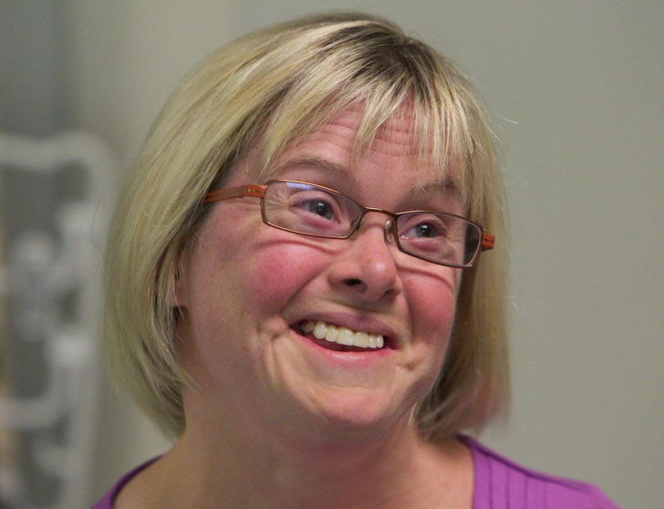 An estimated 95 to 97 percent of the extra chromosome is of maternal origin.
Translocation
Translocation occurs in about 3 to 4 percent of people with Down syndrome. In this type, an extra part of the 21st chromosome gets stuck onto another chromosome. In about half of these situations, one parent carries the extra 21st chromosome material in a "balanced" or hidden form.
Mosaicism
In mosaicism, the person with Down syndrome has an extra 21st chromosome in some of the cells but not all of them. The other cells have the usual pair of 21st chromosomes. About 1 to 2 percent of people with Down syndrome have this type.
Prenatal Testing
Screening tests can identify women at increased risk of having a baby with Down syndrome. These tests have no risks of miscarriage, but can't determine with certainty whether a fetus is affected. Diagnostic tests, on the other hand, are extremely accurate at identifying certain abnormalities in the fetus, but carry a small — generally less than 1 percent — risk of miscarriage. We offer options for both screening and diagnostic testing.
Continue reading
Screening Tests
Sequential Integrated Screening — Sequential integrated screening is offered to all pregnant women by the state of California. This non-invasive screening is performed in two steps.
In the first step, which is performed between 10 and 14 weeks of pregnancy, a blood sample is taken from the mother and a nuchal translucency ultrasound is performed to measure the amount of fluid at the back of the baby's neck. If the blood test is scheduled prior to the ultrasound, we can provide the results at the end of the ultrasound appointment. The results of the blood test, the nuchal translucency measurement and the mother's age are used to estimate the risk for Down syndrome and trisomy 18.
The second step is a maternal blood test between 15 to 20 weeks of pregnancy. When the results of this blood test are combined with the results from the first trimester blood test and nuchal translucency ultrasound, the detection rate for Down syndrome increases.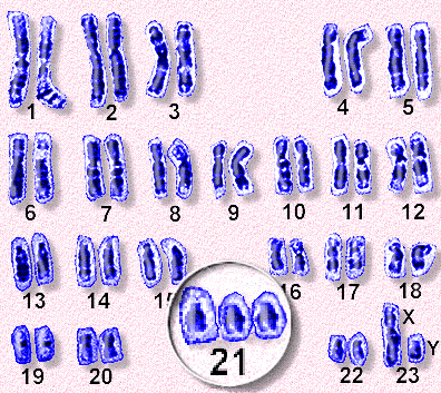 This test also provides a personal risk assessment for having a fetus with trisomy 18, Smith-Lemli-Opitz syndrome, an open neural tube defect or an abdominal wall defect.
Diagnostic Tests
Amniocentesis, chorionic villus sampling (CVS) and ultrasound are the three primary procedures for diagnostic testing.
Amniocentesis — Amniocentesis is used most commonly to identify chromosomal problems such as Down syndrome. When the fetus is known to be at risk, it can detect other genetic diseases like cystic fibrosis, Tay-Sachs disease and sickle cell disease.
An amniocentesis procedure for genetic testing is typically performed between 15 and 20 weeks of pregnancy. Under ultrasound guidance, a needle is inserted through the abdomen to remove a small amount of amniotic fluid. The cells from the fluid are then cultured and a karyotype analysis — an analysis of the chromosomal make-up of the cells — is performed. It takes about two weeks to receive the results of the test.
Amniocentesis detects most chromosomal disorders, such as Down syndrome, with a high degree of accuracy. Testing for other genetic diseases, such as Tay-Sachs disease, is not routinely performed but can be detected through specialized testing if your fetus is known to be at risk. Testing for neural tube defects, such as spina bifida, also can be performed.
There is a small risk of miscarriage as a result of amniocentesis — about 1 in 100 or less. Miscarriage rates for procedures performed at UCSF Medical Center are less than 1 in 350.
Chorionic Villus Sampling (CVS) — Like amniocentesis, chorionic villus sampling is used most commonly to identify chromosomal problems such as Down syndrome. It can detect other genetic diseases like cystic fibrosis, Tay-Sachs disease and sickle cell disease in at-risk fetuses. The main advantage of CVS over amniocentesis is that it is done much earlier in pregnancy, at 10 to 12 weeks rather than 15 to 20 weeks.
CVS involves removing a tiny piece of tissue from the placenta. Under ultrasound guidance, the tissue is obtained either with a needle inserted through the abdomen or a catheter inserted through the cervix. The tissue is then cultured and a karyotype analysis of the chromosomal make-up of the cells is performed. It takes about two weeks to receive the results.
The advantage of CVS over amniocentesis is that the test is performed much earlier in pregnancy, so results are typically available by the end of the third month. A disadvantage is that spinal cord defects cannot be detected. Expanded alpha fetoprotein (AFP) blood testing or ultrasound can be performed later in the pregnancy to screen for spinal cord defects.
There is a small risk of miscarriage as a result of CVS — 1 in 100 or less. Miscarriage rates for procedures performed at UCSF Medical Center are less than 1 in 350.
Ultrasound — The primary purpose of ultrasound is to determine the status of a pregnancy — the due date, size of the fetus and if the mother is carrying multiples. Ultrasound also can provide some information about possible birth defects in a fetus. All patients at UCSF Medical Center undergo a comprehensive ultrasound examination before any invasive tests are performed. Results of the ultrasound are explained at the time of the visit.
In some patients, an ultrasound raises concern of a possible abnormality in the fetus. We have extensive experience in performing and interpreting ultrasounds in pregnancy.
If You Receive a Positive Result
If you receive positive results on a screening test, we recommend that you discuss this with your doctor and a genetic counselor. Options for further diagnostic testing will be explained. The decision as to whether to have invasive genetic testing is up to you.
If a diagnostic test finds a genetic abnormality, the significance of such results should be discussed with experts familiar with the condition, including a medical geneticist and a genetic counselor, as well as your own doctor.
Down syndrome - causes, symptoms of the disease, diagnosis and treatment
I confirm More
- INVITRO
- Library
- Disease Handbook
- Down Syndrome
Trisomy
Brachycephaly
Mental retardation
Speech development
26669 21st of June
IMPORTANT!
The information in this section should not be used for self-diagnosis or self-treatment. In case of pain or other exacerbation of the disease, only the attending physician should prescribe diagnostic tests. For diagnosis and proper treatment, you should contact your doctor.
For a correct assessment of the results of your analyzes in dynamics, it is preferable to do studies in the same laboratory, since different laboratories may use different research methods and units of measurement to perform the same analyzes.
Down syndrome: causes, symptoms, diagnosis and treatment.
Definition
Down's syndrome is a congenital chromosomal anomaly, consisting in the presence of an extra chromosome in the 21st pair (trisomy of the 21st pair of chromosomes). A person has 23 pairs of chromosomes, so an ordinary child has 46 chromosomes, and a child with Down syndrome has 47. Down syndrome is characterized by a special appearance of the patient and a decrease in intellectual abilities. The frequency of this chromosomal anomaly in the population is 1:800 and does not depend on gender, race, family standard of living, or the presence or absence of bad habits in parents.
In Russia, 2,500 children with Down syndrome are born annually.
Causes of Down syndrome
The risk of having a child with Down syndrome for a woman increases from the age of 35 and reaches 1% by the age of 39. Of the total number of newborns with Down's disease, more than 20% are born to mothers over 35 years of age. In addition, risk factors are the presence of hepatitis B or C in the mother, tuberculosis, rubella, Botkin's disease, the father's age is over 45 years, the mother's age is too young (up to 18 years), and closely related marriages.
Classification of Down syndrome
There are three types of Down syndrome:
- trisomy is the most common form of Down syndrome, which is characterized by complete tripling of 21 chromosomes in all cells of the body; this form accounts for 94-95% of all cases of the disease;
- displacement (translocation) of 21 pairs of chromosomes to other chromosomes - occurs in 4% of cases;
- mosaic Down's syndrome (about 2% of cases), when only some cells of the body contain tripled chromosome 21. Patients themselves, as a rule, are no different from healthy ones, but have a high risk of having a child with Down syndrome.
Symptoms of Down syndrome
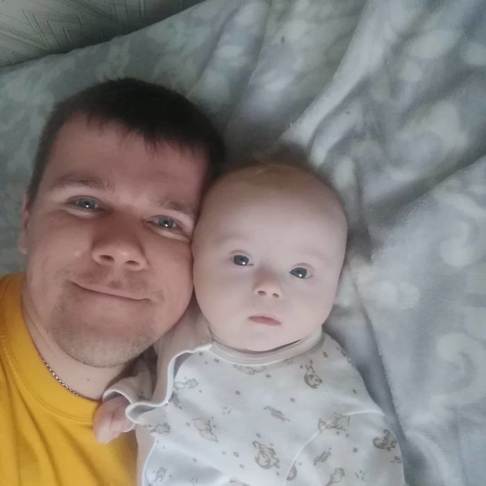
A child with Down syndrome has wide-set eyes with a Mongoloid incision, light pigment spots can be observed on the iris, epicanthus is often present - a vertical fold located between the upper and lower eyelids, partially covering the inner canthus .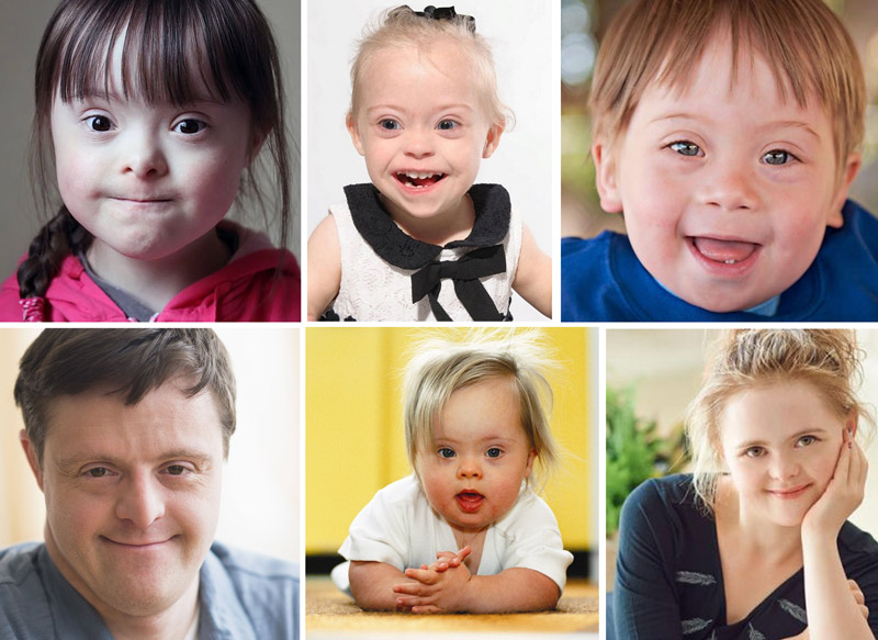 In addition, the distinguishing features are a short nose, a flat bridge of the nose, small auricles, brachycephaly (short and wide, almost round, head), a flat nape, an arched palate. Children often have anomalies of the dentition, underdevelopment of the lower jaw.
The body and limbs are disproportionately formed - the figure is squat, the shoulders are lowered, the limbs are short, there is a skin fold on the neck, the fingers may be shortened due to underdevelopment of the middle phalanges. Children with Down syndrome have a unique pattern of fingers and palms, this does not affect development in any way, but is a diagnostic feature. The feet are normal, but with an increased gap between the first and second toes, the sole often has a deep crease in this place. Most people with Down syndrome have flat feet. Muscle tone is significantly reduced, which affects movements.
Malformations of various organs and systems are diagnosed - the heart, the gastrointestinal tract, hypoplasia of the genital organs, keeled (the sternum protrudes) or funnel-shaped (the sternum is depressed) deformity of the chest.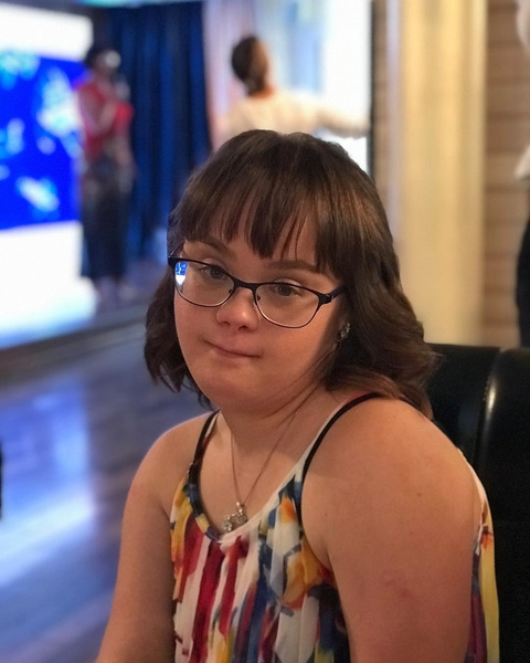
Children with Down syndrome have mental retardation of varying severity. All children with Down syndrome lag behind in psychomotor development - they have reduced emotional activity, they begin to sit, walk, talk later than their peers, their speech is underdeveloped, their vocabulary is poor.
Speech disorders are associated not only with insufficiency of intelligence, but also with frequent hearing impairments and reduced muscle tone.
Despite the lag in intellectual and psycho-emotional development, children with Down syndrome are teachable, although they need more time to master certain knowledge than their peers. They can attend pre-school and school institutions, receive vocational education, be creative, lead a normal life and start families.
Such patients may show affection, benevolence, and curiosity.
Down Syndrome Diagnosis
During pregnancy, a woman can be diagnosed with Down syndrome. Primary (screening) diagnosis includes ultrasound and markers such as free beta hCG and PAPP-A (pregnancy-associated protein A) between the 11th and 13th weeks of pregnancy.
Screening ultrasound 1st trimester of pregnancy (11-13 weeks 6 days)
Investigation necessary to monitor the growth and development of the fetus in the first trimester of pregnancy.
RUB 2,790 Sign up
1st trimester prenatal screening for trisomies, PRISCA-1 (1st trimester biochemical screening - 1st trimester “double test”, risk calculation using PRISCA software)
Synonyms: Prenatal Screening Markers for Down Syndrome; PRISCA-1. Brief description of the study "Prenatal screening for trisomies of the 1st trimester of pregnancy, PRISCA-1)" Test run...
Up to 1 business day
Available with home visit
2 040 RUB
Add to cart
If, according to the results of the research, there is a suspicion of Down's syndrome in the fetus, an invasive procedure is possible - a chorionic villus biopsy, or amniocentesis, when a sample of amniotic fluid is taken.
The decision to conduct an invasive study is made individually in each case, since the procedure is uncomfortable for the mother and has a risk of spontaneous abortion.
There is another screening diagnostic method - non-invasive prenatal testing (NIPT), which allows you to determine the chromosomal abnormalities of the fetus by the mother's blood, due to the detection of DNA fragments of her child in the woman's blood. If a fetus is at high risk of having Down syndrome after NIPT, an invasive examination is still required to confirm the diagnosis.
The second screening is done at 18-20 weeks of gestation and includes an ultrasound and a blood test if not previously done.
Screening ultrasound of the 2nd trimester of pregnancy (18-21 weeks) with Doppler evaluation of blood flow parameters
Study to monitor the course of multiple pregnancy, growth and development of fetuses and the viability of blood circulation.
RUB 3,890 Sign up
2nd trimester prenatal trisomy screening, PRISCA-2 (2nd trimester biochemical screening, 2nd trimester “triple test”, risk calculation using PRISCA software)
The study is performed for screening examination of pregnant women in order to assess the risk of fetal chromosomal abnormalities - trisomy 21 (Down's syndrome), trisomy 1...
Up to 1 business day
Available with house call
2 120 RUB
Add to cart
Third ultrasound screening performed at 30-34 weeks.
Screening ultrasound of the 3rd trimester of pregnancy (30-34 weeks) with Doppler evaluation of blood flow parameters
Ultrasound examination for functional assessment of intrauterine development of the fetus, its estimated height and weight, as well as blood circulation.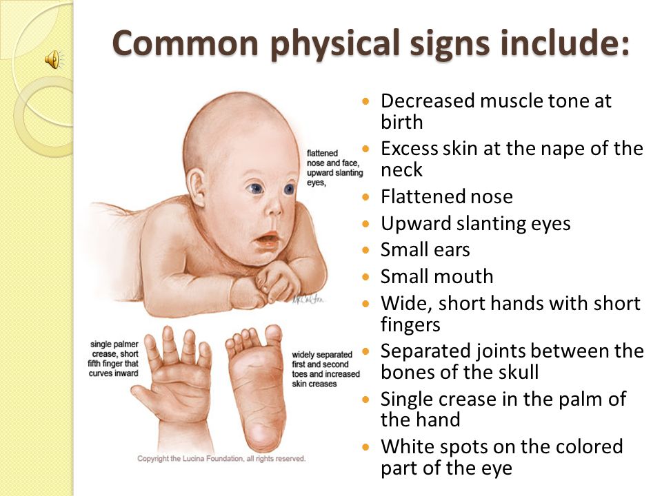
RUB 3 890 Sign up
Which doctors to contact
Prenatal diagnosis of fetal Down syndrome is done by a doctor – obstetrician-gynecologist.
The child is watching pediatrician, if necessary, genetic testing is carried out with a consultation of a geneticist. Further, a patient with Down syndrome should be observed by an ophthalmologist, cardiologist, neurologist, dentist, orthopedist-traumatologist, gynecologist, ENT doctor.
Treatment for Down syndrome
There is no cure for Down syndrome. Patients with Down syndrome usually have a sufficient number of medical problems and conditions that may require care immediately after birth, intermittent treatment, or long-term treatment throughout life. Concomitant malformations are often an indication for surgical treatment.
Children need constant attention and care, physical (exercise) and psychological rehabilitation.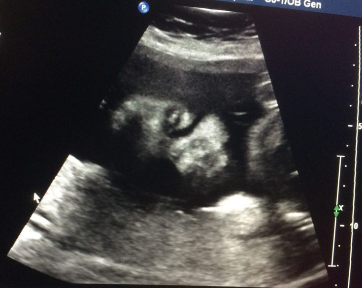 Physiotherapy is prescribed to increase muscle strength, improve posture and balance. Speech therapist helps to solve problems with speech. Many patients are forced to resort to the use of hearing aids, glasses for vision correction, orthodontic constructions, bandages that help move, wear orthopedic shoes. Patients with Down syndrome are prone to obesity, so great attention should be paid to sufficient physical activity and a balanced diet.
In order to correct speech development disorders and related learning difficulties, nootropic drugs are used. Hyperactivity, impulsivity problems, and irritability may be indications for the prescription of psychotropic drugs. In addition, drugs are used to improve metabolic processes and motor activity.
In each age period, the attention of parents and doctors should be directed to the correction of certain conditions.
In the neonatal period (up to 1 month) and in the first year of life, the presence of malformations of the cardiovascular system and the gastrointestinal tract, as well as thyroid function, is assessed. At the age of 1-5 years, control requires sleep disturbances, constipation, instability of the cervical spine, delayed development of motor skills due to reduced muscle tone. At an older age (5-13 years), sleep disorders, constipation, skin lesions, behavioral disorders, learning difficulties, memory development, communication need correction, the period of sexual development begins. Puberty is accompanied by behavioral problems, deficits or delays in cognitive skills and communication, sexual behavior issues, and behavioral changes. In adulthood, sleep problems, constipation, visual and hearing impairments also persist.
Complications
Approximately half of children with Down syndrome are diagnosed with heart defects, the most common are ventricular septal defect, open common atrioventricular canal, tetralogy of Fallot and fibroelastosis. Narrowing of the nasopharynx and oropharynx, Eustachian tube, external auditory canal due to the underdevelopment of the middle part of the face leads to the fact that many children with Down syndrome experience apnea - episodes of complete or partial cessation of breathing during sleep, accompanied by impaired ventilation and a decrease in oxygen levels in blood.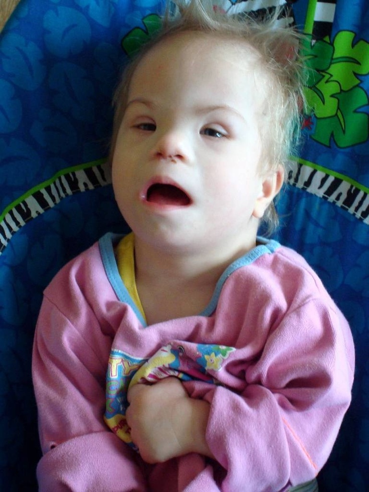
Digestive organs are characterized by regurgitation, bloating, and constipation.
Very high risk of infections, especially of the respiratory tract. The syndrome is often accompanied by eye diseases - congenital cataracts, nystagmus, strabismus, glaucoma, keratoconus, blepharitis and insufficiency of the nasolacrimal ducts. Insufficiency or obstruction of the nasolacrimal canal increases the risk of developing conjunctivitis, lacrimation. Due to frequent otitis media, the outflow of fluid from the middle ear is difficult, which increases the risk of hearing loss. Repeated otitis causes conductive hearing loss and, as a result, impaired speech function.
Children with Down syndrome may develop scoliosis, hip dysplasia, subluxation or dislocation of the hip, and instability of the patella. Due to the abnormal structure of collagen fibers, weakness of the ligamentous apparatus is observed, leading to hypermobility, instability of the joints, and their excessive mobility.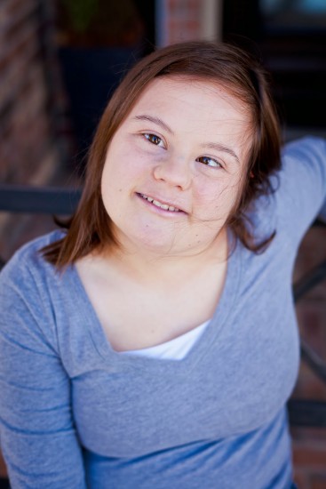
Prevention of Down syndrome
There is no prevention of Down syndrome. Couples planning to have a child are advised not to delay pregnancy until a later age, to undergo prenatal screening and genetic counseling.
Sources:
- Grigoriev K.I. Down syndrome: comorbidity and program goals in the work of a pediatrician with such children // Difficult patient. - 2017. - No. 1–2. – T 15.
- Sapozhnikova T.V. Psychological and pedagogical support for children with Down syndrome and their families in a rehabilitation center: a teaching aid / T.V. Sapozhnikova, N.A. Pershina, N.A. Shchigreva. - Biysk, 2019. - 311 p.
IMPORTANT!
The information in this section should not be used for self-diagnosis or self-treatment. In case of pain or other exacerbation of the disease, only the attending physician should prescribe diagnostic tests. For diagnosis and proper treatment, you should contact your doctor.
For a correct assessment of the results of your analyzes in dynamics, it is preferable to do studies in the same laboratory, since different laboratories may use different research methods and units of measurement to perform the same analyzes.
Recommendations
-
Pulmonary embolism (PE, pulmonary thromboembolism)
1060 November 23
-
Epidermophytosis
374 November 22
-
Liver dystrophy
5991 November 18th
Show 9 more0003
Convulsions
Hypopigmentation
Mental retardation
Phenylketonuria
Phenylketonuria: causes, symptoms, diagnosis and treatment.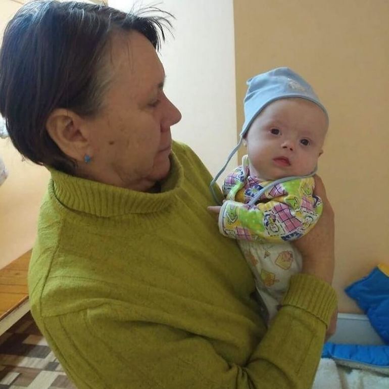
More
Mental retardation
Convulsions
Muscular ataxia
Apnea
Scoliosis
Epilepsy
Rett syndrome
Rett syndrome: causes, symptoms, diagnosis and treatment.
More
Trisomy
Late pregnancy
Underdevelopment of the brain
Hypertelorism
Cleft lip
Wolf mouth
Patau syndrome
Patau syndrome: causes, symptoms, diagnosis and treatment.
More
Pain in the knee
Edema
Osteochondrosis
Inflammation of the joint
Osgood-Schlatter disease
Osgood-Schlatter disease: causes, symptoms, diagnosis and treatment.
More
Osteoporosis
Obesity
Fracture of the femoral neck
Fracture of the femoral neck: causes, symptoms, diagnosis and treatment.
More
Nothing found
Try changing your request or select a doctor or service from the list.
Doctor not found
Try changing your query or select doctor from the list
Medical office not found
Try changing your request or select medical office from the list
Therapist Traumatologist-orthopedist Endocrinologist Urologist Gynecologist Ultrasound doctor Cardiologist Pediatrician
Nothing found
Try changing your query
Thank you!
You have successfully made an appointment
Detailed information has been sent to your e-mail
Subscribe to our newsletters
Enter e-mail
I consent to processing of personal data
Subscribe
Ultrasound diagnosis of Down syndrome and other chromosomal abnormalities
- Fetal ultrasound is the main method for diagnosing the likelihood of Down syndrome
- Basic ultrasound markers of Down syndrome
- Signs of Down syndrome on ultrasound in the second trimester of pregnancy
- How else can you determine the risk of having a baby with Down syndrome
Down's syndrome is a relatively common congenital pathology caused by the presence of an extra chromosome in 21 pairs.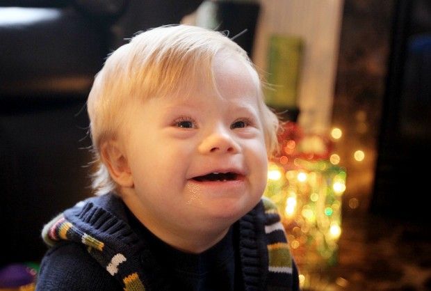 Down syndrome is also called trisomy for 21 pairs of chromosomes. Of all the chromosomal abnormalities, this pathology is the most well-known for a wide range of people.
Down's syndrome is associated with many significant malformations. Such as, for example, congenital heart defects, duodenal atresia (a condition where part of the small intestine does not develop), an increased risk of developing acute leukemia (leukemia). About 50% of children with this condition are born with heart defects, the most common of which is a ventricular septal defect. Another disease that is often observed in such children is Hirschsprung's disease - an aganglionic area of the large intestine, leading to the development of intestinal obstruction.
Screening tests during pregnancy are used to assess the risk of having a baby with Down syndrome. These tests are painless and non-invasive (do not cause damage to the tissues of the pregnant woman and fetus), but do not accurately judge the presence or absence of congenital pathology. Nevertheless, their widespread use makes it possible to suspect risks and recommend invasive procedures. Invasive diagnostic methods include interventions performed under ultrasound guidance - amniocentesis, chordocentesis and chorionic villus sampling.
Fetal ultrasound is the main way to diagnose the likelihood of Down syndrome
Fetal ultrasound to detect developmental abnormalities is called ultrasound screening. Most often, ultrasound screening is combined with biochemical blood tests of a pregnant woman, and such a study is called combined screening. Depending on the choice of criteria, the probability of detecting Down syndrome in the second trimester is 60-91%. The use of color Doppler technology for ultrasound screening makes it possible to detect defects in the fetal heart and increases the accuracy of diagnosis.
Major ultrasound markers of Down syndrome
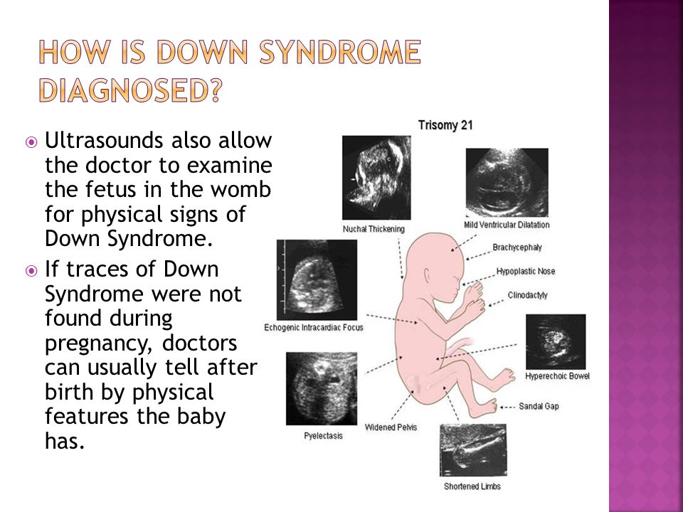
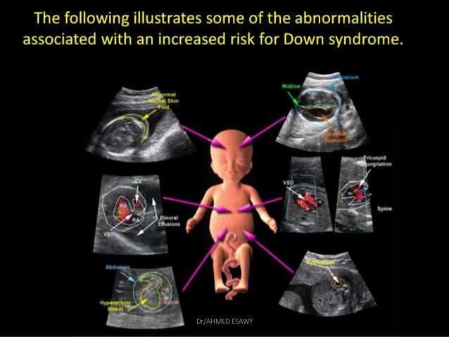
Ultrasound markers are the ultrasound measurements most likely to be associated with a high chance of having Down syndrome in the fetus. None of these markers is specific, that is, inherent only in this pathology. The following indicators are taken into account: the thickness of the collar space (75% of sensitivity), the absence of the nasal bone (58%), heart defects, short femurs and humeri, hyperechoic intestines, choroid plexus cysts, echogenic foci in the heart, signs of duodenal atresia . Of course, none of these markers is an absolute sign of a chromosomal abnormality.
Measuring the thickness of the nuchal space
The nuchal space (cervical fold) is the area between the folds of fetal tissues, which is transparent on ultrasound, located below the occiput in the area of neck formation. In children with chromosomal abnormalities, such as Down's syndrome or trisomy 18, fluid accumulates in this place. The measurement of this space is carried out from 11 to 14 weeks during the ultrasound of pregnancy in the first trimester. Measurements obtained at this time are the most significant for predicting the individual risk of developing fetal chromosomal abnormalities. The thickness of the collar space more than 3 mm is taken as an indicator of an increased likelihood of genetic defects in the fetus. The increase in the collar space does not mean that the fetus necessarily has Down syndrome, but if we take into account the gestational age of the fetus on which measurements were taken, the age of the mother and biochemical screening indicators, the probability of a correct diagnosis becomes higher than 80%. Although nuchal thickness screening is one of the most important tests for detecting chromosomal abnormalities, there are biases. The error arises due to violations of the measurement technique. This applies not only to measurements of the collar space, but also to the nasal bone. There are errors due to the human factor. The correct measurement technique and the experience of the ultrasound specialist are very important.
Fetal Pelvis and Brain Measurements
In fetuses with trisomy 21, ultrasound may show cerebellar hypoplasia (reduction in size) and a reduction in the frontal lobe. These facts are used to assess the likelihood of Down syndrome. The combination of a shortened transverse size of the cerebellum and a decrease in the fronto-thalamic distance is of greater importance for the diagnosis of this chromosomal anomaly than measuring each distance of intracerebral structures separately.
In a fetus with Down's syndrome during ultrasound, the length of the iliac bones is clearly reduced and the angle between these bones is increased. This can be measured most clearly at the level of the middle of the sacrum in cross section.
Signs of Down's syndrome during ultrasound in the second trimester of pregnancy
When performing ultrasound of the fetus in the second trimester of pregnancy, abnormalities such as a violation of the formation of bones of the skeleton, expansion of the collar space by more than 5 mm, the presence of heart defects, expansion of the renal pelvis (pyeloectasia ), echogenicity of the intestine, cysts of the choroid plexus of the brain.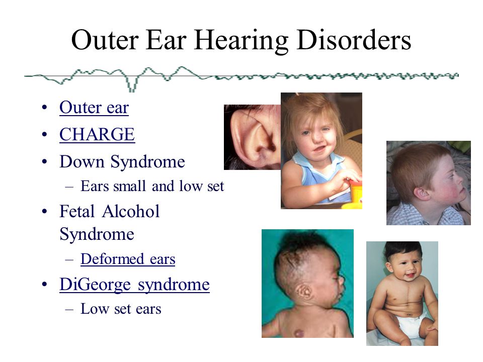 Moreover, only heart defects, disorders of skeletal formation and expansion of the collar space are independent risk factors.
The use of only one of the measurements in assessing the probability of the presence of chromosomal abnormalities of the fetus, for example, determining the thickness of the collar space or measuring the iliac angle, is less accurate than taking into account the totality of measurements.
Another way to determine the risk of having a baby with Down syndrome
In addition to ultrasound signs, biochemical screening in the first and second trimester is used to determine the degree of risk. In this case, the blood of a pregnant woman is examined. The best results are obtained by combining ultrasonic and biochemical signs. If the combination of factors is threatening, a decision may be made to conduct invasive methods for diagnosing Down syndrome and other chromosomal abnormalities. Invasive techniques consist in obtaining particles of the skin or blood of the fetus, followed by genetic analysis.