Can you see fertilized egg on ultrasound
How Early Can a Healthy Pregnancy Be Seen on an Ultrasound Scan?
Skip to content- View Larger Image
Many women look forward to their first ultrasound during pregnancy to catch a glimpse of their growing child. It is a beautiful and monumental moment for many families, and that is why at Carreras Medical Center, we offer early pregnancy ultrasounds. Ultrasounds are performed early on during pregnancy to ensure that your baby is growing and developing in the most healthy way possible. The innovative technology allows us to look inside your uterus to see what’s going on.
Ultrasounds are extremely safe and give you insight into gestational age, due dates, and viability. Your comfort is the most important thing during an early pregnancy ultrasound. Be sure to come to the appointment relaxed, knowing that you are taking a step towards your prenatal health. Be sure to ask any questions you may have and to describe how you have been. Let’s jump right into uncovering everything you need to know about your two-week pregnant ultrasound and beyond.
As soon as the stick turns blue, you may wonder how early you can see pregnancy on ultrasound. We recognize this exciting sign quite often. You may find yourself googling, “how soon can an ultrasound detect pregnancy,” or calling up your doctor asking, “how early can you see pregnancy on ultrasound?”
Many women want to rule out complications by having an early pregnancy sonogram, and they can. However, pregnancy tests are more sensitive and can actually detect pregnancy much sooner than an ultrasound. You typically have a one-week window between your pregnancy test and before your pregnancy is visible on a scan. Although, remember that this is the very early stages of pregnancy, and your baby is still teeny-tiny on the scan.
The soonest an ultrasound can detect a pregnancy is 17 days after ovulation. Ovulation is the moment when the egg is released from the ovary. When you are to become pregnant, the egg is fertilized. Just four days after a missed period, a typical early pregnancy looks like a small dot. After about two weeks of pregnancy, you can see your future baby as an embryo.
Two-weeks Pregnant UltrasoundAfter about two weeks, you can see your baby as an embryo on an early pregnancy sonogram. The baby will resemble a small bubble. Your child is still tiny, and the resemblance of a baby is still too young to see. But, after 12-17 days, you can detect a heartbeat on the embryo. Once we can detect your child’s heartbeat, the risk of miscarriage becomes lower. From your two weeks pregnancy ultrasound, your baby will develop rapidly.
You do not need to take any special steps to prepare for your ultrasound. The main thing to remember is to come to the appointment with a full bladder for better imaging results. When you come in for your appointment, it is essential to feel relaxed. We suggest wearing loose-fitting clothes. A loose top can make it easier to perform the ultrasound. However, we may provide you with a light gown.
The most important thing we can suggest is to relax and enjoy the moment. Comfort is key to maintaining a relaxed environment. Your two-week pregnant ultrasound will be one of the soonest methods to detect pregnancy and get ahead of your prenatal care.
Throughout your ultrasound journey, you can unveil pregnancy milestones, monitor your baby’s heartbeat, and determine an accurate expectation for your due date. An ultrasound will also reveal if you are carrying multiples.
What to ExpectDuring an early pregnancy ultrasound, we will peek inside your womb to see how your baby is growing and developing. Anticipation is a common theme among pregnancy. We wonder how our baby will look, we are curious about gender, and overall we want to ensure the delivery of a healthy child.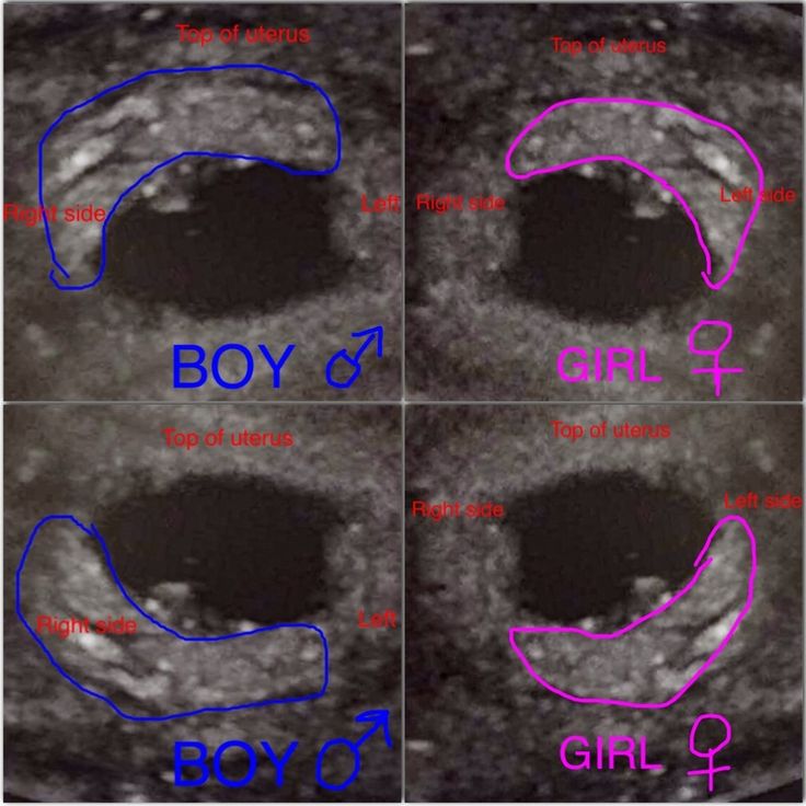 Regular ultrasounds and prenatal care are the best way to detect and treat complications if they should arise. Ultrasounds can give us insight into our child’s due date and primarily well-being.
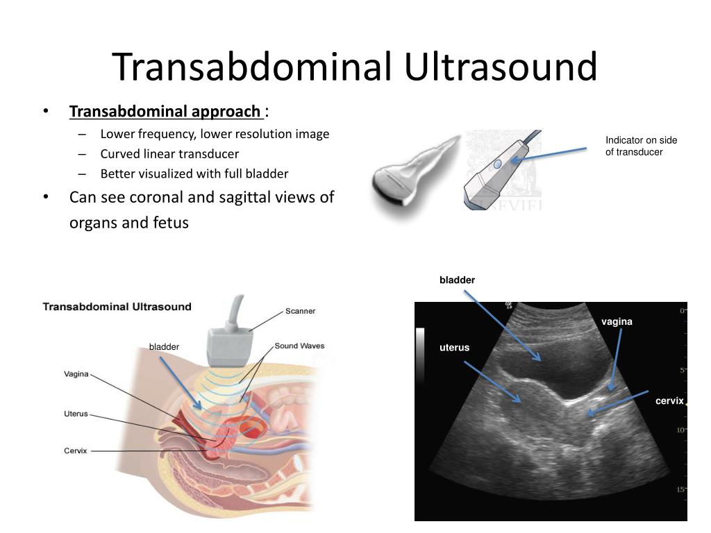
An ultrasound can be done in two different ways. The first way we can do an ultrasound is transabdominal. A transabdominal ultrasound is administered over your belly and is the more known of the two. The other ultrasound is transvaginal, meaning into your vagina. This ultrasound will be done if it is very early on in your pregnancy. A transvaginal ultrasound will produce authentic images of your still tiny baby.
Your doctor may recommend a transvaginal ultrasound for various reasons:
- If you had undergone infertility problems before pregnancy
- To detect pregnancy the earliest possible
- If you are experiencing pelvic pain
- If you are suspected of having an ectopic pregnancy
- If you are experiencing irregular bleeding
A transabdominal ultrasound is the type of ultrasound we all probably know, heard of, and possibly seen on television. Your healthcare professional will put a cool jelly over your stomach to take a peek inside of your womb. When you come in for an ultrasound on your belly, you should have a full bladder. A full bladder will increase visibility by tilting your uterus up and turning your intestines out of the viewpoint.
Once you have some gel on your tummy, the technician will use a transducer to see inside your stomach. The transducer is a handheld device that will release sound waves to form an image of your child inside your uterus. If you look over to the side during your ultrasound, you will see a video of your baby on the screen. The moment of seeing your healthy child on an ultrasound is a moment you will hold dear and remember forever.
During an ultrasound, you should not feel any discomfort. You will only feel the light pressure applied by the technician and experience the sensation of it gliding across your stomach.
Did you know that an ultrasound is one of the safest methods of diagnostic testing? You may be wondering about how safe digital imaging around your womb is. CT Scans are not safe for pregnant women, so why can I see inside my womb? However, ultrasounds are incredibly secure because they do not use ionizing radiation. Ultrasounds are used for other types of diagnostic testing as well.
The scan should take anywhere from 30 minutes to one hour. The technician will use light pressure while using the transducer on your belly.
Early Pregnancy SonogramAt about three weeks, an early pregnancy ultrasound can detect your baby’s head and body. Your child’s spine, brain, arm, and legs will begin to develop and be visible at about four weeks. At around eight to nine weeks gestation, the heart rate will continue to speed up.
Experience Expert CareAt Carreras Medical Center, we have an on-site ultrasound facility with state-of-the-art equipment and care professionals. Our services are at your convenience with expert care to ensure you receive the best experience possible.
If you think you are pregnant, please do not hesitate to give us a call. We are here to answer any and all of your questions and get you scheduled for an ultrasound right away.
Patience is key: Understanding the timing of early ultrasounds | Your Pregnancy Matters
×
What can we help you find?Refine your search: Find a Doctor Search Conditions & Treatments Find a Location
Appointment New Patient Appointment
or Call214-645-8300
MedBlog
Your Pregnancy Matters
November 20, 2018
Your Pregnancy Matters
Robyn Horsager-Boehrer, M. D. Obstetrics and Gynecology
The excitement newly pregnant women have to see how their baby is doing via an ultrasound can send them to an Ob/Gyn quickly. However, it’s important that they’re patient when their doctor recommends waiting until they are six weeks pregnant for their first ultrasound. Here’s why.
The first step women usually take after learning they’re pregnant is visiting with an Ob/Gyn. During this visit, an ultrasound is frequently done to confirm early pregnancy.
But an ultrasound doesn’t immediately show what women might expect. It’s typically not until a woman is six weeks pregnant that any part of the fetus is visible, which allows the doctor to determine whether a pregnancy will be viable.
Because of this, it’s important that women understand what information their ultrasound can and cannot provide at certain times during their pregnancy. And they should be prepared to wait to learn more about their baby, if it’s too soon.
4 stages of early pregnancy and what we might see on ultrasound
An ultrasound is a routine part of prenatal care at six to nine weeks. The ultrasounds we might do prior to that, and the information those exams would reveal, generally occur in four stages:
- Stage One: If performed around the time a women’s menstrual period is expected, this ultrasound typically shows a fluffy, thick lining of the uterus that’s preparing for the fertilized egg to implant.
- Stage Two: This is usually at four to five weeks after a pregnant woman’s last period. The ultrasound commonly shows a small collection of fluid within the lining of the uterus that represents the early development of the gestational sac.

- Stage Three: This is usually about five and a half weeks after a pregnant woman’s last period. The ultrasound typically shows a gestational sac and within it we can see a 3-5 mm bubble-like structure, which is the yolk sac.
- Stage Four: Approximately six weeks after a pregnant woman’s last period, we can see a small fetal pole, one of the first stages of growth for an embryo, which develops alongside the yolk sac.
While these are the expected times to see the developing pregnancy with an ultrasound, not all pregnancies develop along the same timeline. Just like pregnancy tests, if there’s variability in the length of the menstrual cycle or when fertilization takes place, then what we see on ultrasound can change. It’s important that this ultrasound is performed vaginally for high-quality pictures.
If the first ultrasound doesn’t show a developing baby with a heartbeat, when should the next one be scheduled?
Newly pregnant women get anxious if we don’t see both a fetus and a heartbeat on the first ultrasound and frequently want to come back soon after for another look. But it takes time to move through the early stages of pregnancy. And repeating an ultrasound still won’t be able to reassure the patient that the fetus is alive and growing, if we do it too soon.
The general recommendations are to wait two weeks if we only see a gestational sac and at least 11 days if a gestational and yolk sac are seen without a fetal pole. I prefer to wait two weeks for the next ultrasound in both of these scenarios. If we can see an early fetal pole but can’t see cardiac movement, then we repeat an ultrasound in one week. I know waiting is hard – but in my experience, it is much better to wait and get a definitive report on the status of your pregnancy than potentially have to come back multiple times.
Pregnancy loss can occur during any stage of pregnancy, though it’s most common in the first trimester. If we do an ultrasound and the length of the baby is more than 7mm, we should always see movement of the fetal heart. If we don’t, we know the pregnancy is not going to develop.
"It’s important that women understand the right time to visit their Ob/Gyn for an ultrasound, so they can know with more certainty what to expect during the exam."
– Robyn Horsager-Boehrer, M.D.
When might earlier ultrasounds be recommended?
If you have a history of tubal pregnancy, the most common being an ectopic pregnancy, or when the fertilized egg implants outside the uterus, or develop symptoms that are concerning for the possibility of a tubal pregnancy, your doctor might perform an ultrasound sooner. In this instance, the doctor is looking for any evidence of a pregnancy within the uterus, so a gestational sac with a yolk sac is reassuring and means that the chances of a tubal pregnancy are low. They also will likely want to take a close look at the fallopian tubes to see if there is any sign of a mass in the tube that might represent a growing tubal pregnancy.
Although it is hard to be patient, understanding the early timeline for a developing pregnancy will help you understand when first trimester ultrasounds should be performed.
Related reading: Helping you understand scary (but often harmless) pregnancy ultrasound findings
Stay on top of health care news. Subscribe to our blog today.
More in: Your Pregnancy Matters
Your Pregnancy Matters
- Shivani Patel, M.D.
November 22, 2022
Your Pregnancy Matters
- Robyn Horsager-Boehrer, M.
 D.
D.
November 15, 2022
Your Pregnancy Matters
- Robyn Horsager-Boehrer, M.D.
November 7, 2022
Mental Health; Your Pregnancy Matters
- Robyn Horsager-Boehrer, M.
 D.
D.
October 11, 2022
Prevention; Your Pregnancy Matters
- Robyn Horsager-Boehrer, M.D.
October 4, 2022
Mental Health; Your Pregnancy Matters
- Meitra Doty, M.
 D.
D.
September 27, 2022
Your Pregnancy Matters
- Robyn Horsager-Boehrer, M.D.
September 20, 2022
Men's Health; Women's Health; Your Pregnancy Matters
- Yair Lotan, M.
 D.
D.
September 6, 2022
Your Pregnancy Matters
August 29, 2022
More Articles
© 2022 The University of Texas Southwestern Medical Center
Member of Southwestern Health Resources
What should the corpus luteum look like during pregnancy on ultrasound, and when should you worry?
Ultrasound corpus luteum during examination of the ovaries
Ultrasound diagnosis can sometimes show the presence of a corpus luteum in the patient's ovary. Many girls, seeing such an entry in the diagnosis, begin to panic, but this is not scary. It is worth finding out what the corpus luteum looks like on ultrasound and what kind of gland it is, as well as in what cases it can appear and what it means.
What is the corpus luteum?
This gland appears in the body of a sexually mature woman every month in the absence of diseases from the granulosa cells of the dominant follicle and theca. The gland responsible for the production of progesterone is found in one of the ovaries after ovulation. The corpus luteum is responsible for an adequate supply of the hormone to the body during pregnancy. The name appeared due to the fact that the cavity of the gland is filled with a yellow substance.
The course and progression of pregnancy is accompanied by an increase in the hormone progesterone in the body of a woman. It is this function that is assigned to the corpus luteum, because this gland will play its role until the formation of the placenta, which will provide the body with the hormone to the right extent. If you do not know what the corpus luteum looks like on ultrasound, you should read the information below.
Why does the corpus luteum appear and develop?
A scar begins to form near the rupture site from which the mature egg came out. Next to this scar, you can find a temporary gland. Its functioning allows the egg to enter the fallopian tube, where it can be fertilized, and then descend into the uterus, where the fetus will develop. All this time is the right moment for research, when you can see the corpus luteum on ultrasound.
If a woman does not become pregnant, and the reproductive cell is not fertilized, menstruation occurs and the corpus luteum, which is visible on ultrasound, begins to degenerate into a white scar, after which, after a certain time, it completely disappears. This process is completely physiological and cyclic, it happens every month.
Ultrasound preparation
You need to prepare for the procedure, depending on what type of study was prescribed by the doctor. So, during a transabdominal examination, it will be necessary to drink 1-1.5 liters of water or tea in advance so that the bladder is full. In a transvaginal examination, on the contrary, it should be emptied. The day before the examination, it is necessary to exclude from the diet any gas-producing foods. Gas-filled intestinal cavities will significantly impair the passage of ultrasound and affect the accuracy of the results.
It often happens that this method of research shows that the corpus luteum is absent on ultrasound. This is evidence of insufficient or complete absence of the production of the necessary hormone by the woman's body. In this case, a woman will not be able to get pregnant and bear a child, since it is progesterone that is responsible for all the processes.
How is an ultrasound done?
There are two ways to examine the corpus luteum on ultrasound during or without pregnancy - using transabdominal and transvaginal examinations. Depending on what type of diagnosis is assigned to a woman, both the preparation for ultrasound and the course of this study differ.
Transabdominal examination
It is an examination of organs through the abdominal wall. To do this, a woman needs to drink a liter and a half of liquid in advance so that the bladder is as full as possible. After the patient enters the office, the doctor will tell her to expose her stomach and lie down on the couch. The doctor will apply a special gel to the abdomen and proceed to the diagnosis.
Transvaginal examination
It is necessary to examine the corpus luteum after ovulation on ultrasound using the transvaginal method in a different way. The woman undresses below the waist, lies down on the couch, her legs slightly bent. A condom is put on a special sensor of the device, after which it is inserted into the vagina. Normally, the complete or almost complete absence of discomfort is considered. The bladder in this case must be emptied immediately before the procedure.
What does the corpus luteum look like on ultrasound?
The corpus luteum on ultrasound looks like an additional structure located in one of the ovaries. During the examination, the doctor fixes the position of the corpus luteum and its dimensions and notes on which ovary it is located, which allows us to say about the presence of diseases. Enlargement of the gland and its constant presence may indicate that the corpus luteum is affected by a cyst.
Dimensions of the corpus luteum
The normal size of the gland is 11-25 mm. The magnitude indicator changes as the woman's menstrual cycle continues. If the size of the gland is too small or large, and the structure is heterogeneous, this may indicate the likelihood of deviations in the functioning of the organs. The reason for this phenomenon may be a delay in menstruation or the occurrence of a cyst. In the event that the corpus luteum is enlarged on ultrasound, but a fetal egg is not visible in the uterus, the doctor can refer the woman to a blood test. Such a study will allow you to find out the level of hCG and determine the presence of a fetus. This can occur in the first weeks of pregnancy, when the fetus is difficult to detect, and only blood changes indicate its presence.
Yellow body during pregnancy
It is a mistake to assume that the detection of a corpus luteum on ultrasound is an accurate indication of the onset of pregnancy. Regardless of whether the egg was fertilized, the gland will still appear. The presence of a corpus luteum on ultrasound can only indicate that ovulation has occurred. The corpus luteum is not a guarantee of fertilization of the egg.
Such a phenomenon can report a possible pregnancy only when the gland does not decrease, although the cycle has already come to an end.
Now you know what to do if an ultrasound shows a corpus luteum and what it means.
Ultrasound monitoring of folliculogenesis
When a married couple faces the problem of infertility, the doctor prescribes many studies, including monitoring of folliculogenesis for a woman. It allows you to see how ovulation goes and whether it goes at all. Folliculometry (a study using an ultrasound machine of the female reproductive organs throughout the cycle) allows you to see violations of the growth of follicles.
A follicle is a fluid-filled cavity in which an egg matures. Folliculogenesis is a complex and multi-stage process that precedes ovulation. During ovulation, the follicle bursts, and an egg ready for fertilization comes out of it. It is the monitoring of folliculogenesis that will enable the gynecologist to determine the optimal days for conceiving a child or possible causes of infertility.
When to do?
The female menstrual cycle lasts an average of 28 days. The day of the cycle is counted from the first day of the last menstruation. The first ultrasound of folliculogenesis, if a woman has a normal cycle, is recommended for 8-10 days.
In cases where the woman's menstrual cycle is regular, but shorter or longer (from 23 to 35 days), the first ultrasound examination is scheduled 4-5 days before the expected ovulation. The doctor determines the number of folliculogenesis monitoring sessions individually, but usually 2-3 visits to the ultrasound room are enough.
When the cycle is irregular, the first monitoring of folliculogenesis is usually started 3-5 days after the end of menstruation. At the same time, a study of both the follicles and the endometrium is also carried out in order to more accurately determine which factors provoke a violation of folliculogenesis. Monitoring helps to make the correct diagnosis and subsequently prescribe effective treatment. The number of sessions with an irregular menstrual cycle for each patient is individual.
What will the monitoring of folliculogenesis show?
Ultrasound Monitoring - Ultrasound monitoring of how changes occur in the uterus and ovaries during the menstrual cycle.
On the 8-10th day of the classical menstrual cycle, one dominant follicle is visible on the screen of the ultrasound machine, which has reached a diameter of 12-15 mm against the background of other, much smaller follicles. In rare cases, there may be two or more dominant follicles. Every day the follicle increases in size, on the day of ovulation it can reach 18-25 mm in diameter.
In ultrasound, attention is drawn not only to folliculogenesis, but also to the endometrium. When ovulation occurs, the thickness of the endometrium, which has a three-layer structure, reaches 10-12 mm. The body then releases luteinizing hormone, which promotes ovulation. In this case, a small amount of follicular fluid is poured into the abdominal cavity.
During an ultrasound of folliculogenesis, the following signs indicate that ovulation has occurred:
- the presence of a mature follicle on the eve of ovulation;
- disappearance or gradual reduction of the dominant follicle, destruction of its walls;
- after normal ovulation, free fluid appears in the Douglas space of the abdominal cavity;
- instead of a mature follicle, a corpus luteum appears.
It should be noted that sometimes even the visualization of a mature follicle and the presence of a corpus luteum instead of it after a week does not give an absolute guarantee that ovulation was complete.

