Can a pelvic ultrasound detect pregnancy
Vaginal ultrasound | Pregnancy Birth and Baby
beginning of content3-minute read
Listen
A vaginal ultrasound is an ultrasound scan taken by a probe inserted into the vagina. It gives a clear picture of the fetus, cervix and placenta. It is also called an internal ultrasound or a transvaginal ultrasound.
What does a vaginal ultrasound test for?
A vaginal ultrasound lets the doctor or ultrasound technologist (sonographer) see and measure the fetus. You might even discover if you are pregnant with twins or triplets.
It also allows the doctor or sonographer to look at your vagina, placenta, cervix, fallopian tubes, uterus and ovaries.
A vaginal ultrasound can be used as well as, or instead of, the standard abdominal ultrasound in which the probe is put on your belly.
Why is a vaginal ultrasound recommended?
A vaginal ultrasound will be recommended when your doctor or midwife wants a clearer diagnostic image than the one they can get through a standard abdominal ultrasound.
When is it used during pregnancy?
A vaginal ultrasound can confirm you are pregnant as it can detect the heartbeat very early in your pregnancy. It can also record the location and size of the fetus and determine if you are pregnant with 1 baby or more.
A vaginal ultrasound can also be used to diagnose problems or potential problems, including to:
- detect an ectopic pregnancy
- measure the cervix to determine the risk of a premature birth, which will allow any necessary interventions
- detect abnormalities in the placenta or cervix
- to determine the source of any bleeding
How is a vaginal ultrasound done?
A vaginal ultrasound is done by your doctor or sonographer in a hospital, clinic or consulting room. It uses a probe that is slightly wider than your finger.
You’ll probably be asked to empty your bladder (pee) before the ultrasound starts. If you’re using a tampon, you’ll need to take it out.
You’ll be asked to take off the lower half of your clothing and lie back on the examination table with your knees bent. There might be stirrups, or your hips might be slightly raised.
The probe is covered by a sheath, which is then covered in lubricating gel. It will be inserted slowly for about 5 to 8cm into your vagina. That usually doesn’t hurt, but you will feel pressure and it can be uncomfortable.
The probe will be moved around to get the best view of what is being examined. The examination usually takes 15 to 30 minutes.
If you are not comfortable having a male perform the examination, you can ask for a female sonographer. You can also ask for a female health worker to accompany you for support, or have a family member with you.
If you live in Western Australia, you must give written consent to a vaginal ultrasound.
When will I get the results?
Often you can see the ultrasound images on a monitor while you have your scan. If your specialist is there, they might discuss the results with you straight away.
If a specialist isn’t there, the sonographer is usually not allowed to discuss what they see with you. Your doctor or midwife will see the images after they have been processed. It usually takes a day or two to get the results.
Are there any risks involved?
Vaginal ultrasounds are safe for you and your baby, as long as your waters haven’t broken.
If you are allergic to latex, let the sonographer know so they can use a latex-free sheath on the probe.
There are no after-effects of the procedure, so you can get back to your normal activities, including driving yourself home if you wish.
Sources:
Australasian Society for Ultrasound in Medicine (Guidelines for the performance of first trimester ultrasound), Inside Radiology (Transvaginal ultrasound), Royal Australian and New Zealand College of Obstetricians and Gynaecologists (Screening in early pregnancy for adverse perinatal outcomes), The Royal Women's Hospital (Bleeding in early pregnancy)Learn more here about the development and quality assurance of healthdirect content.
Last reviewed: June 2020
Back To Top
Related pages
- Pregnancy checkups, screenings and scans
- Ultrasound scans during pregnancy
This information is for your general information and use only and is not intended to be used as medical advice and should not be used to diagnose, treat, cure or prevent any medical condition, nor should it be used for therapeutic purposes.
The information is not a substitute for independent professional advice and should not be used as an alternative to professional health care. If you have a particular medical problem, please consult a healthcare professional.
Except as permitted under the Copyright Act 1968, this publication or any part of it may not be reproduced, altered, adapted, stored and/or distributed in any form or by any means without the prior written permission of Healthdirect Australia.
Support this browser is being discontinued for Pregnancy, Birth and Baby
Support for this browser is being discontinued for this site
- Internet Explorer 11 and lower
We currently support Microsoft Edge, Chrome, Firefox and Safari. For more information, please visit the links below:
- Chrome by Google
- Firefox by Mozilla
- Microsoft Edge
- Safari by Apple
You are welcome to continue browsing this site with this browser. Some features, tools or interaction may not work correctly.
What To Expect, Purpose & Results
Overview
What is an ultrasound in pregnancy?
A prenatal ultrasound (or sonogram) is a test during pregnancy that checks on the health and development of your baby. An obstetrician, nurse midwife or ultrasound technician (sonographer) performs ultrasounds during pregnancy for many reasons. Sometimes ultrasounds occur to check on your baby and make sure they’re growing properly. Other times your pregnancy care provider orders an ultrasound after they detect a problem.
During an ultrasound, sound waves are sent through your abdomen or vagina by a device called a transducer. The sound waves bounce off structures inside your body, including your baby and your reproductive organs. Then, the sound waves transform into images that your provider can see on a screen. It doesn’t use radiation, like X-rays, to see your baby.
Even though prenatal ultrasounds are safe, you should only have them when it’s medically necessary. If there’s no reason for an ultrasound (for example, if you just want to see your baby), your insurance company might not pay for it.
Prenatal ultrasounds may be called fetal ultrasounds or pregnancy ultrasounds. Your provider will talk to you about when you can expect ultrasounds during pregnancy based on your health history.
Why is a fetal ultrasound important during pregnancy?
An ultrasound is one of the few ways your pregnancy care provider can see and hear your baby. It can help them determine how far along you are in pregnancy, if your baby is growing properly or if there are any potential problems with the pregnancy. Ultrasounds may occur at any time in pregnancy depending on what your provider is looking for.
What can be detected in a pregnancy ultrasound?
A prenatal ultrasound does two things:
- Evaluates the overall health, growth and development of the fetus.
- Detects certain complications and medical conditions related to pregnancy.
In most pregnancies, ultrasounds are positive experiences and pregnancy care providers don’t find any problems. However, there are times this isn’t the case and your provider detects birth disorders or other problems with the pregnancy.
Reasons why your provider performs a prenatal ultrasound are to:
- Confirm you’re pregnant.
- Check for ectopic pregnancy, molar pregnancy, miscarriage or other early pregnancy complications.
- Determine your baby’s gestational age and due date.
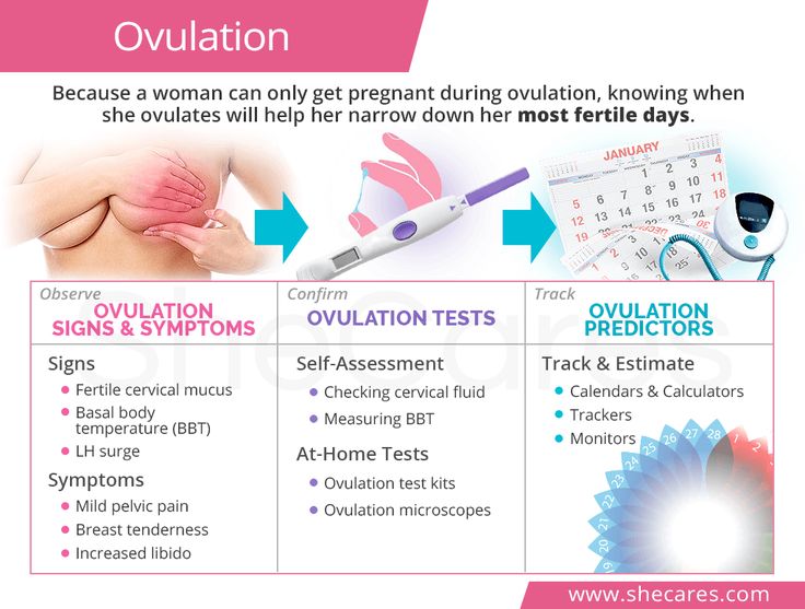
- Check your baby’s growth, movement and heart rate.
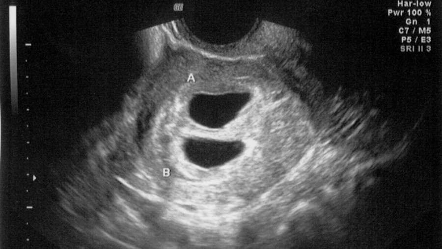
- Look for multiple babies (twins, triplets or more).
- Examine your pelvic organs like your uterus, ovaries and cervix.
- Examine how much amniotic fluid you have.
- Check the location of the placenta.
- Check your baby’s position in your uterus.
- Detect problems with your baby’s organs, muscles or bones.
Ultrasound is also an important tool to help providers screen for congenital conditions (conditions your baby is born with). A screening is a type of test that determines if your baby is more likely to have a specific health condition. Your provider also uses ultrasound to guide the needle during certain diagnostic procedures in pregnancy like amniocentesis or CVS (chorionic villus sampling).
An ultrasound is also part of a biophysical profile (BPP), a test that combines ultrasound with a nonstress test to evaluate if your baby is getting enough oxygen.
How many ultrasounds do you have during your pregnancy?
Most pregnant people have one or two ultrasounds during pregnancy. However, the number and timing vary depending on your pregnancy care provider and if you have any health conditions. If your pregnancy is high risk or if your provider suspects you or your baby has a health condition, they may suggest more frequent ultrasounds.
When do you have your first prenatal ultrasound?
The timing of your first ultrasound varies depending on your provider. Some people have an early ultrasound (also called a first-trimester ultrasound or dating ultrasound). This can happen as early as seven to eight weeks of pregnancy. Providers do an early ultrasound through your vagina (transvaginal ultrasound). Early ultrasounds do the following:
- Confirm pregnancy (by detecting a heartbeat).
- Check for multiple fetuses.
- Measure the size of the fetus.
- Help confirm gestational age and due date.
Some providers perform your first ultrasound closer to 12 weeks of pregnancy.
20-week ultrasound (anatomy scan)
You can expect an ultrasound around 18 to 20 weeks in pregnancy. This is known as the anatomy ultrasound or 20-week ultrasound. During this ultrasound, your pregnancy care provider can see your baby’s sex (if your baby is in a good position for viewing their genitals), detect birth disorders like cleft palate or find serious conditions related to your baby’s brain, heart, bones or kidneys. If your pregnancy is progressing well and with no complications, your 20-week ultrasound may be your last ultrasound during pregnancy. However, if your provider detects a problem during your 20-week ultrasound, they may order additional ultrasounds.
How soon can you see a baby on an ultrasound?
Pregnancy care providers can detect an embryo on an ultrasound as early as six weeks into the pregnancy. An embryo develops into a fetus around the eighth week of pregnancy.
If your last menstrual period isn’t accurate, it’s possible that it may be too early to detect a fetal heart rate.
Which ultrasound is most important during pregnancy?
All ultrasounds during pregnancy are important. Your pregnancy care provider uses ultrasound to tell them important information about your pregnancy.
Test Details
What are the two main types of pregnancy ultrasounds?
The two main types of pregnancy ultrasound are transvaginal ultrasound and abdominal ultrasound. Both use the same technology to produce images of your baby. Your pregnancy care provider performs a transvaginal ultrasound by placing a wand-like device inside your vagina. They perform an abdominal ultrasound by placing a device on the skin of your belly.
Transvaginal ultrasound
During a transvaginal ultrasound, your pregnancy care provider places a device inside your vaginal canal (similar to how you place a tampon). In early pregnancy, this ultrasound helps to detect a fetal heartbeat or determine how far along you are in your pregnancy (gestational age). Images from a transvaginal ultrasound are clearer in early pregnancy as compared to abdominal ultrasound.
Abdominal ultrasound
Your pregnancy care provider performs an abdominal ultrasound by placing a transducer directly on your skin.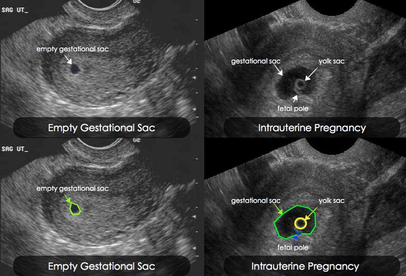 Then, they move the transducer around your belly (abdomen) to capture images of your baby. Sometimes slight pressure has to be applied to get the best views. Providers use abdominal ultrasounds after about 12 weeks of pregnancy.
Traditional ultrasounds are 2D. More advanced technologies like 3D or 4D ultrasound can create better images. This is helpful when your provider needs to see your baby’s face or organs in greater detail. Not all providers have 3D or 4D ultrasound equipment or specialized training to conduct this type of ultrasound.
Your provider may recommend other types of ultrasounds. Examples of additional ultrasounds are:
- Doppler ultrasound: This type of ultrasound checks how your baby’s blood flows through its blood vessels. Most Doppler ultrasounds occur later in pregnancy.
- Fetal echocardiogram: This type of ultrasound looks at your baby’s heart size, shape, function and structure. Your provider may use it if they suspect your baby has a congenital heart condition, if you had another child that had a heart condition or if you have certain health conditions that warrant taking a closer look at the heart.
How do I prepare for the test?
There’s no special preparation for an ultrasound. Some pregnancy care providers ask that you come with a full bladder and don’t use the restroom before the test. This helps them view your baby better on the ultrasound. You can bring a support person, but bringing children is discouraged as this is an important test that requires complete focus.
You may be asked to change into a hospital gown, but this isn’t usually required for abdominal ultrasounds. If your provider is performing a transvaginal ultrasound in your first trimester, you’ll put on a hospital gown or undress from the waist down.
What should I expect during a prenatal ultrasound?
You’ll lie on a padded examining table during the test. Most ultrasounds occur in a dimly lit room, which helps your ultrasound technician (or sonographer) see the screen. Your sonographer applies a small amount of water-soluble gel to the skin of your belly. The gel doesn’t harm your skin or stain your clothes, but it may feel cold. This gel helps transmit sound waves more efficiently.
Next, the sonographer places a transducer on the skin of your abdomen. The transducer sends sound waves into your body, which reflect off internal structures, including your baby. The sound waves that reflect back create pictures on a screen. Your sonographer uses these images to take important measurements such as your baby’s head circumference and length. You may see them making lines on the screen or clicking a button to “freeze” certain angles.
There’s virtually no discomfort during a prenatal ultrasound. You may feel mild discomfort if you have to pee. The ultrasound test takes about 30 minutes to complete.
If you have a transvaginal ultrasound, the process is only different in that the transducer is inside your vagina and not on your belly.
What should I expect after a pregnancy ultrasound?
If you had an abdominal ultrasound, your sonographer wipes the gel off your belly. They may print off some ultrasound pictures for you to take home with you.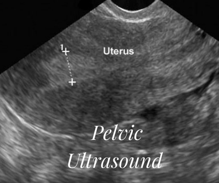
In most cases, your sonographer won’t discuss the results of your test with you. If your obstetrician performs your ultrasound, they may discuss what they see as they go along.
If a sonographer performs your ultrasound, an obstetrician will look at the images, then discuss their findings with you at your next appointment. Most practices schedule your appointment right after your ultrasound so you get your results the same day.
What are the risks of prenatal ultrasounds?
Studies have shown ultrasounds are safe during pregnancy. There are no harmful side effects to you or your baby.
Is it safe to do an ultrasound every month during pregnancy?
While ultrasounds are safe for you and your baby, most major medical associations recommend that pregnancy care providers should only do ultrasounds when the tests are medically necessary. If your ultrasounds are normal and your pregnancy is uncomplicated or low risk, repeat ultrasounds aren’t necessary.
Results and Follow-Up
What results do you get on a pregnancy ultrasound?
Your ultrasound results will be normal or abnormal.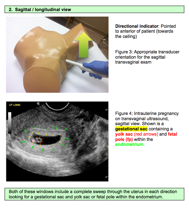 A normal result means your pregnancy care provider didn’t find any problems and that your baby is growing and developing normally. An abnormal result means your provider noticed something irregular. If they do, your provider will order additional ultrasounds or diagnostic tests to determine if something is wrong.
Occasionally, the ultrasound is incomplete if there’s difficulty seeing all the structures needed for that particular ultrasound. Your baby’s position or movement sometimes makes it difficult to see everything your provider needs to see. If this is the case, you’ll need a repeat ultrasound and they’ll try again.
There are some limitations to ultrasounds, so your provider may not find certain abnormalities until after birth.
What are reasons you need more ultrasounds during pregnancy?
There are several reasons your pregnancy care provider may order additional ultrasounds during your pregnancy. Some of these reasons include:
- Problems with your ovaries, uterus, cervix or other pelvic organs.

- Your baby is measuring small for their gestational age or your provider suspects IUGR (intrauterine growth restriction).
- Problems with the placenta like placenta previa or placental abruption.
- You’re pregnant with twins, triplets or more.
- Your baby is breech.
- You have too much amniotic fluid (polyhydramnios).
- You have too little amniotic fluid (oligohydramnios).
- You have a condition like gestational diabetes or preeclampsia.
- Your baby has a congenital disorder.
Normal results on pregnancy ultrasounds can vary. Generally, a normal result means your baby appears healthy and your provider didn’t find any issues.
Why do some pregnancy providers schedule ultrasounds differently?
The number of ultrasounds you’ll have and when you have them can vary between providers. Every practice operates differently and some providers do things differently based on your health history or symptoms.
When does a pregnancy ultrasound determine sex?
Your baby’s sex isn’t visible on an ultrasound until about 18 to 20 weeks.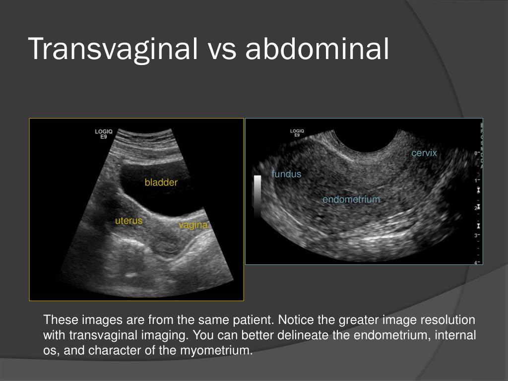 Be sure to tell your pregnancy care provider whether or not you want to know the sex of your baby before your ultrasound.
A note from Cleveland Clinic
An ultrasound during pregnancy can be both exciting and terrifying. Your pregnancy care provider uses ultrasound to get a better idea of how your baby is growing and developing. There are different types of ultrasounds, and the exact timing may vary depending on your provider. Most pregnant people have two ultrasounds — one in the first trimester and one in the second trimester. However, if there’s a potential complication or medical reason for more ultrasounds, your provider will order more as a precaution. Talk to your provider about the ultrasound schedule during pregnancy and what you can expect.
Ultrasound diagnostics (ultrasound of the pelvic organs)
Ultrasound examination (ultrasound) - recognition of pathological changes in organs and tissues of the body using ultrasound. Ultrasound is based on the principle of echolocation - the reception of signals sent and then reflected from the interfaces of tissue media with different acoustic properties.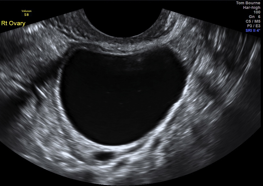
Ultrasound of the pelvic organs is performed in order to visually determine the presence of a particular pathology in a woman (or a fetus with obstetric ultrasound) by echographic signs.
Ultrasound of the pelvic organs can be performed with an abdominal probe (through the abdomen) or vaginal (vaginal). In the pelvis of a woman, ultrasound examines the uterus, fallopian tubes, vagina, ovaries and bladder.
- Uterus: The position, shape, main dimensions of the uterus and the structure of its walls are determined.
In addition, median uterine structures are examined separately: the uterine cavity and endometrium (M-echo). In a non-pregnant woman, the uterine cavity is slit-like. The endometrium - the functional inner layer - changes during the menstrual cycle. - Ovaries: The position in relation to the uterus, the size, size of the follicles and the corpus luteum (the formation that remains in place of the follicles after the release of the egg from the ovary) is assessed.
A comparison is made with the phase of the menstrual cycle.
If formations are found in the ovaries, they are also described (shape, structure, size). - The presence of free fluid is also determined (normally, after the release of the egg from the ovary, it is in a small amount) and the presence of tumor formations in the pelvic cavity.
- In addition to the structure of the uterus and ovaries, the state of the bladder is assessed during ultrasound (if it is sufficiently filled).
Advantages of ultrasound diagnostics
Ultrasound examination is carried out quickly, the ultrasound method is clear, economical and easy, can be used repeatedly and with minimal effort to prepare for the study. It is reliably confirmed that ultrasound is absolutely safe even for a pregnant woman.
Indications for ultrasound of the pelvic organs
The method of ultrasound examination is widely used for suspected gynecological diseases, pregnancy, to monitor the treatment and cure of the patient.
- Ultrasound of the uterus can diagnose early pregnancy.
- Ultrasound of the pelvis in women should be performed for menstrual irregularities (delayed menstruation, early onset of menstruation, bleeding in the middle of the cycle), with heavy or scanty menstruation, in the absence of menstruation, with various vaginal discharges, with pain in the lower abdomen, with the appearance of discharge during menopause.
- With the help of gynecological ultrasound, various diseases are detected: from inflammatory gynecological diseases to benign and malignant tumors of the uterus and ovaries (including endometriosis, salpingo-oophoritis, ovarian cysts, endometritis, etc.).
- Ultrasound of the uterus enables early diagnosis of uterine fibroids.
- Pelvic ultrasound is widely used to monitor the ovarian follicular apparatus in the treatment of infertility and pregnancy planning.
- Ultrasound examination of the pelvis is prescribed when taking contraceptive and hormonal drugs, in the presence of an intrauterine contraceptive ("spiral") to control and prevent complications.
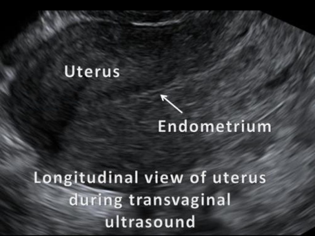
- Ultrasound during pregnancy (obstetric ultrasound) allows you to monitor the normal development of the fetus and timely detect pathology.
- In urology, pelvic ultrasound is necessary to identify the causes of urinary disorders, urinary incontinence and pathology of the urethra (urethra).
Contraindications for pelvic ultrasound
There are no contraindications for ultrasound.
Preparing for ultrasound of the pelvic organs
When visiting the ultrasound diagnostics room to remove the remaining gel from the skin after the examination, you must have a towel or napkin with you, as well as a diaper on which you will lie down for the examination.
In non-pregnant women, routine gynecological ultrasonography is done on a full bladder unless otherwise instructed by a physician. To ensure maximum accuracy and reliability of the results, it is necessary to strictly adhere to the established rules for preparing for ultrasound of the pelvic organs:
- transabdominal (through the abdomen) gynecological ultrasound requires bladder preparation: drink 1-1.
 5 liters of non-carbonated liquid 1 hour before the procedure and do not urinate until the examination;
 - for transvaginal (through the vagina) gynecological ultrasound, special preparation is not required, the study is performed with an empty bladder;
- obstetric ultrasound (ultrasound during pregnancy) is performed with a moderately full bladder (drink 2 glasses of liquid 1 hour before the procedure).
When examining the organs of the genitourinary system (bladder, prostate, uterus, ovaries), it is necessary to drink 0.5 liters of liquid 1-1.5 hours before the examination or not urinate for 2 hours. This is necessary in order to fill the bladder, which pushes the examined organs.
A prerequisite for a successful ultrasound is an empty intestine and the absence of gases in it. Therefore, preparation for ultrasound should be started in advance: it is important to follow a diet with restriction of foods that cause constipation or gas formation 2-3 days before the upcoming ultrasound. It is recommended to exclude from the diet foods that cause increased gas formation (black bread, fruits, raw vegetables, confectionery, milk). Enzyme preparations are recommended: festal, panzinorm, enzistal, creon, etc. Cleansing enemas are not recommended, as they often increase gas formation. In addition, you can take activated charcoal, espumizan, dill water. If you are constipated, it is recommended that you take a laxative, especially if you need a rectal probe test.
Ultrasound is performed on an empty stomach (last meal 8-12 hours before the examination) and immediately after a bowel movement.
Examination of the mammary glands, uterus and appendages is recommended in the first half or middle of the menstrual cycle.
Folliculogenesis examination performed at 5; 9; 11-14 and 15 days of the menstrual cycle.
The accuracy of the results obtained largely depends on how you prepare for the ultrasound.
In emergency cases, ultrasound is performed without preparation, but its effectiveness is lower.
How an ultrasound of the pelvic organs is performed
You lie down on the couch (after spreading the diaper) with your head towards the doctor (to the ultrasound machine) and expose your stomach and lower abdomen. The ultrasound doctor will lubricate the ultrasound transducer with gel (for a transvaginal ultrasound, put a condom on the transducer and lubricate it with gel) and drive the transducer over you, occasionally pressing to view the pelvic organs from a different angle. The procedure is absolutely painless, except for the diagnosis of acute inflammatory processes of the pelvic organs. An ultrasound examination takes from 10 to 20 minutes, depending on the purpose of the examination.
Complications of pelvic ultrasound
After ultrasound, complications are not observed, but transvaginal ultrasound during pregnancy, especially in early pregnancy, is performed only after assessing the risk to the fetus.
Interpretation of the results of ultrasound of the pelvic organs
Only an experienced doctor can correctly decipher the results of ultrasound
What ultrasound of the pelvic organs can detect
bicornuate, saddle-shaped, unicornuate, doubling of the uterus).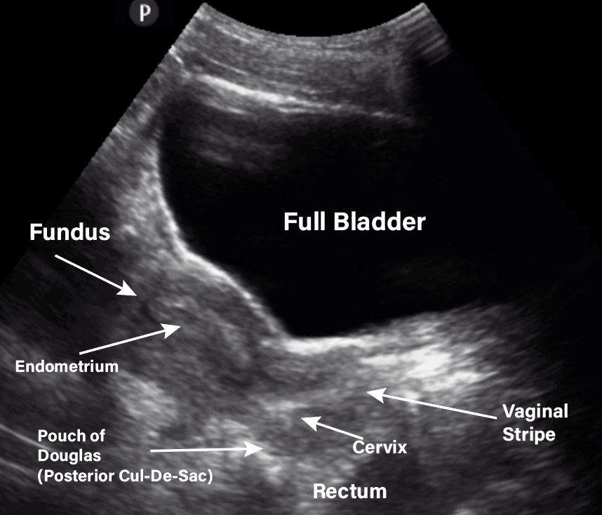
The presence of congenital malformations can cause infertility, increase the risk of preterm birth, spontaneous abortion, intrauterine fetal death, malposition of the fetus and impaired labor activity.
Endometriosis: Endometriosis is a pathological process characterized by the extension of the endometrium outside the uterine cavity (walls of the uterus, ovaries, peritoneum, etc.). An ultrasound of the pelvic organs reveals internal endometriosis or adenomyosis (growth of the endometrium into the wall of the uterus) and endometrioid ovarian cysts.
Diagnosis of endometriosis is important for predicting the possibility of pregnancy (endometriosis can be the cause of infertility), its bearing.
Uterine fibroids: Uterine fibroids are benign tumors of the female reproductive system. Ultrasound determines the presence, number, location and size of myomatous nodes. In addition, ultrasound allows you to monitor the dynamics of their growth rates. Because ultrasound is done several times a year. Diagnosis of fibroids is extremely important in preparing for conception, since the presence of fibroids can affect the course of pregnancy.
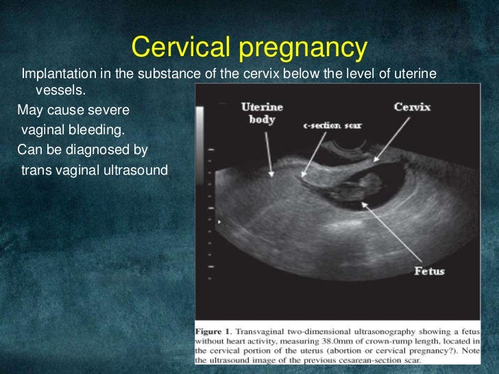
Pregnancy diagnosis: Ultrasound can diagnose pregnancy from 3 to 4 weeks. Small terms of pregnancy are determined only with the help of a transvaginal sensor, a device with good resolution. Various types of ectopic pregnancy are diagnosed (tubal - the fetal egg is attached to the fallopian tube, cervical - the fetal egg is attached to the cervix, ovarian - the fetal egg is attached to the ovary), which allows the woman to maintain her health.
Intrauterine contraception: Ultrasound monitors the insertion and removal of an intrauterine contraceptive. timely detect incorrect location, partial or complete prolapse of the IUD from the uterine cavity, ingrowth of parts of the contraceptive into the uterine wall. If you are planning a pregnancy, then after removing the intrauterine contraceptive, the doctor will recommend that you do an ultrasound.
Hyperplastic processes of the endometrium (hyperplasia, polyps, malignant tumors of the endometrium), ovarian masses are also detected.
Preventive ultrasound of the pelvic organs
For preventive purposes, healthy women need to have an ultrasound of the pelvic organs once every 1-2 years, and after the age of 40 - once a year in order to detect hidden pathology. Preventive ultrasound of the pelvic organs is usually performed in the first phase of the cycle (5-7th day from the onset of menstruation).
First and second ultrasound for early pregnancy
Today, neither doctors nor expectant mothers can imagine how a pregnancy can pass without an ultrasound examination. With the help of ultrasound, you can determine the presence of pregnancy, track how the baby develops in utero, and prepare for his birth. Modern achievements have made it possible to make the survey completely safe, very accurate and informative already at an early stage.
How is ultrasound performed during pregnancy
- At the beginning, when the fetus is still small, ultrasound is performed transvaginally using a transducer inserted into the vagina. Thus, a high accuracy of diagnostics is achieved, it becomes possible to detect the slightest deviations in the state of the expectant mother and baby in time. This examination is performed on an empty bladder at any time of the day.
- In the later stages, the fetus is also clearly visible during abdominal examination. For this examination, it is better that the bladder is full, for which it is enough for a pregnant woman to drink up to half a liter of liquid half an hour before the examination. If the indicated volume of liquid is a burden for the expectant mother, doctors do not insist on it.
At what time is
- The first ultrasound during pregnancy is performed at 12-14 weeks. At this time, it is important to determine where and how the fetal egg was fixed, how harmoniously it develops, whether there are any deviations in it, whether there is a threat of miscarriage in the early stages.
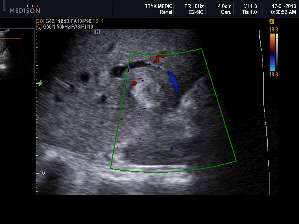
- The second ultrasound is performed at 20-24 weeks. It allows you to assess the compliance with the norms of the size of the fetus, the level of development and proportionality of individual parts of his body, makes it possible to scan the heartbeat. At this stage, you can already determine the sex of the child. At the same time, the doctor during this period is interested in the condition of the uterus, placenta and the volume of amniotic fluid of the woman.
- A third routine pregnancy test is needed at 30-34 weeks. With its help, height and weight are determined, and even the one that will be approximately at birth. The examination reveals the degree of development of internal organs, the presence of deviations in them. Necessarily with the help of Doppler sonography, the fetal heart rate is studied. Along with this, the reserve of the placenta is evaluated, its ability to provide the baby with all the necessary substances, as well as the volume of amniotic fluid.


