Brain development womb
When Does a Fetus Develop a Brain?
Pregnancy is an exciting time full of rapid change and development for both you and your baby. While the growth happening on the outside is clear to everyone (hello, growing belly!), it’s the development we can’t see that is truly fascinating.
Your fetus will begin the process of developing a brain around week 5, but it isn’t until week 6 or 7 when the neural tube closes and the brain separates into three parts, that the real fun begins.
Around week 5, your baby’s brain, spinal cord, and heart begin to develop. Your baby’s brain is part of the central nervous system, which also houses the spinal cord. There are three key components of a baby’s brain to consider. These include:
- Cerebrum: Thinking, remembering, and feeling occurs in this part of the brain.
- Cerebellum: This part of the brain is responsible for motor control, which allows the baby to move their arms and legs, among other things.
- Brain stem: Keeping the body alive is the primary role of the brain stem. This includes breathing, heartbeat, and blood pressure.
The first trimester is a time of rapid development and separation of the various parts of the brain, according to Kecia Gaither, MD, MPH, double board certified in obstetrics and gynecology and maternal-fetal medicine, and director of perinatal services at NYC Health + Hospitals/Lincoln.
Within 4 weeks, the rudimentary structure known as the neural plate develops, which Gaither says is considered the precursor to the nervous system. “This plate elongates and folds on itself forming the neural tube — the cephalad portion of the tube becomes the brain, while the caudal portion elongates to eventually become the spinal cord,” she explains.
The neural tube continues to grow, but around week 6 or 7, Gaither says it closes, and the cephalad portion (aka the rudimentary brain) separates into three distinct parts: front brain, midbrain, and hindbrain.
It’s also during this time that neurons and synapses (connections) begin to develop in the spinal cord. These early connections allow the fetus to make its first movements.
During the second trimester, Gaither says the brain begins to take command of bodily functions. This includes specific movements that come from the hindbrain, and more specifically, the cerebellum.
One of the first notable developments, sucking and swallowing, are detectable around 16 weeks. Fast-forward to 21 weeks, and Gaither says baby can swallow amniotic fluid.
It’s also during the second trimester that breathing movements begin as directed by the developing central nervous system. Experts call this “practice breathing” since the brain (and more specifically, the brain stem) is directing the diaphragm and chest muscles to contract.
And don’t be surprised if you feel some kicking during this trimester. Remember the cerebellum or the part of the brain responsible for motor control? Well, its directing the baby’s movements, including kicking and stretching.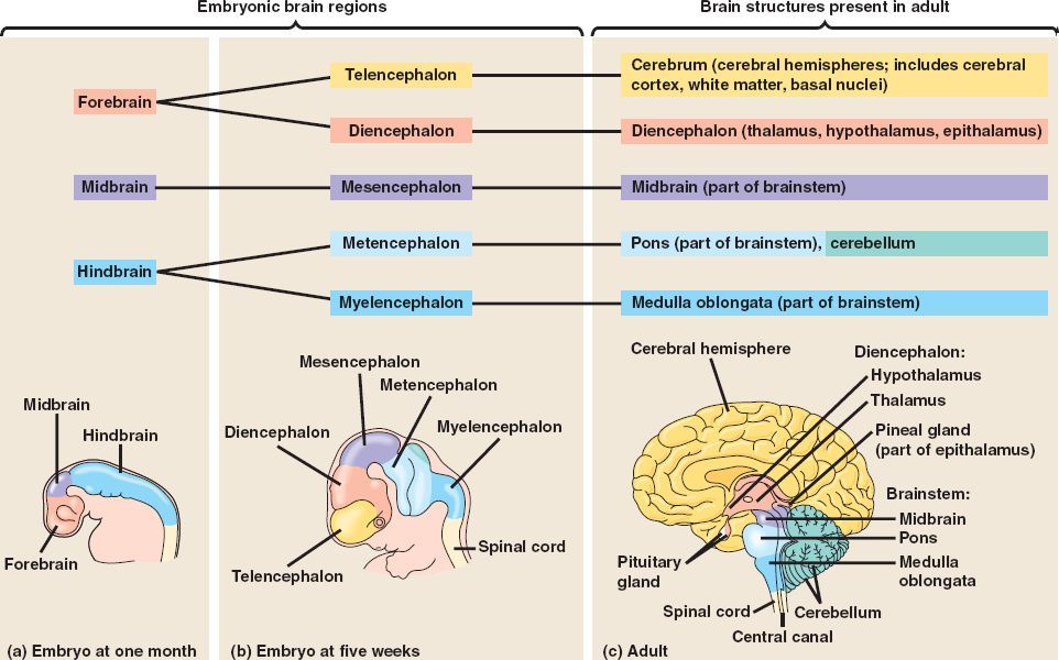
Gaither points out that a fetus can begin to hear during the late second trimester, and a sleep pattern emerges as the brainwaves from the developing hypothalamus become more mature.
By the end of the second trimester, Gaither says the fetal brain looks structurally much like the adult brain with the brain stem almost entirely developed.
The third trimester is full of rapid growth. In fact, as your baby continues to grow, so does the brain. “All the convoluted surfaces of the brain materialize, and the halves (right brain and left brain) will separate,” explains Gaither.
The most notable part of the brain during this final trimester is the cerebellum — hence, the kicking, punching, wiggling, stretching, and all of the other movements your baby is performing.
Share on PinterestIllustration by Alyssa Kiefer
While it may feel like you have control over nothing for the next 9 months, you do have a say in the foods you eat. Healthy brain development starts before pregnancy.
According to the Centers for Disease Control and Prevention, a healthy diet that includes folic acid, both from foods and dietary supplements, can promote a healthy nervous system.
“There are a number of defects along the baby’s brain and spinal cord that can occur when there is an abnormality occurring within the first weeks of brain development,” says Gaither. This may include anencephaly or spina bifida.
Gaither says two supplements in particular are involved with fetal brain development:
Folic acidFolic acid (vitamin B9, specifically) supports fetal brain and spinal development. Not only does it play a role forming the neural tube, but Gaither says it’s also involved in the production of DNA and neurotransmitters, and it’s important for the production of energy and red blood cells.
Gaither recommends taking at least 400 to 600 micrograms of folic acid daily while you’re trying to conceive, and then continue with 400 micrograms daily during pregnancy.
“If you’ve had a child with a neural tube defect, then 4 grams daily in the preconceptual period is advised,” says Gaither.
Foods rich in folate/folic acid include dark green leafy vegetables, flaxseed, and whole grains.
Omega-3 fatty acids
Also important for fetal brain development are omega-3 fatty acids. “The brain has a high fat content, and the omegas are helpful in the deposition of the fat in not only the brain, but the eyes as well,” explains Gaither.
Omegas are also helpful in the neural synapse development or nerve connections to each other.
Foods rich in omega 3-fatty acids include salmon, walnuts, and avocados.
Fetal brain development starts before you may even realize you’re pregnant. That’s why it’s important to start on a prenatal vitamin that contains folic acid right away. If you’re not pregnant, but thinking about having a baby, add a prenatal vitamin to your daily routine.
The brain begins to form early in the first trimester and continues until you give birth.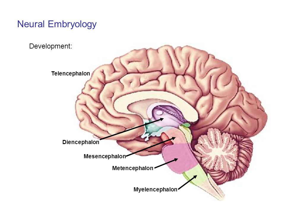 During pregnancy, fetal brain development will be responsible for certain actions like breathing, kicking, and the heartbeat.
Talk to your doctor if you have any questions about your pregnancy, fetal brain development, or how to nurture the baby’s developing brain.
When Does a Fetus Develop a Brain?
Pregnancy is an exciting time full of rapid change and development for both you and your baby. While the growth happening on the outside is clear to everyone (hello, growing belly!), it’s the development we can’t see that is truly fascinating.
Your fetus will begin the process of developing a brain around week 5, but it isn’t until week 6 or 7 when the neural tube closes and the brain separates into three parts, that the real fun begins.
Around week 5, your baby’s brain, spinal cord, and heart begin to develop. Your baby’s brain is part of the central nervous system, which also houses the spinal cord. There are three key components of a baby’s brain to consider.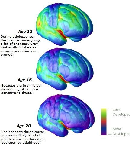 These include:
- Cerebrum: Thinking, remembering, and feeling occurs in this part of the brain.
- Cerebellum: This part of the brain is responsible for motor control, which allows the baby to move their arms and legs, among other things.
- Brain stem: Keeping the body alive is the primary role of the brain stem. This includes breathing, heartbeat, and blood pressure.
The first trimester is a time of rapid development and separation of the various parts of the brain, according to Kecia Gaither, MD, MPH, double board certified in obstetrics and gynecology and maternal-fetal medicine, and director of perinatal services at NYC Health + Hospitals/Lincoln.
Within 4 weeks, the rudimentary structure known as the neural plate develops, which Gaither says is considered the precursor to the nervous system. “This plate elongates and folds on itself forming the neural tube — the cephalad portion of the tube becomes the brain, while the caudal portion elongates to eventually become the spinal cord,” she explains.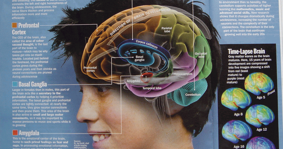
The neural tube continues to grow, but around week 6 or 7, Gaither says it closes, and the cephalad portion (aka the rudimentary brain) separates into three distinct parts: front brain, midbrain, and hindbrain.
It’s also during this time that neurons and synapses (connections) begin to develop in the spinal cord. These early connections allow the fetus to make its first movements.
During the second trimester, Gaither says the brain begins to take command of bodily functions. This includes specific movements that come from the hindbrain, and more specifically, the cerebellum.
One of the first notable developments, sucking and swallowing, are detectable around 16 weeks. Fast-forward to 21 weeks, and Gaither says baby can swallow amniotic fluid.
It’s also during the second trimester that breathing movements begin as directed by the developing central nervous system. Experts call this “practice breathing” since the brain (and more specifically, the brain stem) is directing the diaphragm and chest muscles to contract.
And don’t be surprised if you feel some kicking during this trimester. Remember the cerebellum or the part of the brain responsible for motor control? Well, its directing the baby’s movements, including kicking and stretching.
Gaither points out that a fetus can begin to hear during the late second trimester, and a sleep pattern emerges as the brainwaves from the developing hypothalamus become more mature.
By the end of the second trimester, Gaither says the fetal brain looks structurally much like the adult brain with the brain stem almost entirely developed.
The third trimester is full of rapid growth. In fact, as your baby continues to grow, so does the brain. “All the convoluted surfaces of the brain materialize, and the halves (right brain and left brain) will separate,” explains Gaither.
The most notable part of the brain during this final trimester is the cerebellum — hence, the kicking, punching, wiggling, stretching, and all of the other movements your baby is performing.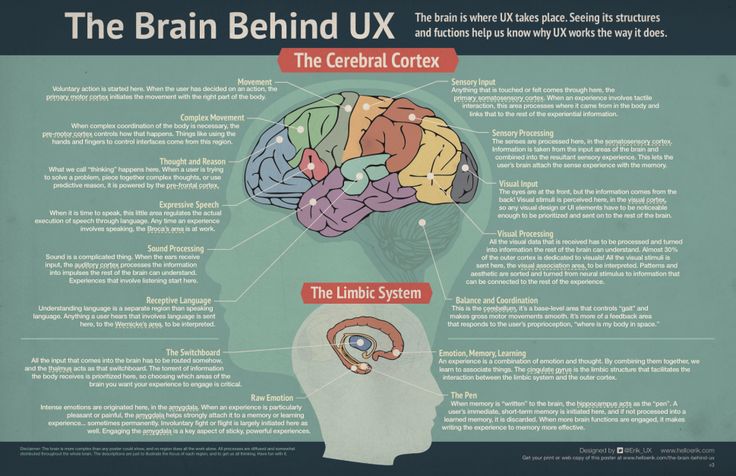
Share on PinterestIllustration by Alyssa Kiefer
While it may feel like you have control over nothing for the next 9 months, you do have a say in the foods you eat. Healthy brain development starts before pregnancy.
According to the Centers for Disease Control and Prevention, a healthy diet that includes folic acid, both from foods and dietary supplements, can promote a healthy nervous system.
“There are a number of defects along the baby’s brain and spinal cord that can occur when there is an abnormality occurring within the first weeks of brain development,” says Gaither. This may include anencephaly or spina bifida.
Gaither says two supplements in particular are involved with fetal brain development:
Folic acidFolic acid (vitamin B9, specifically) supports fetal brain and spinal development. Not only does it play a role forming the neural tube, but Gaither says it’s also involved in the production of DNA and neurotransmitters, and it’s important for the production of energy and red blood cells.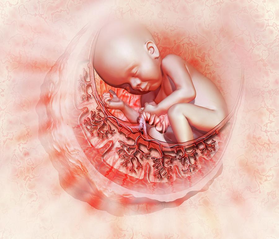
Gaither recommends taking at least 400 to 600 micrograms of folic acid daily while you’re trying to conceive, and then continue with 400 micrograms daily during pregnancy.
“If you’ve had a child with a neural tube defect, then 4 grams daily in the preconceptual period is advised,” says Gaither.
Foods rich in folate/folic acid include dark green leafy vegetables, flaxseed, and whole grains.
Omega-3 fatty acids
Also important for fetal brain development are omega-3 fatty acids. “The brain has a high fat content, and the omegas are helpful in the deposition of the fat in not only the brain, but the eyes as well,” explains Gaither.
Omegas are also helpful in the neural synapse development or nerve connections to each other.
Foods rich in omega 3-fatty acids include salmon, walnuts, and avocados.
Fetal brain development starts before you may even realize you’re pregnant. That’s why it’s important to start on a prenatal vitamin that contains folic acid right away.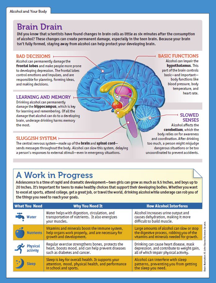 If you’re not pregnant, but thinking about having a baby, add a prenatal vitamin to your daily routine.
The brain begins to form early in the first trimester and continues until you give birth. During pregnancy, fetal brain development will be responsible for certain actions like breathing, kicking, and the heartbeat.
Talk to your doctor if you have any questions about your pregnancy, fetal brain development, or how to nurture the baby’s developing brain.
Scientists have discovered that the uterus affects cognitive abilities - HEROINE
There are more and more studies and experiments that show that our thinking depends not only on the brain, but on the whole body as a whole. And a University of Arizona study has found a new link between the uterus and women's cognitive abilities.
It was previously known that hormones produced in the reproductive system affect the brain. For example, a lack of estrogen can lower magnesium levels in the brain, which in turn can lower serotonin levels and lead to mood swings and even depression.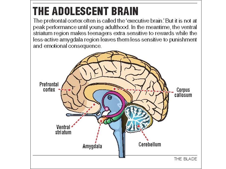
But most of the research has focused on the connection between the ovary and the brain, and very few scientists have been interested in the uterus outside of reproduction. Moreover, some endocrinology textbooks describe the non-pregnant uterus as "dormant" and "useless". But university researchers Heather Bemonte-Nelson and Stephanie Kobele pointed out that evolution could not allow one organ or system of the body to perform only one single function.
To find out what additional functions the uterus might have, Bimonte-Nelson and Kobele divided laboratory rats into four groups: some had their ovaries removed, some had their uterus removed, some had both removed, and the fourth group was not operated on. . Six weeks later, they trained the rats to navigate a maze and tested how well they could find specific platforms.
We believe there is an associated uterine-ovarian-brain triad that may affect brain functions such as memory.
Rats that had only their uterus removed looked for platforms where there were none. Rats from other groups did not experience a similar problem - even those who had their ovaries and uterus removed. The researchers also found that these rats had different levels of hormones in their bloodstream than those that did not receive surgery.
The ovaries of rats that survived hysterectomy remained the same as those of other rats, suggesting that it was the removal of the uterus itself that led to these differences.
These data support findings from a previous neurodegenerative disease study showing that women who had a hysterectomy had a higher risk of developing dementia.
Surgical removal of the uterus alone may impair some types of memory in the short term, two months after surgery. These results show the unique negative effect of hysterectomy alone on memory and indicate that the uterus is part of a system that communicates with the brain for cognitive development.
Operations performed on rats were intentionally performed in the same manner as those performed on humans, so the study may have implications for people considering hysterectomy.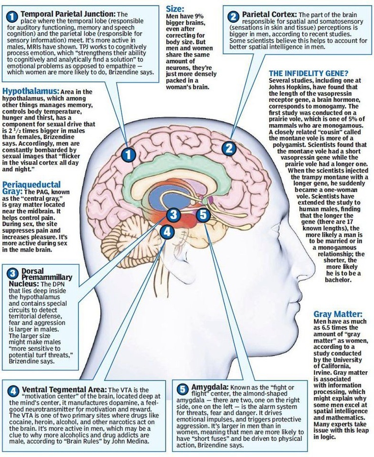
We hope that this study will inspire others to explore the complexity of women's health throughout life.
According to Beamonte-Nelson, these results could be a starting point from which we can begin to ask more questions about how our reproductive system affects the brain outside of conception.
Add to favorites
Share
Related articles:
when women stop carrying and giving birth to children
Elizabeth Console news editor
Researchers believe that in the future, humanity will begin to develop an embryo outside the uterus: it will be able to go through the process from fertilization to birth without human intervention. Hi-Tech looks into how childbirth and pregnancy will change in the future, and what will happen if people stop using their bodies to raise a child.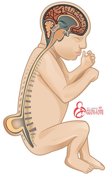
Read "High Tech" in
First an incubator, and then a biobag and an artificial uterus - this is how technologies that preserve the fetus and help it develop evolve.
How did the idea come about not to bear children, but to raise them separately from the mother's body?
Obstetrician Stéphane Tarnier came up with the first idea to create a human incubator in 1880: outwardly, the design resembled a wooden box, in which there was a special cell for storing containers with warm water. They called him a kuvez. A simple device has reduced infant mortality by almost half.
After 80 years, more serious experiments began in this area. Biologists wanted to replicate the functions of the human uterus, which bears a fetus. For this, embryos of rabbits, lambs and goats were used. The researchers decided that it would be cheaper to transport valuable biomaterial from one continent to another.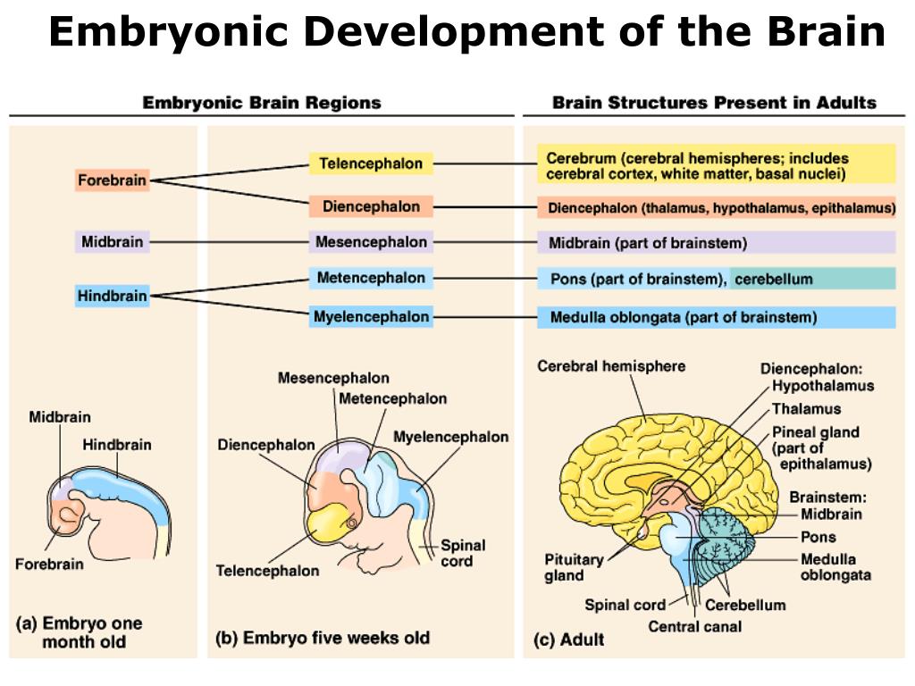
Fertilized sheep eggs were implanted in female rabbits who were sent from England to South Africa at a cost of only $8 per "passenger". On the spot, the embryos underwent another transplant - now into sheep's wombs. A few months later, several lambs were born.
But the idea did not end with success - the fruits died from infection, system failures and many other factors.
How do modern incubators work?
Modern obstetricians actively use incubators; not a single obstetric facility can do without them. Such incubators can monitor the parameters of the body, track even minor changes in the child's condition and temperature around the clock.
Babies can stay in the incubator for up to three to four months. The device simulates conditions that are as similar as possible to those in utero. The main thing is to create the optimal temperature to maintain heat exchange. Do not overheat or overcool the child.
What is an artificial womb and a biobag?
Whatever the attitude towards pregnancy and childbirth, the advent of artificial uterus technology, in other words, ectogenesis, can completely change it. This technology promises many medical benefits: women who have pregnancy complications will be able to transfer the fetus to an artificial womb and reduce the risks to themselves and the baby. And premature embryos will be able to continue their development in an artificial uterus and be born on time.
Researchers have created a biobag and an artificial womb to continue the life of the fetus outside the womb. Both devices work in a similar way.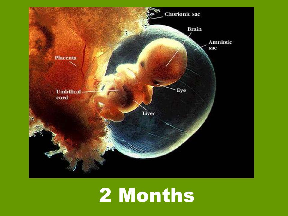 Usually this is a prototype of an artificial uterus, which was used as an ectopic device, where the life of premature fetuses was supported.
In 2017, US researchers published a paper claiming to have successfully tested an artificial womb. She raised premature lambs for four weeks. By design, this is a plastic bag that is filled with an analogue of amniotic fluid, in other words, amniotic fluid. The fetus receives oxygen and other necessary substances through the umbilical cord.
Scientists transferred premature lambs that were 15-17 weeks of development into the system. Normal pregnancy in sheep lasts 21 weeks.
Are there similar studies in this area?
Yes, scientists from Israel have advanced the furthest in this direction: in 2021, they raised mouse embryos - with a working heart and brain - from five-day-old embryos consisting of only 250 cells.
The researchers grew mouse embryos in glass vials filled with nutrient fluid. The composition of this liquid included blood serum taken from the human umbilical cord. The glass vials were constantly shaken to prevent the embryos from sticking to the walls and becoming deformed. The ventilation apparatus supplied the embryos with an oxygen mixture under pressure. Before, scientists have never been able to grow embryos to such a state without a living uterus.
Researchers are now considering how to conduct a similar experiment with a human embryo.
In 2019, scientists from the Netherlands said they would need another 10 years to create the first artificial womb.
What is the problem with artificial womb and biobag?
It is not yet possible to state that an artificial uterus can be used for premature babies. One of the limitations is the level of brain formation.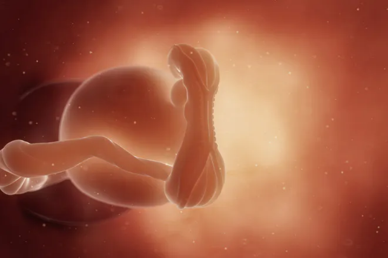 Early experiments with lambs assumed a higher level of brain development, so in theory it would be much more difficult to create an artificial womb for a child than for an animal. Also, the lamb and the child are of different sizes, so the device will not be universal.
What will artificial rearing of children lead to?
Improvements in the biobag and artificial womb will make it possible to grow embryos from the earliest stages. This will make the natural process of pregnancy unnecessary.
Scientists say that this format may be even safer for the baby: for example, the contact of the fetus with alcohol and drugs that a pregnant woman can take is excluded. The need for surrogate motherhood will also disappear.
Canadian-Russian programmer Vitalik Buterin, co-founder of the Ethereum crypto project, proposed to save women from the “heavy burden of pregnancy” with the help of synthetic wombs.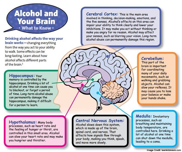 He cited a 2018 Vox article that reports a marked decline in women's incomes after having their first child:
Disparities in economic success between men and women are far larger once marriage+children enter the picture. Synthetic wombs would remove the high burden of pregnancy, significantly reducing the inequality.https://t.co/Zpin8tTlR6
— vitalik.eth (@VitalikButerin) January 18, 2022
It is not yet known how such a gestation procedure will affect the fetus, perhaps a certain cocktail of hormones, sounds from the outside, touching the stomach form a healthy child, and inside the artificial uterus he will lose it. Based on experimental animals (for example, lambs), it is difficult to track the factors that critically affect the future person.
Read more
Hydrogen-fueled hypersonic aircraft develops speeds up to Mach 12.