Body of uterus function
uterus | Definition, Function, & Anatomy
uterus
See all media
- Related Topics:
- fallopian tube cervix puerperium bicornate uterus duplex uterus
See all related content →
uterus, also called womb, an inverted pear-shaped muscular organ of the female reproductive system, located between the bladder and the rectum. It functions to nourish and house a fertilized egg until the fetus, or offspring, is ready to be delivered.
The uterus has four major regions: the fundus is the broad curved upper area in which the fallopian tubes connect to the uterus; the body, the main part of the uterus, starts directly below the level of the fallopian tubes and continues downward until the uterine walls and cavity begin to narrow; the isthmus is the lower, narrow neck region; and the lowest section, the cervix, extends downward from the isthmus until it opens into the vagina. The uterus is 6 to 8 cm (2.4 to 3.1 inches) long; its wall thickness is approximately 2 to 3 cm (0.8 to 1.2 inches). The width of the organ varies; it is generally about 6 cm wide at the fundus and only half this distance at the isthmus. The uterine cavity opens into the vaginal cavity, and the two make up what is commonly known as the birth canal.
Britannica Quiz
Human Body: Fact or Fiction?
How deep is your body of knowledge about the inner workings of humans? Test it with this quiz.
Lining the uterine cavity is a moist mucous membrane known as the endometrium. The lining changes in thickness during the menstrual cycle, being thickest during the period of egg release from the ovaries (see ovulation). If the egg is fertilized, it attaches to the thick endometrial wall of the uterus and begins developing. If the egg is unfertilized, the endometrial wall sheds its outer layer of cells; the egg and excess tissue are then passed from the body during menstrual bleeding.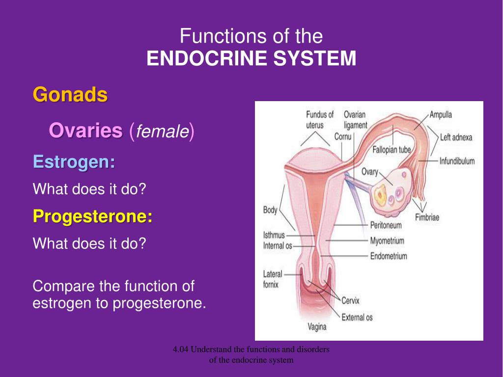 The endometrium also produces secretions that help keep both the egg and the sperm cells alive. The components of the endometrial fluid include water, iron, potassium, sodium, chloride, glucose (a sugar), and proteins. Glucose is a nutrient to the reproductive cells, while proteins aid with implantation of the fertilized egg. The other constituents provide a suitable environment for the egg and sperm cells.
The uterine wall is made up of three layers of muscle tissue. The muscle fibres run longitudinally, circularly, and obliquely, entwined between connective tissue of blood vessels, elastic fibres, and collagen fibres. This strong muscle wall expands and becomes thinner as a child develops inside the uterus. After birth, the expanded uterus returns to its normal size in about six to eight weeks; its dimensions, however, are about 1 cm (0.4 inch) larger in all directions than before childbearing. The uterus is also slightly heavier and the uterine cavity remains larger.
The uterus of a female child is small until puberty, when it rapidly grows to its adult size and shape. After menopause, when the female is no longer capable of having children, the uterus becomes smaller, more fibrous, and paler. Some afflictions that may affect the uterus include infections; benign and malignant tumours; malformations, such as a double uterus; and prolapse, in which part of the uterus becomes displaced and protrudes from the vaginal opening. Uterus transplantation, in which a uterus from a healthy female is transplanted into the affected woman, has been considered a potential form of treatment in extreme cases of uterine disease or absence of the uterus; the first birth of a healthy infant to a uterus transplant recipient occurred in 2014.
The Editors of Encyclopaedia Britannica This article was most recently revised and updated by Kara Rogers.
fallopian tube | Anatomy & Function
uterus
See all media
- Key People:
- Gabriel Fallopius
- Related Topics:
- Rubin’s test ampulla tubae uterinae infundibulum fimbria of the fallopian tube isthmus of the fallopian tube
See all related content →
fallopian tube, also called oviduct or uterine tube, either of a pair of long narrow ducts located in the human female abdominal cavity that transport male sperm cells to the egg, provide a suitable environment for fertilization, and transport the egg from the ovary, where it is produced, to the central channel (lumen) of the uterus.
Each fallopian tube is 10–13 cm (4–5 inches) long and 0.5–1.2 cm (0.2–0.6 inch) in diameter. The channel of the tube is lined with a layer of mucous membrane that has many folds and papillae—small cone-shaped projections of tissue. Over the mucous membrane are three layers of muscle tissue; the innermost layer has spirally arranged fibres, the middle layer has circular fibres, and the outermost sheath has longitudinal fibres that end in many fingerlike branches (fimbriae) near the ovaries, forming a funnel-shaped depository called the infundibulum. The infundibulum catches and channels the released eggs; it is the wide distal (outermost) portion of each fallopian tube. The endings of the fimbriae extend over the ovary; they contract close to the ovary’s surface during ovulation in order to guide the free egg. Leading from the infundibulum is the long central portion of the fallopian tube called the ampulla. The isthmus is a small region, only about 2 cm (0.8 inch) long, that connects the ampulla and infundibulum to the uterus.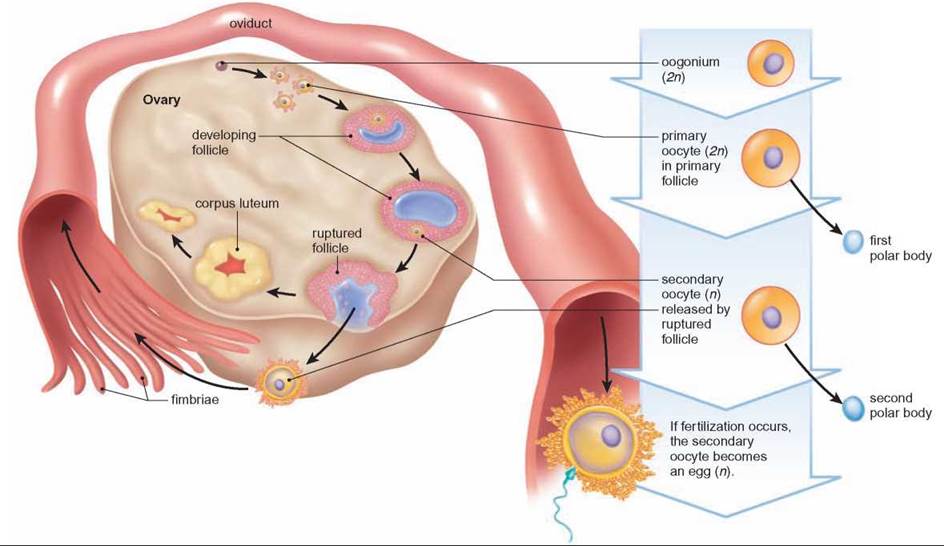 The final region of the fallopian tube, known as the intramural, or uterine, part, is located in the top portion (fundus) of the uterus; it is a narrow tube continuous with the isthmus, and it leads through the thick uterine wall to the uterine cavity, where fertilized eggs normally attach and develop. The channel of the intramural duct is the narrowest part of the fallopian tube.
Britannica Quiz
Human Body: Fact or Fiction?
How deep is your body of knowledge about the inner workings of humans? Test it with this quiz.
The mucous membrane lining the fallopian tube gives off secretions that help to transport the sperm and the egg and to keep them alive. The major constituents of the fluid are calcium, sodium, chloride, glucose (a sugar), proteins, bicarbonates, and lactic acid. The bicarbonates and lactic acid are vital to the sperm’s use of oxygen, and they also help the egg to develop once it is fertilized. Glucose is a nutrient for the egg and sperm, whereas the rest of the chemicals provide an appropriate environment for fertilization to occur.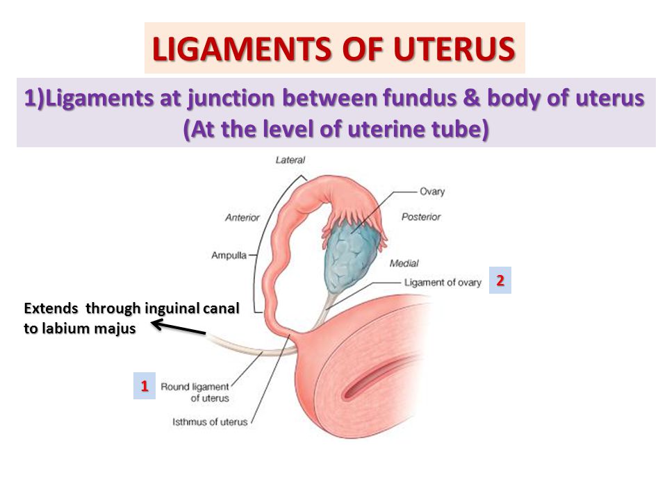
Besides the cells that secrete fluids, the mucous membrane contains cells that have fine hairlike structures called cilia; the cilia help to move the egg and sperm through the fallopian tubes. Sperm deposited in the female reproductive tract usually reach the infundibulum within a few hours. The egg, whether fertilized or not, takes three to four days to reach the uterine cavity. The swaying motions of the cilia and the rhythmic muscular contractions (peristaltic waves) of the fallopian tube’s wall work together while moving the egg or sperm.
Abnormalities of or damage to the fallopian tubes can affect a woman’s fertility. If the tubes are blocked or damaged, for example, sperm are unable to reach the egg, or the fertilized egg may be prevented from traveling to the uterus. Abnormalities in fallopian tube anatomy and function have various causes, including pelvic infection (e.g., pelvic inflammatory disease), endometriosis, and congenital defects.
The Editors of Encyclopaedia Britannica This article was most recently revised and updated by Kara Rogers.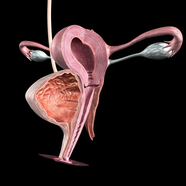
51. Structure and functions of the uterus
The structure and functions of the uterus.
Uterus, uterus , — unpaired muscular pear-shaped organ for the development of the embryo in the case fertilization of the egg, and removal of the fetus during childbirth. In the uterus emit: bottom, reversed up and forward; body - middle part triangular shape and lower - neck. Cervix lower the end is connected to the vagina. Place transition of the uterine body to the cervix narrow and is called isthmus uterus. Part of neck uterus that enters the vagina is called vaginal part, and outside it supravaginal part. B The uterus is divided into two surfaces: anterior - cystic, and back - intestinal, a also two edges: right and left.
Uterine cavity , small, triangular forms. Top from the sides open into it the fallopian tubes.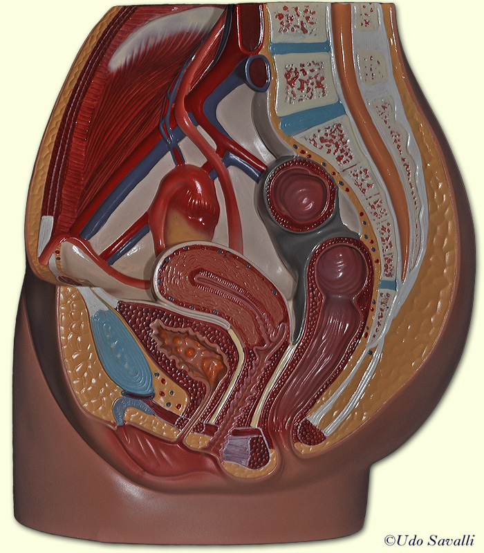 Below the uterine cavity goes to channel necks, which opens into the vagina with a hole uterus. It is limited two lips: front, and back. Neonatal the uterus is cylindrical, and the cervix longer than in adult women. Body uterus develops rapidly the onset of puberty. Topography. The uterus is located in the pelvis between bladder anteriorly and straight gut - behind. The fundus of the uterus is turned forward, its front surface is down and forward, and the back - up and back. The body of the uterus forms with the cervix open anterior bend. Large part of the uterus up to the level of the cervix is in the peritoneal cavity of the small pelvis, so loops of thin or sigmoid colon.
The structure of the uterus. The wall of the uterus consists of three shells: 1) inner mucous, containing many uterine glands; 2) medium, most thick, muscular,), having three layers: longitudinal, circular, longitudinal; 3) outdoor serous, Mucous lining of the uterus in mature women changes cyclically due to menstruation.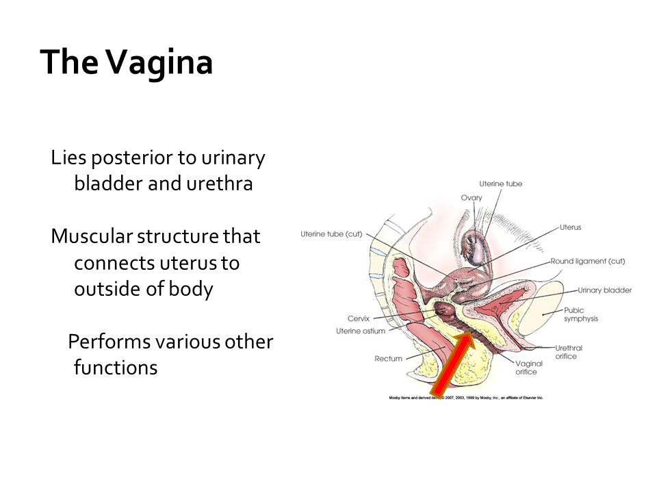 . After the end menstruation mucosa quickly is being restored. Serous membrane - peritoneum - covers the fundus of the uterus, as well as its anterior and posterior surfaces. front she covers the uterus to the level of the cervix and passes to the bladder vesicouterine deepening. Rear peritoneum lines the posterior surface uterus and posterior vaginal fornix and passes nor the rectum, forming recto-uterine deepening. On the edges the peritoneum passes to the pelvic wall, forming wide uterine ligament, between the leaves of which are periuterine fiber, and fallopian tube, uterine artery, powerful uterine venous plexus and nervous plexus. From the upper corners of the uterus go to the deep inguinal ring round bundles uterus. Through the inguinal channel they reach the symphysis, where and are running out.
The size of the uterus varies depending on on the age of the woman, the number of births and pregnancies.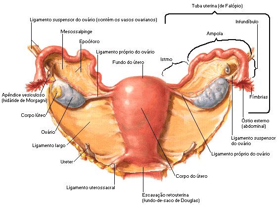 So, in a nulliparous women its length is 7-8 cm, width - 5 cm, weight does not exceed 50 g. Thanks to structural features, during pregnancy, the uterus is able to stretch up to 32 cm long and up to 20 cm wide.
Uterine menstruation occurs in the uterus cycle - a cycle of changes in the endometrium.
1 phase - desquamation. There is a rejection functional layer of the endometrium due to a sharp spasm of the spiral arteries mucous membranes that have muscle pulp at a level between basal and functional layers of the endometrium. Spasm due to low levels of progesterone and an increase in estrogen levels. Goes enzymatic degradation by proteolytic enzymes secreted leukocytes that migrate to mucosa at the end of the secretion phase. Phase lasts 3-5 days and is clinically expressed secretion of dark blood with mucus, without pieces of tissue from the female genital tract.
Phase 2 - regeneration. Regeneration starts from 2-3 days of the cycle.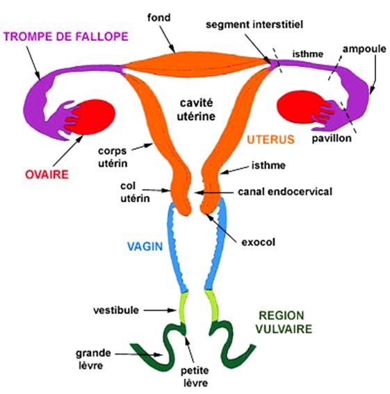 Gradually discharge from the genital tract stops. After rejection, a wound is formed surface that is epithelialized due to the donets tubular glands of the basal layer.
3 phase - proliferation, Complete restoration of the functional layer, glands, blood vessels, nerves. This phase ends by the 12th day of the cycle and lasts 8 days. Phases 2 and 3 are under the influence estrogen.
4 phase - secretions. Under the influence of the growing concentration of progesterone in epithelial accumulates in endometrial glands glycogen, glycoprotein lipids and all necessary for nutrition and implantation blastocysts. Getting ready to receive fertilized egg. When pregnancy occurs decidual transformation occurs endometrium. By the 20-21st day of the cycle, the endometrium ready to receive the blastocyst.
Uterus / Female anatomy - Alexander Nikitin. Tel: +7(926)278-67-02
The uterus is a muscular organ that resembles a bag. The uterus is a fruit-bearing place.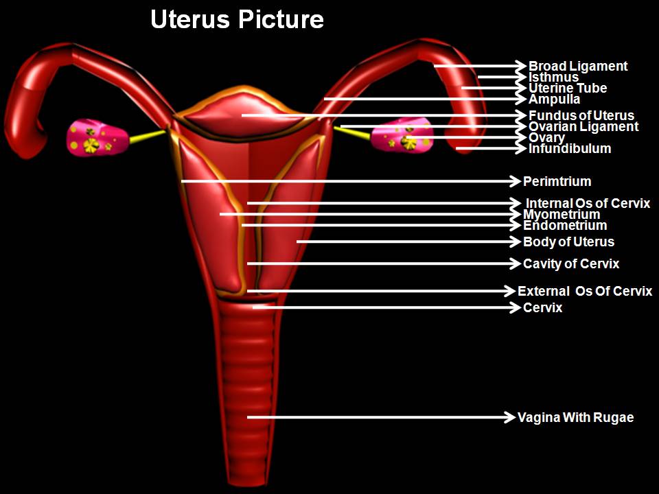 Her task is to bear the fetus and push it out at the right time. The uterus is anatomically divided into the body and the cervix. The cervix has a cylindrical shape, half located in the vaginal cavity. During pregnancy, the cervix is closed and holds the fetus in the uterus. During childbirth, the cervix gradually opens and releases the fetus from the uterus.
The body of the uterus has three layers:
- endometrium
- myometrium
- serous
Endometrium is the inner lining of the uterus. The endometrium is an epithelium (layer of cells) whose task is to receive a fertilized egg. For a better understanding, I compare the endometrium with fertile soil in which a seed (fertilized egg) has fallen. Like a sprout in the ground, pregnancy begins its development in the endometrium.
The endometrium consists of two layers: basal and functional.
The basal layer is the inner layer. From the cells of the basal layer after menstruation, a functional layer is formed.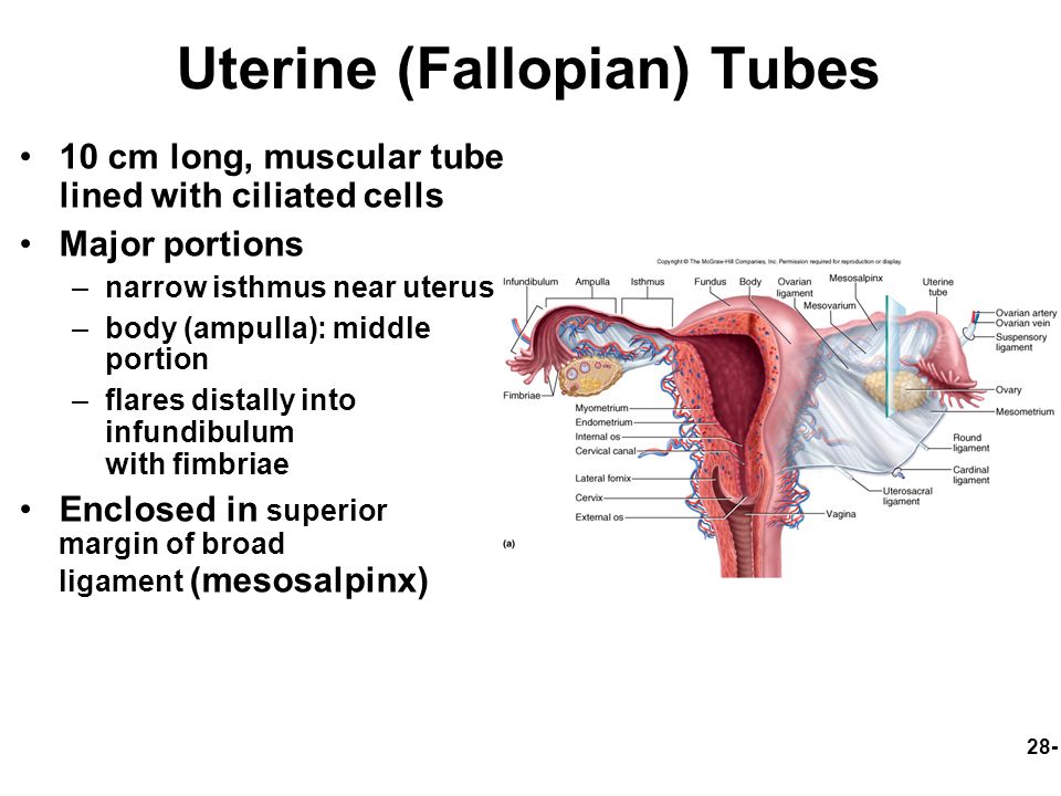
The functional layer is formed in the intervals between menstruation and is rejected during it. In the functional layer of the endometrium, pregnancy begins to develop.
To give an understanding of the function of the endometrium, you need to talk about the woman's menstrual cycle. The menstrual cycle begins with menstruation, but menstruation is the rejection of the endometrium, so it is more logical to start talking about the endometrium from the stage of its new growth, that is, immediately after menstruation.
Normally, menstruation lasts 3-5 days. Then the cells of the basal layer of the endometrium give rise to new cells - the cells of the functional layer. Under the influence of estrogens (ovarian hormones), the functional layer becomes lush, glands form in it. The endometrium prepares for the acceptance (implantation) of a fertilized egg. By the middle of the cycle (day 14, ovulation time), the functional layer of the endometrium is formed.
In the second phase of the menstrual cycle (after ovulation), the endometrial glands begin to function, that is, to secrete substances that contribute to the development of pregnancy in it. This happens under the influence of the hormone of the second phase of the menstrual cycle - progesterone. It is also called the pregnancy hormone. For a better understanding, here I will give a comparison. The endometrium in the first phase of the menstrual cycle can be compared to plowed and loosened soil. In the second phase - with soil that is well fertilized. Endometrial glands under the influence of progesterone saturate the endometrium, make it loose, juicy, nutritious. Such an endometrium creates good conditions for the development of pregnancy.
If pregnancy does not occur, then under hormonal influence, the functional layer of the endometrium is rejected. The bulk of the endometrium falls out through the vagina, a small part - through the fallopian tubes into the pelvic cavity. The process of rejection of the endometrium, accompanied by bleeding, is called menstruation.
Summary. The menstrual cycle is a cyclical change in a woman's body. The menstrual cycle normally lasts 28 days.
